. 2004 Nov 2;101(44):15621-6.
doi: 10.1073/pnas.0401502101. Epub 2004 Oct 25.

AtTPK4, an Arabidopsis tandem-pore K+ channel, poised to control the pollen membrane voltage in a pH- and Ca2+-dependent manner

Affiliations

AtTPK4, an Arabidopsis tandem-pore K+ channel, poised to control the pollen membrane voltage in a pH- and Ca2+-dependent manner

D Becker et al. Proc Natl Acad Sci U S A. .

Abstract

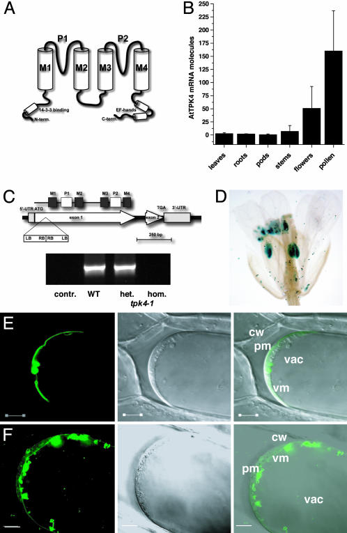
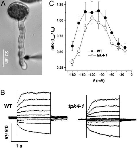
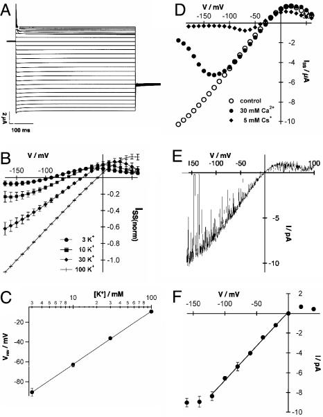
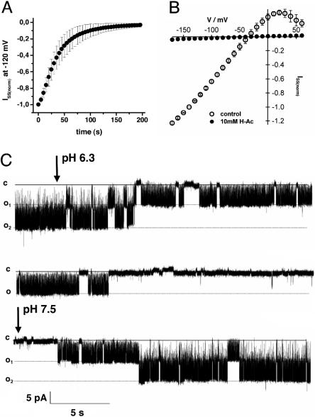
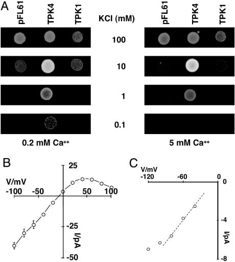
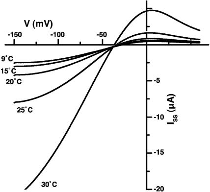
The Arabidopsis tandem-pore K(+) (TPK) channels displaying four transmembrane domains and two pore regions share structural homologies with their animal counterparts of the KCNK family. In contrast to the Shaker-like Arabidopsis channels (six transmembrane domains/one pore region), the functional properties and the biological role of plant TPK channels have not been elucidated yet. Here, we show that AtTPK4 (KCO4) localizes to the plasma membrane and is predominantly expressed in pollen. AtTPK4 (KCO4) resembles the electrical properties of a voltage-independent K(+) channel after expression in Xenopus oocytes and yeast. Hyperpolarizing as well as depolarizing membrane voltages elicited instantaneous K(+) currents, which were blocked by extracellular calcium and cytoplasmic protons. Functional complementation assays using a K(+) transport-deficient yeast confirmed the biophysical and pharmacological properties of the AtTPK4 channel. The features of AtTPK4 point toward a role in potassium homeostasis and membrane voltage control of the growing pollen tube. Thus, AtTPK4 represents a member of plant tandem-pore-K(+) channels, resembling the characteristics of its animal counterparts as well as plant-specific features with respect to modulation of channel activity by acidosis and calcium.


Figures

Fig. 1.
Fig. 1.
Expression and localization of AtTPK4. (A) Proposed topology of the tandem-pore K+ channels. According to the ARAMEMNON database (25), four transmembranes (M1-M4) and two pore regions (P1 and P2) are predicted for Arabidopsis TPKs. (B) Quantitative real-time RT-PCR on cDNA, derived from tissues indicated, revealed pronounced mRNA abundance of AtTPK4 transcripts in Arabidopsis flowers. Among the flower organs tested (not shown), pollen exhibited highest AtTPK4 expression. Data represent mean values of n ≥ 3 ± SD. (C) Identification of a T-DNA insertion mutant for AtTPK4. Schematic view of the T-DNA insertion in tpk4-1 (SALK 000212) and RT-PCR data obtained on pollen mRNA by using AtTPK4 full-length primers. contr., without reverse transcriptase; het., heterozygous plant; hom., homozygous plant. (D) Promoter-GUS staining of transgenic T2 plants revealed AtTPK4 expression in pollen. (E and F) Transient expression of AtTPK4::mgfp4 (E) and AtTPK1::mgfp4 (F) fusion proteins in onion epidermal cells. Cells were plasmolyzed with 0.5 M KNO3 and investigated. Laser-scanning image of GFP fluorescence (Left), transmitted light (Center), and overlay of both images (Right). Cw, cell wall; pm, plasma membrane; vm, vacuolar membrane; vac, vacuole. (Scale bars: 10 μm.)
Fig. 2.
Fig. 2.
Pollen tube membrane voltage recordings. (A) Pollen tubes growing on agar plates were impaled with double-barreled electrodes at or below the emerging tip. (B) Voltage-clamp recordings from pollen tubes of WT plants (Left) and the tpk4-1 mutant (Right). The plasma membrane was clamped from a holding voltage of -100 mV stepwise to test voltages ranging from -180 to 0 mV. (C) Relative contribution of instantaneous currents (Iinst, sampled 30-50 ms after start of the test voltage) to the steady state currents (ISS). Data points represent mean ± SE, n ≥ 7.
Fig. 3.
Fig. 3.
Biophysical properties of AtTPK4 in Xenopus oocytes. (A) Whole-cell currents of AtTPK4-expressing oocytes in response to 10-mV voltage steps from +60 to -200 mV perfused with the standard solution containing 30 mM KCl, 70 mM NaCl, 0.5 mM CaCl2, 0.5 mM MgCl2, and 10 mM Mes/Tris (pH 5.6). (B) I-V relationship of AtTPK4-mediated steady-state currents at varying external K+ concentrations. External solutions were composed of the indicated K+ concentrations, 0.5 mM CaCl2, 0.5 mM MgCl2, and 10 mM Mes/Tris (pH 5.6). NaCl was used to maintain the ionic strength. Currents were normalized to -150 mV in the 100-mM KCl solution. Data points represent mean ± SD, n = 4. Note the deviation from linearity at positive membrane voltages. (C) K+ selectivity of AtTPK4-mediated currents was obtained from the reversal voltages in B, and a slope of 62.5 ± 2.2 mV for a 10-fold change in external K+ concentration was calculated. (D) Voltage-dependent block of AtTPK4 currents by Cs+ and Ca2+ as indicated. The solutions were composed of 30 mM KCl, 1 mM CaCl2, 1 mM MgCl2, and 10 mM Tris/Mes (pH 7.5). (E) I-V relation of single channels during a 1,000-ms voltage ramp from +100 to -160 mV, starting from a holding voltage of 0 mV. Recordings were performed in the cell-attached mode of the patch-clamp technique in the presence of 100 mM KCl and 10 mM Mes/Tris (pH 5.6) in the pipette and bath solution. (F) I-V plot of single channel recordings of AtTPK4. Slope conductances were determined between -120 and 0 mV to be 68.7 ± 4 pS (mean ± SD, n = 4). Solutions and configuration were as in E.
Fig. 5.
Fig. 5.
Acidosis-mediated inhibition of AtTPK4 currents. (A) Time-dependent inhibition of AtTPK4-mediated whole-cell currents in oocytes after cytoplasmic acidification by bath perfusion with 10 mM sodium acetate. Shown are normalized steady-state currents at -120 mV. The bath solution was composed of 30 mM KCl, 1 mM CaCl2, 1.5 mM MgCl2, 10 mM sodiumacetate, and 10 mM Mes/Tris (pH 5.6). (B) The I-V analysis of AtTPK4-mediated steady-state currents in response to bath perfusion with sodium acetate reveals the acidosis-mediated inhibition of AtTPK4 currents. Data points represent mean ± SD, n = 3. Steady-state currents were normalized to -140 mV in the absence of sodium-acetate. Control solution was 30 mM KCl, 1 mM CaCl2, 1.5 mM MgCl2, and 10 mM Mes/Tris (pH 5.6). (C) Single-channel recordings in the inside-out configuration at -100 mV at pH 7.5 and pH 6.3 show the reduction in channel open probability (Po). Bath perfusion with the solution at pH 6.3 starts after the first arrow. Recovery in the bath solution at pH 7.5 is shown after the second arrow. Bath solution was composed of 100 mM KCl and 10 mM Tris/Mes (pH 7.5) or 10 mM Mes/Tris (pH 6.3). The pipette solution contained 100 mM KCl and 10 mM Tris/Mes (pH 7.5).
Fig. 4.
Fig. 4.
Functional analysis of AtTPK4 in yeast. (A) AtTPK4 complements the potassium-dependent growth defect of the Δtrk1trk2tok1 triple deletion mutant (17). Yeast triple deletions transformed with the empty pFL61 plasmid or with the pFL61 plasmid carrying either the AtTPK4 or the AtTPK1 gene, respectively. Although all strains grew perfectly well in 100 mM KCl at low (0.2 mM, Left) and high (5 mM, Right)Ca2+, mutants harboring the empty plasmid or expressing the vacuolar channel AtTPK1 did not grow at lower KCl concentrations. In contrast, AtTPK4-expressing mutants were able to restore yeast growth even at submillimolar K+ concentrations when calcium was low (0.2 mM), whereas, in the presence of high calcium (5 mM), AtTPK4-expressing yeast mutants exhibited a K+- and Ca2+-dependent growth phenotype. (B) Time-averaged electrical currents recorded from the plasmalemma of AtTPK4-expressing yeast cells. Current recordings were performed in the outside-out configuration, and the mean currents of 2.5-s traces, corrected for a 200-pS leak, are plotted vs. the applied membrane voltage. Data represent mean ± SEM (n = 3; see Fig. 9). (C) From the I-V plot, a maximum open channel conductance of 77 pS could be determined between -40 mV and -80 mV (dotted linear regression line). Solutions in B and C were as indicated in Methods.
Fig. 6.
Fig. 6.
AtTPK4 activation by heat. Whole-cell currents of AtTPK4-expressing oocytes recorded at different temperatures. The I-V curves were obtained by a -150-mV voltage pulse for 100 ms followed by a 700-ms voltage ramp from -150 to +60 mV.

References

    1. Czempinski, K., Zimmermann, S., Ehrhardt, T. & Mueller-Roeber, B. (1997) EMBO J. 16, 2565-2575. - PMC - PubMed
    1. Czempinski, K., Zimmermann, S., Ehrhardt, T. & Mueller-Roeber, B. (1997) EMBO J. 16, 6896. - PMC - PubMed
    1. Very, A. A. & Sentenac, H. (2002) Trends Plant Sci. 7, 168-175. - PubMed
    1. Derst, C. & Karschin, A. (1998) J. Exp. Biol. 201, 2791-2799. - PubMed
    1. Goldstein, S. A., Bockenhauer, D., O'Kelly, I. & Zilberberg, N. (2001) Nat. Rev. Neurosci. 2, 175-184. - PubMed

Publication types

MeSH terms

Associated data