. 2004 Nov 9;101(45):16004-9.
doi: 10.1073/pnas.0407192101. Epub 2004 Oct 25.

Antigen-independent memory CD8 T cells do not develop during chronic viral infection

Affiliations

Antigen-independent memory CD8 T cells do not develop during chronic viral infection

E John Wherry et al. Proc Natl Acad Sci U S A. .

Abstract

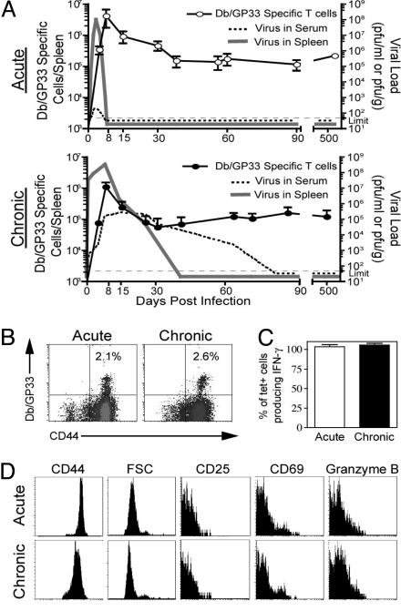
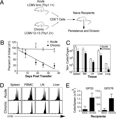
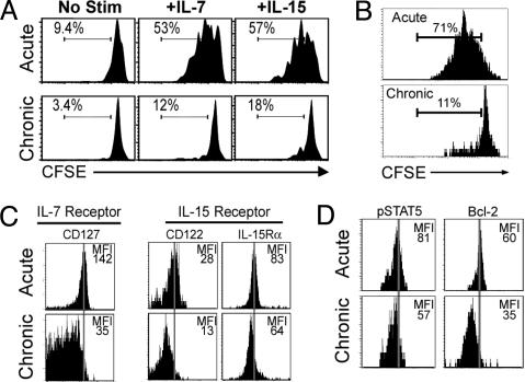
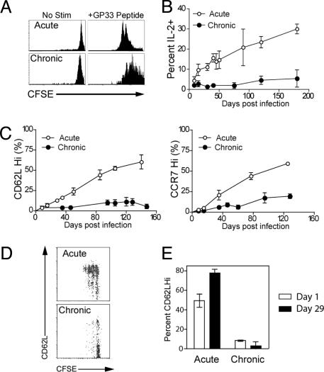
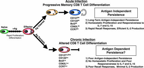
Memory T cells can persist for extended periods in the absence of antigen, and long-term T cell immunity is often seen after acute infections. Paradoxically, there have been observations suggesting that T cell memory may be antigen-dependent during chronic infections. To elucidate the underlying mechanisms we have compared memory CD8 T cell differentiation during an acute versus chronic infection by using the mouse model of infection with lymphocytic choriomeningitis virus. We found that during a chronic infection virus-specific CD8 T cells failed to acquire the cardinal memory T cell property of long-term antigen-independent persistence. These chronically stimulated CD8 T cells were unable to undergo homeostatic proliferation, responded poorly to IL-7 and IL-15, and expressed reduced levels of the IL-7 and IL-15 receptors, thus providing a possible mechanism for the inability of these cells to persist long term in the absence of antigen. In striking contrast, virus-specific memory CD8 T cells that developed after an acute lymphocytic choriomeningitis virus infection could persist without antigen, were capable of self-renewal because of homeostatic proliferation, responded efficiently to IL-7 and IL-15, and expressed high levels of receptors for these two cytokines. Thus, memory CD8 T cells generated after acute infections are likely to have a competitive advantage over CD8 T cells that develop during chronic infections. These findings raise concerns about using vaccines that may persist and also suggest that there may be limitations and challenges in designing effective immunological interventions for the treatment of chronic infections and tumors.


Figures

Fig. 1.
Fig. 1.
Characterization of acute memory and chronic memory CD8 T cells. (A) The kinetics of viral infection (dashed line, serum; solid line, spleen) and Db/GP33-specific CD8 T cell responses after LCMV Armstrong and LCMV clone 13 infection. (B) Db/GP33 tetramer staining of CD8 T cells from the spleen after acute or chronic infection (days 500 and 825 p.i., respectively; similar results were observed between days 30 and 800). (C) Intracellular cytokine staining for IFN-γ after 5 h of GP33 peptide stimulation at day 180 acute or chronic infection. Data are the percent of Db/GP33+ CD8 T cells that produced IFN-γ. Similar results were observed at other time points after day 100 p.i. (data not shown). (D) The phenotype of LCMV Db/GP33+ CD8 T cells day 140 after acute or chronic LCMV infection. Plots were gated on Db/GP33+ CD8 T cells. Similar results were observed at multiple time points after day 100 of chronic infection. Similar results were observed for Db/GP276+ CD8 T cells (data not shown).
Fig. 2.
Fig. 2.
Chronic memory CD8 T cells do not persist in the absence of antigen. (A) Experimental design for adoptive transfers. Acute memory (after day 30 p.i.) and chronic memory (after day 120 p.i.) CD8 T cells were purified, mixed to contain equal numbers of Db/GP33+ CD8 T cells, and transferred to naïve mice. Acute memory (Thy1.1+) and chronic memory CD8 T cells (Thy1.2+) were tracked independently in the same recipient. Similar results were obtained when acute and chronic memory CD8 T cells were transferred into separate groups of naïve mice (data not shown). (B) Recipients were bled longitudinally, and the frequency of Db/GP33+ acute and chronic memory CD8 T cells in the PBMC is expressed as a percentage of their initial frequency ≈36 h posttransfer. Data represent three to eight mice per group in five independent experiments, and the difference between acute and chronic memory CD8 T cells is significant (P < 0.05) on days 18, 25, and 35 posttransfer. We have fit a straight line to the curve, but it is also possible that this T cell loss is biphasic with some of the chronic memory CD8 T cells waning faster than others. (C) The number of transferred Db/GP33+ acute and chronic memory CD8 T cells was quantified on day 35 in the indicated tissues by tetramer staining (n = 2 mice per group and is representative of five independent experiments). Dotted line indicates the limit of detection. (D) Acute memory (day 150) and chronic memory (day 120) CD8 T cells were labeled with CFSE before adoptive transfer, and homeostatic proliferation was assessed 35 days later. Histograms are gated on acute memory (Db/GP33+Thy1.1+) or chronic memory CD8 T cells (Db/GP33+Thy1.2+) isolated from the indicated tissues of naïve recipient mice. Data are representative of five independent experiments with two to three mice each. For BD similar results were observed for Db/GP276+ CD8 T cells (data not shown). (E) Chronic memory CD8 T cells (day 150) were purified and adoptively transferred into naïve or chronically infected mice (≈130 days p.i.). On day 56 significant differences were observed between the number of donor Db/GP33+ and Db/GP276+ chronic memory CD8 T cells in the spleen determined by tetramer staining (GP33, P = 0.02; GP276, P = 0.01). Data are representative of two independent experiments with two to three mice per group.
Fig. 3.
Fig. 3.
Chronic memory CD8 T cells respond poorly to IL-7 and IL-15. (A) Acute memory (day 180) or chronic memory (day 240) CD8 T cells were CFSE-labeled and tested for responsiveness to IL-7 and IL-15 in vitro (5 ng/ml each). Division was assessed after 60 h. Numbers indicate the percent of D/bGP33+ CD8 T cells that had divided at least once. (B) Acute memory (day 140) and chronic memory (day 115) CD8 T cells were purified and labeled with CFSE, and equal numbers of Db/GP33+ cells were cotransferred into naïve recipients. IL-15 was injected i.p. daily (5 μg per injection), and division of Db/GP33+ CD8 T cells was analyzed on day 5. (C) IL-7 and IL-15 receptors on acute memory (day 150) and chronic memory (day 120) CD8 T cells. Histograms are gated on Db/GP33+ CD8 T cells. (D) Staining for intracellular pSTAT5 and Bcl-2 expression in the same populations as in C. For all panels similar results were observed for the LCMV Db/GP276+ CD8 T cell population (data not shown). The lower expression of cytokine receptors (except CD132 and IL-15Ra) and intracellular molecules by chronic memory Db/GP33+ CD8 T cells was statistically significant (Fig. 6).
Fig. 4.
Fig. 4.
Impaired memory CD8 T cell differentiation during chronic infection. (A) CFSE-labeled acute memory (day 240) or chronic memory (day 240) CD8 T cells were stimulated in vitro with GP33 peptide, and division of Db/GP33+ CD8 T cells was assessed after 60 h. (B) The ability to produce IL-2 after in vitro stimulation with GP33 peptide was assessed at the indicated times by intracellular cytokine staining. Percentage indicates the percent of Db/GP33+ CD8 T cells from the spleen that produced IL-2; n = 2–7 for all points. The difference between acute and chronic infection is significant (P < 0.05) after day 8. (C) CD62L and CCR7 expression by Db/GP33+ CD8 T cells after acute versus chronic infection; n = 2–7 for all points. The difference between acute and chronic infection is significant (P < 0.05) after day 8 for CD62L and day 15 for CCR7. (D) Acute memory (day 100) and chronic memory (days 140–160) CD8 T cells were purified and transferred to naïve mice. Thirty-four days later, division and phenotype of CFSE-labeled, Db/GP33+ CD8 T cells and acute or chronic memory CD8 T cells were analyzed by costaining for CD62L. Results shown are for the spleen. Similar results were observed for lymph nodes, PBMC, and liver (data not shown). (E) CD62L expression on Db/GP33+ acute and chronic memory CD8 T cells in the PBMC after adoptive transfer to naïve recipients. There was a significant increase in the percentage of CD62LHi acute memory CD8 T cells (P < 0.01) after 34 days in naïve adoptive hosts, but no significant change in the percentage of CD62LHi chronic memory. Similar results were observed in the spleen (data not shown). n = two to three per group, and data are representative of four independent experiments. Similar results were observed for CCR7 and CD127 expression (data not shown). For all panels, similar results were observed for the Db/GP276+ CD8 T cells (data not shown).
Fig. 5.
Fig. 5.
Model of CD8 T cell differentiation during acute versus chronic infections. During the first week of infection naïve antigen-specific CD8 T cells undergo antigen-driven proliferation and differentiation into effector CD8 T cells. If antigen is cleared (i.e., acute infection) 5–10% of CD127Hi effector CD8 T cells (14) survive and undergo further antigen-independent differentiation, resulting in the generation of memory CD8 T cells that have acquired three defining memory T cell properties: (i) long-term antigen-independent persistence, (ii) homeostatic proliferation in response to IL-7 and IL-15, and (iii) rapid recall responses including vigorous antigen-driven proliferation, secretion of cytokines (IFN-γ, tumor necrosis factor α, and IL-2), and acquisition of cytotoxicity (, –14) (Upper). In contrast, if antigen persists past the effector stage and a chronic infection ensues (Lower) the antigen-independent phase of memory CD8 T cell differentiation does not occur and the resulting CD8 T cells do not optimally develop the three main memory T cell properties.

Similar articles

Cited by

References

    1. Kaech, S. M., Wherry, E. J. & Ahmed, R. (2002) Nat. Rev. Immunol. 2, 251–262. - PubMed
    1. Grayson, J. M., Zajac, A. J., Altman, J. D. & Ahmed, R. (2000) J. Immunol. 164, 3950–3954. - PubMed
    1. Jameson, S. C. (2002) Nat. Rev. Immunol. 2, 547–556. - PubMed
    1. Becker, T. C., Wherry, E. J., Boone, D., Murali-Krishna, K., Antia, R., Ma, A. & Ahmed, R. (2002) J. Exp. Med. 195, 1541–1548. - PMC - PubMed
    1. Goldrath, A. W., Sivakumar, P. V., Glaccum, M., Kennedy, M. K., Bevan, M. J., Benoist, C., Mathis, D. & Butz, E. A. (2002) J. Exp. Med. 195, 1515–1522. - PMC - PubMed

Publication types

MeSH terms