. 2004 Nov 9;101(45):15905-10.
doi: 10.1073/pnas.0403668101. Epub 2004 Oct 25.

EosFP, a fluorescent marker protein with UV-inducible green-to-red fluorescence conversion

Affiliations

EosFP, a fluorescent marker protein with UV-inducible green-to-red fluorescence conversion

Jörg Wiedenmann et al. Proc Natl Acad Sci U S A. .

Abstract

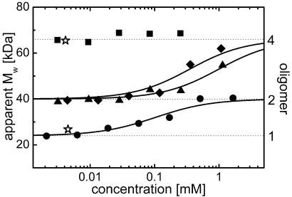
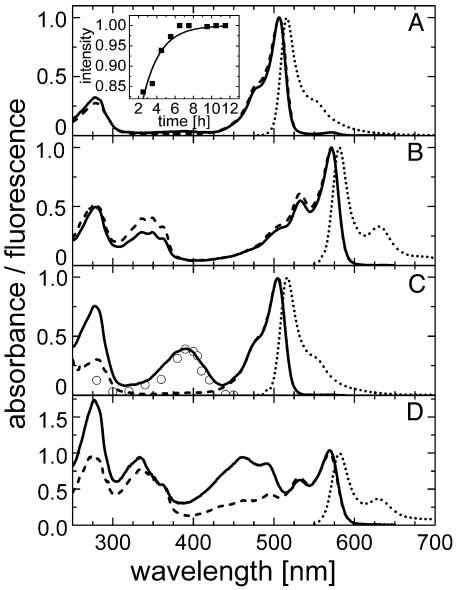
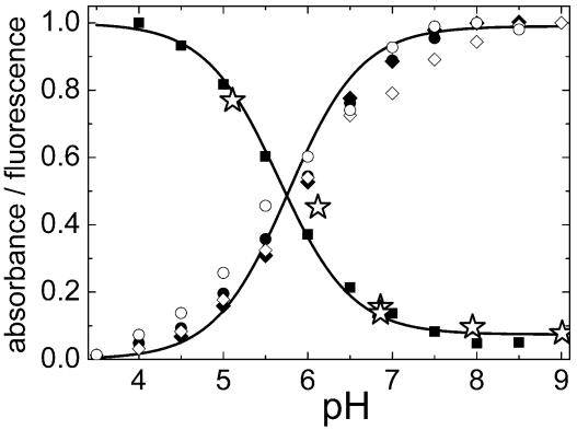
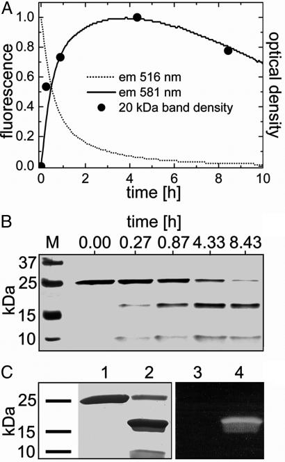
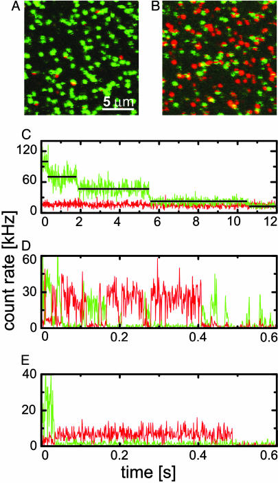
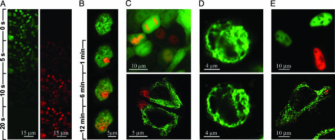
A gene encoding a fluorescent protein from the stony coral Lobophyllia hemprichii has been cloned in Escherichia coli and characterized by biochemical and biophysical methods. The protein, which we named EosFP, emits strong green fluorescence (516 nm) that changes to red (581 nm) upon near-UV irradiation at approximately 390 nm because of a photo-induced modification involving a break in the peptide backbone next to the chromophore. Single-molecule fluorescence spectroscopy shows that the wild type of EosFP is tetrameric, with strong Forster resonance coupling among the individual fluorophores. We succeeded in breaking up the tetramer into AB and AC subunit dimers by introducing the single point mutations V123T and T158H, respectively, and the combination of both mutations yielded functional monomers. Fusion constructs with a variety of proteins were prepared and expressed in human cells, showing that normal biological functions were retained. The possibility to locally change the emission wavelength by focused UV light makes EosFP a superb marker for experiments aimed at tracking the movements of biomolecules within the living cell.


Figures

Fig. 1.
Fig. 1.
Concentration dependence of the apparent MW in size-exclusion chromatography experiments on EosFP and mutants. Fitted binding isotherms, calculated by assuming saturation levels at 40 and 66 kDa, are shown as solid lines. Data for the monomeric EGFP and tetrameric DsRed are included for comparison (⋆). ▪, wild type; ♦, d1EosFP; ▴, d2EosFP; •, mEosFP.
Fig. 2.
Fig. 2.
Spectra of the green and red states of EosFP at pH 7 and pH 5.5. Solid lines, absorbance; dashed lines, excitation; dotted lines, emission spectra. (A and C) Green species at pH 7 (A) and pH 5.5 (C). Excitation (emission) spectra were measured with emission (excitation) set to 520 (490) nm. ○, conversion yields scaled to the absorbance. (Inset) In vitro chromophore maturation at 27°C determined from the absorbance at 506 nm (solid line, exponential fit). (B and D) Red species at pH 7 (B) and pH 5.5 (D). Excitation (emission) spectra were measured with emission (excitation) set to 590 (560) nm.
Fig. 3.
Fig. 3.
Normalized pH dependencies of absorbance, emission, and photoconversion rate. All data follow a Henderson–Hasselbalch relation with pK of 5.8 ± 0.1 (solid lines). Green species: •, absorbance at 506 nm; ▪, absorbance at 390 nm; ○, fluorescence at 516 nm; ⋆, relative green-to-red conversion rate. Red species: ♦, absorbance at 572 nm; ⋄, fluorescence at 581 nm.
Fig. 4.
Fig. 4.
Time dependence of green-to-red photoconversion monitored by a combination of SDS/PAGE and fluorescence. (A) Fluorescence from green (dotted line) and red (solid line) species. •, Densities of the 20-kDa band in B. (B) SDS gel of marker bands (lane M) and EosFP protein after irradiation for a certain time, showing the correlation between the appearance of the 20- and 8-kDa bands and the red fluorescence. (C) Ponceau staining of the green (lane 1) and red (lane 2) form of EosFP after SDS/PAGE separation and transfer to a nitrocellulose membrane. With 366-nm excitation, red fluorescence is observed for the 20-kDa band (lane 4) but not for the 25-kDa band (lane 3).
Fig. 5.
Fig. 5.
Single-molecule spectroscopy of EosFP immobilized on a BSA surface. Confocal scan images were taken with 488-nm excitation (A) and additional 400-nm irradiation (B). (C) Fluorescence emission traces from an individual EosFP tetramer (488-nm excitation) showing sequential bleaching of the four subunits. (D) An individual EosFP tetramer (488-nm excitation plus 400-nm irradiation) showing green-to-red switching and sequential bleaching. (E) mEosFP (488-nm excitation plus 400-nm irradiation) showing single-step green-to-red conversion and bleaching.
Fig. 6.
Fig. 6.
Cellular applications of the green-to-red photoconvertible protein EosFP. (A) The fusion protein mNotch-1-IC–EosFP, localized in the nuclei of stably transfected HEK293 cells, is converted from green to red under the microscope. (B) Tracking of movement of the fusion protein RBP-2N–d2EosFP in the nucleus by localized photoconversion, observed after 0.5-sec irradiating with 400-nm light (1 μW) in the single-molecule setup. (C) HEK293 cells cotransfected with cDNA of EGFP and RBP-2N–d2EosFP. Chromatin association of RBP-2N is clearly visible after photoconversion. (Upper) EGFP highlights the cell morphology. (Lower) HeLa cells expressing cytokeratin 18-d2EosFP, local conversion. (D) HEK293 cell expressing cytokeratin 18-d2EosFP. (Upper) One-photon excitation at 488 nm. (Lower) Improved image quality with two-photon excitation by 808-nm Ti:sapphire laser pulses. (E) Functional expression at 28°C and local photoconversion of mEosFP fusions in HeLa cells. (Upper) Nuclear localization of mNotch-1-IC–mEosFP. (Lower) Mitochondrial localization of mt-mEosFP.

References

    1. Prasher, D. C., Eckenrode, V. K., Ward, W. W., Prendergast, F. G. & Cormier, M. J. (1992) Gene 111, 229–233. - PubMed
    1. Chalfie, M., Tu, Y., Euskirchen, G., Ward, W. W. & Prasher, D. C. (1994) Science 263, 802–805. - PubMed
    1. Tsien, R. Y. (1998) Annu. Rev. Biochem. 67, 509–544. - PubMed
    1. Wiedenmann, J., Röcker, C. & Funke, W. (1999) in Verhandlungen der Gesellschaft für Ökologie, ed. Pfadenhauer, J. (Springer, Heidelberg), Vol. 29, pp. 497–503.
    1. Wiedenmann, J. (1997) in Offenlegungsschrift DE 197 18 640 A1 (German Patent and Trade Mark Office, Munich), pp. 1–18.

Publication types

Associated data