. 2004 Nov 2;101(44):15772-7.
doi: 10.1073/pnas.0401596101. Epub 2004 Oct 25.

Facile isolation and the characterization of human retinal stem cells

Affiliations

Facile isolation and the characterization of human retinal stem cells

Brenda L K Coles et al. Proc Natl Acad Sci U S A. .

Abstract

This study identifies and characterizes retinal stem cells (RSCs) in early postnatal to seventh-decade human eyes. Different subregions of human eyes were dissociated and cultured by using a clonal sphere-forming assay. The stem cells were derived only from the pars plicata and pars plana of the retinal ciliary margin, at a frequency of approximately 1:500. To test for long-term self-renewal, both the sphere assay and monolayer passaging were used. By using the single sphere passaging assay, primary spheres were dissociated and replated, and individual spheres demonstrated 100% self-renewal, with single spheres giving rise to one or more new spheres in each subsequent passage. The clonal retinal spheres were plated under differentiation conditions to assay the differentiation potential of their progeny. The spheres were produced all of the different retinal cell types, demonstrating multipotentiality. Therefore, the human eye contains a small population of cells (approximately equal to 10,000 cells per eye) that have retinal stem-cell characteristics (proliferation, self-renewal, and multipotentiality). To test the in vivo potential of the stem cells and their progeny, we transplanted dissociated human retinal sphere cells, containing both stem cells and progenitors, into the eyes of postnatal day 1 NOD/SCID mice and embryonic chick eyes. The progeny of the RSCs were able to survive, migrate, integrate, and differentiate into the neural retina, especially as photoreceptors. Their facile isolation, integration, and differentiation suggest that human RSCs eventually may be valuable in treating human retinal diseases.


Figures
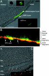
Fig. 3.
Fig. 3.
Transplantation into the mouse eye. (A) The EGFP+ human RSCs and progeny are able to survive and integrate into the neural retina and the RPE. The green fluorescence is primarily located in the photoreceptor inner and outer segments. (Scale bar: 250 μm.) (Insets) The same human RSC sphere (210-μm diameter) derived from an EGFP–lentiviral-labeled stem cell in phase contrast and under GFP fluorescence (GFP+ve). The pigmentation of some of the cells in the sphere masks the fluorescence of the cells, making the ubiquitously green-labeled sphere appear mottled. (B) The EGFP+ human RSCs and progeny (green) are capable of making Rom1+ (red) photoreceptor cells. The long arrow points to some isolated Rom1+ outer segments of the photoreceptor layer double-labeled with EGFP (yellow). The arrowheads indicate EGFP+/Rom1- cells located in the RPE layer. (Scale bar: 50 μm.) (C) Transplanted human RSCs and progeny that differentiated as RPE cells labeled with a human-specific RPE marker (bestrophin). (Inset) A magnified fluorescence image of the bestrophin staining in the RPE layer. The bestrophin+ cells are EGFP-, indicating that the lentivirus has been silenced in these human donor cells. INL, inner nuclear layer; ONL, outer nuclear layer; PRL, photoreceptor layer.
Fig. 1.
Fig. 1.
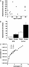
Primary sphere formation and self-renewal. (A) Retinal sphere numbers appear to be grossly similar across postnatal ages in humans when the dissociated cells of the ciliary margin are plated at 20 cells per μl. (B) Ciliary margin cells proliferate in 7 days to form clonal spheres that can arise without the presence of exogenous growth factors, but growth factors significantly increase the number of cells capable of forming a clonal sphere in vitro.(C) The spheres are capable of long-term self-renewal as demonstrated by monolayer passaging of single clonal spheres that arose from a single stem cell. Each line represents the expansion of a RSC from three different donor eyes.
Fig. 2.
Fig. 2.
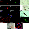
Multipotentiality. The progeny of single spheres formed clonally from human RSCs differentiate to form all of the cell types of the retina when plated on adherent substrates. The nuclei of all cells are stained with the blue dye Hoechst 33328 in B, C, and EG. (AH) These cell types include RPE cells (RPE65+) (A), undifferentiated cells (nestin+) (B), retinal progenitors/amacrine [Pax6+](C), photoreceptor cells (Rho4D2+)(D), retinal ganglion cells (neurofilament-M+) (E), horizontal cell (calbindin+) (F), photoreceptor cells (Rom1+)(G), and bipolar cell (Chx10+)(H). (Scale bars: 20 μm.) (IL) Monolayer culture differentiation. In the presence of 2% FBS and EGF, the cells differentiated into neural cells. Several neuronal markers were observed: syntaxin (I), MAP2 (J), and calbindin (K). (L) These cells were also capable of differentiating along a glial lineage as indicated by vimentin staining when plated in the presence of EGF and 10% FBS. (Magnification: A and B, ×200; CI, ×400.) (M) Upon addition of 10% FBS, the clonal sphere cells spread out of the retinal sphere (remnant of sphere indicated by an arrow), leading to a monolayer culture. (N) Monolayer culture stained with nestin (red).
Fig. 4.
Fig. 4.
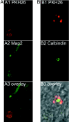
Transplantation into the chick eye. Human cells labeled with the cell membrane dye PKH26 (red) were injected into the vitreous of the eye and collected at different time points. (A) Labeled human cells were injected into the vitreous at embryonic day 3.5 and collected at embryonic day 12. (Top)A human cell incorporated into the ganglion cell layer (GCL) of the retina (red). (Middle) This cell also expressed the RGC marker MAP2 (green). (Bottom) The differential interference contrast overlay. (B) Labeled human cells were injected at embryonic day 4 and collected at embryonic day 10. A human cell (Top, red) expressing the horizontal cell marker, calbindin (Middle, green) was also detected near the ganglion cell layer of the developing chick retina. (Bottom) The differential interference contrast overlay.

References

    1. Tropepe, V., Coles, B. L. K., Chiasson, B. J., Horsford, D. J., Elia, A. J., McInnes, R. R., van der Kooy, D. (2000) Science 287, 2032-2036. - PubMed
    1. Ahmad, I., Tang, L. & Pham, H. (2000) Biochem. Biophy. Res. Commun. 270, 517-521. - PubMed
    1. Yang, P., Seiler, M. J., Aramant, R. B. & Whittemore, S. R. (2002) Exp. Neurol. 177, 326-331. - PubMed
    1. Jensen, A. M. & Raff, M. C. (1997) Dev. Biol. 188, 267-279. - PubMed
    1. Vescovi, A. L., Gritti, A., Galli, R. & Parati, E. A. (1999) J. Neurotrauma 16, 689-693. - PubMed

Publication types