. 2004 Nov 2;101(44):15793-8.
doi: 10.1073/pnas.0403485101. Epub 2004 Oct 25.

Conformational activation of Ca2+ entry by depolarization of skeletal myotubes

Affiliations

Conformational activation of Ca2+ entry by depolarization of skeletal myotubes

Gennady Cherednichenko et al. Proc Natl Acad Sci U S A. .

Abstract

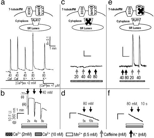
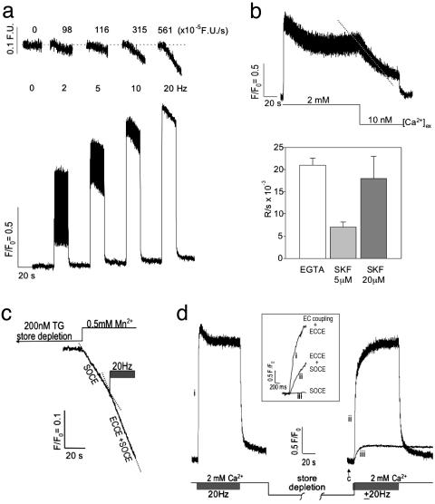
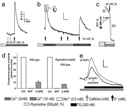
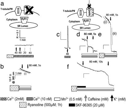
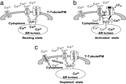
Store-operated Ca(2+) entry (SOCE) occurs in diverse cell types in response to depletion of Ca(2+) within the endoplasmic/sarcoplasmic reticulum and functions both to refill these stores and to shape cytoplasmic Ca(2+) transients. Here we report that in addition to conventional SOCE, skeletal myotubes display a physiological mechanism that we term excitation-coupled Ca(2+) entry (ECCE). ECCE is rapidly initiated by membrane depolarization. Like excitation-contraction coupling, ECCE is absent in both dyspedic myotubes that lack the skeletal muscle-type ryanodine receptor 1 and dysgenic myotubes that lack the dihydropyridine receptor (DHPR), and is independent of the DHPR l-type Ca(2+) current. Unlike classic SOCE, ECCE does not depend on sarcoplasmic reticulum Ca(2+) release. Indeed, ECCE produces a large Ca(2+) entry in response to physiological stimuli that do not produce substantial store depletion and depends on interactions among three different Ca(2+) channels: the DHPR, ryanodine receptor 1, and a Ca(2+) entry channel with properties corresponding to those of store-operated Ca(2+) channels. ECCE may provide a fundamental means to rapidly maintain Ca(2+) stores and control important aspects of Ca(2+) signaling in both muscle and nonmuscle cells.


Figures

Fig. 1.
Fig. 1.
RyR1 and α1sdhpr are both necessary for engaging ECCE. (a) Response of WT myotubes (n = 25 cells) to 5-sec stimulation with caffeine (caf) or K+ in the presence of [Ca2+]o and to K+ immediately after reducing the [Ca2+]o concentration to 7 μM. PM, plasma membrane. (b) Depolarization (2, 4, and 8 sec) of primary myotubes (n = 25) resulted in an extremely rapid entry of Mn2+ as determined by monitoring the quench of fura-2 fluorescence. The response of fura-2 to depolarization exhibited an overshoot in quench, which is likely the consequence of slight deviations from excitation (ex) at 360 nm, indicating that Ca2+ was released during EC coupling. The average rates of baseline quench and that induced by depolarization are indicated by dashed traces i (horizontal) and ii (vertical), respectively, and are summarized in Table 1. (c) Response of dyspedic myotubes (n = 25) to 10 sec of stimulation with caffeine or K+ in the presence of [Ca2+]o. (d) Dyspedic myotubes (n = 56) did not produce detectable quench of fura-2 fluorescence upon K+ depolarization for 5 or 10 sec. (e) Response of dysgenic myotubes (n = 35) to 10 sec of stimulation with K+ or caffeine in the presence of [Ca2+]o. (f) Dysgenic myotubes (n = 45) did not produce detectable quench of fura-2 fluorescence upon K+ depolarization. The very slow basal rate of Mn2+ entry in quiescent cells was linear over several minutes and comparable across all cell types. (Scale: R represents a 340- to 380-nm excitation ratio for fluorescence emission at 510 nm; F.U. are at an excitation of 360 nm.)
Fig. 2.
Fig. 2.
Electrical-field stimulation induces ECCE. (a)Ca2+ transients produced in primary myotubes (n = 15) in response to trains of electrical stimuli at 2, 5, 10, and 20 Hz (25-msec duration, 3 V) in the presence of 2 mM [Ca2+]o.(Upper) The rate of Mn2+ entry is proportional to the stimulus frequency monitored by the quench in fura-2 fluorescence. (Lower) Ca2+ transients exhibit an initial peak that decrements to a S-S level that is sustained for the duration of the pulse train. (b) (Upper) Removal of external Ca2+ results in the loss of the S-S response (dashed trace). (Lower) Likewise, SKF-96365 produces a decay of the S-S phase of the response with a rate that depends on the concentration of this SOCC blocker. The decay of the S-S phase subsequent to buffering [Ca2+]o with EGTA or brief addition of SKF-96365 is summarized (mean and SE from n = 8, 3, and 3 myotubes, respectively). The decay rate of the S-S phase observed in the presence of 20 μM SKF-96365 (SKF) was not significantly different from that measured when [Ca2+]o was removed from primary myotubes (P > 0.05). Field stimuli are at 3 V and 25 ms in duration at 20 Hz, and Ca2+ transients were recorded with fluo-4. (c) Quench of fura-2 fluorescence in response to Mn2+ entry from the external medium subsequent to complete store depletion with 200 nM TG was ascribable to SOCE. A pulse train initiated after measurement of the SOCE component resulted in an ≈2-fold increase in the rate of quench (ECCE plus SOCE). The trace shown is an average from three myotubes. See the text for rates. (d) The responses of WT myotubes to a pulse train before and after store depletion (without the use of SERCA blocker TG) using fluo-4 gave a rapid rate of Ca2+ rise (time to peak, <100 msec). After an initial pulse train in the presence of 2 mM [Ca2+]o, aCa2+-free solution was applied and the cells were challenged four to six times (30 sec each) with 20 mM caffeine. The last caffeine challenge failed to produce measurable Ca2+ release from the SR (arrow). Rapid substitution of 2 mM [Ca2+]o in the presence (trace ii) or absence (trace iii) of a train of electrical pulses resulted in a significantly different initial rates of rise for cytoplasmic Ca2+. (Inset) An expanded time scale of the initial rates of Ca2+ entry for EC coupling plus ECCE (trace i), ECCE plus SOCE (trace ii), and SOCE (trace iii). Rates are given in the text. Data were acquired at 20 Hz by using photometry. Data shown is an average of three myotubes and is representative of data acquired from at least 10 cells in separate culture wells and on separate days.
Fig. 3.
Fig. 3.
ECCE persists after inactivation of RyR1 by ryanodine pretreatment. (a) Response of WT (wt) myotubes pretreated with ryanodine (n = 33) to 5 sec of stimulation with caffeine or K+ in the presence of [Ca2+]o.(b) Response of myotubes pretreated with ryanodine (n = 16) to stimulation by K+ for 5 sec in the absence and presence of [Ca2+]o. (Inset) Illustrated is the sustained response of myotubes pretreated with ryanodine to K+ for 5 sec by showing an expanded time axis of the transient region indicated within the box. (c) Myotubes pretreated with ryanodine (n = 45) exhibited large Mn2+ entry in response to depolarization as monitored by the quench of fura-2 fluorescence. The average quench rates indicated by dashed traces i and ii are summarized in Table 1. (d) Inhibitory effect of SOCC blockers SKF-96365 (20μM, 1- to 2-min exposure) and 2-APB (100 μM, 5-min exposure) on a 5-sec, 40 mM, K+-induced Mn2+ quench in control (ctrl; n = 18) and ryanodine-pretreated (500 μM, 1 hr; n = 17) WT myotubes. The absolute peak amplitudes of Mn2+ quench were 18.2 ± 0.6 and 100 ± 5.7 (in arbitrary F.U.) for naïve and ryanodine-pretreated cells, respectively. (Scale: R represents a 340- to 380-nm excitation ratio; F.U. are at an excitation of 360 nm.) (e) Representative responses of control (n = 11) and ryanodine-pretreated (n = 27) WT primary myotubes challenged with 200 nM TG in the absence of [Ca2+]o.
Fig. 4.
Fig. 4.
Role of DHPR in ECCE. (a) Response of primary dysgenic myotubes pretreated with ryanodine (n = 35) to 5 sec of stimulation with K+ or caffeine in the presence of [Ca2+]o.(b) Dysgenic myotubes pretreated with ryanodine (n = 47) did not produce additional quench of fura-2 fluorescence upon depolarization for 5 sec with 80 mM K+. The mean rate of quench indicated by dashed trace i is given in Table 1. (c) Response of dysgenic myotubes transfected with SkEIIIKdhpr (n = 30) to 5 sec of stimulation with K+ in the absence of [Ca2+]o. Cells pretreated with ryanodine (500 μM) failed to respond to caffeine but produced a large Ca2+ entry upon depolarization ascribable to ECCE (data not shown). (d) Dysgenic myotubes transfected with SkEIIIKdhpr (n = 42) produced transient entry of Mn2+ upon being depolarized for 5 sec, as determined by monitoring the quench in fura-2 fluorescence. (e) Dysgenic myotubes transfected with SkEIIIKdhpr and pretreated with ryanodine exhibited a basal quench in the absence of a K+ bolus (n = 40; rate measured from region indicated by dashed trace i) (Table 1). Depolarization by addition of K+ dramatically enhanced rate of Mn2+ entry (dashed trace ii; see Table 1). (f) Mn2+ entry was reversibly blocked by SKF-96365 in dysgenic myotubes expressing SkEIIIKdhpr and pretreated with ryanodine (n = 20). (Scale: R represents a 340- to 380-nm exitation ratio; F.U. are at an excitation of 360 nm.)
Fig. 5.
Fig. 5.
Proposed model for ECCE. (a) Resting state. (b) Activated state. α1Sdhpr initially senses plasma membrane depolarization (ΔEM), which triggers concurrent signals to RyR1 and possibly SOCC. The signal to RyR1 induces a conformational change (represented by changing from a trapezoid in the resting state to a pentagon in the activated state) such that RyR1 engages SOCC to activate [Ca2+]o influx (gray arrow) into the cytoplasm. The Ca2+ is then resequestered into the SR lumen to replenish the Ca2+ released from the SR by means of RyR1 (black arrow) during EC coupling. (c) Depleted state showing the activation of conventional SOCE.

References

    1. Ma, J. & Pan, Z. (2003) Front. Biosci. 8, D242-D255. - PubMed
    1. Putney, J. W., Broad, L. M., Braun, F.-J., Lievremont, J.-P. & Bird, G. S. J. (2001) J. Cell Sci. 114, 2223-2229. - PubMed
    1. Launikonis, B. S., Barnes, M. & Stephenson, D. G. (2003) Proc. Natl. Acad. Sci. USA 100, 2941-2944. - PMC - PubMed
    1. Kiselyov, K., Shin, D. M., Wang, Y., Pessah, I. N., Allen, P. D. & Muallem, S. (2000) Mol. Cell 6, 421-431. - PubMed
    1. Kiselyov, K., Xu, X., Mozhayeva, G., Kuo, T., Pessah, I. N., Mignery, G., Zhu, Z., Birnbaumer, L. & Muallem, S. (1998) Nature 396, 478-482. - PubMed

Publication types

MeSH terms

LinkOut - more resources