

Tissue-based assay for ornithine decarboxylase to identify patients likely to respond to difluoromethylornithine
- PMID: 15505341
- PMCID: PMC3957822
- DOI: 10.1369/jhc.4A6358.2004
Tissue-based assay for ornithine decarboxylase to identify patients likely to respond to difluoromethylornithine
Abstract
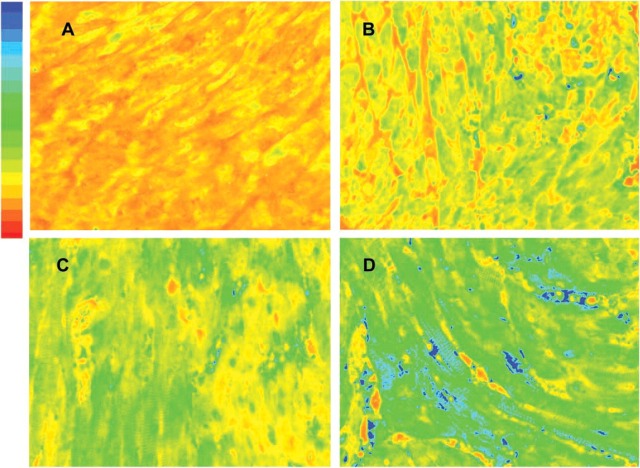
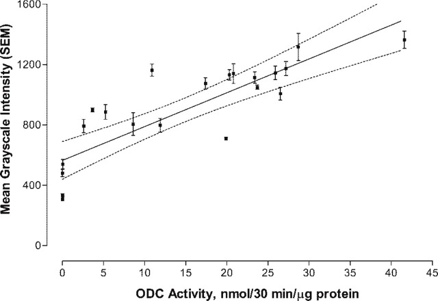
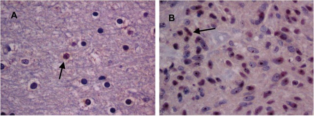
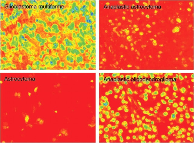
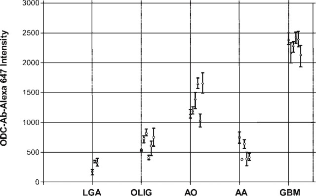
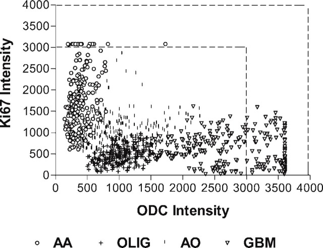
In a previous publication, we showed that a clinical trial of DL-alpha-difluoromethyl ornithine (DFMO), in combination with PCV (procarbazine, CCNU, vincristine) increased survival of patients with anaplastic gliomas (WHO III) but not glioblastoma multiforme (WHO IV). We believe that treatment outcome (survival) is inversely related to tumor ornithine decarboxylase (ODC) levels. To prove this, we needed to develop an assay to quantify ODC levels in formalin-fixed tumor tissues, which would enable a retrospective study of tumor biopsy specimens from the landmark clinical trial. We developed an assay using a specific polyclonal antibody coupled to an Alexa fluorescent dye. Transgenic MHC-ODC mice with differing levels of ODC in heart muscle were used to establish the relationship between mean gray-scale intensity and enzymatic ODC activity. We found a direct relationship between mean gray-scale intensity of the ODC antibody coupled to Alexa 647 dye and enzymatic activity. Preliminary analysis of a human glioma tissue array shows that tumor-specific variations in levels of ODC can be semiquantitated. We show that mean gray-scale intensity of astrocytoma:glioblastoma is 1:6 and of anaplastic astrocytoma:glioblastoma is 1:4. We also compared the intensity of antibody to Ki67 coupled with phycoerythrin simultaneously in cells but failed to see a relationship that crossed histologies. We conclude that we can measure levels of ODC in formalin-fixed tumor tissue using an antibody to ODC coupled to Alexa 647 dye, and this will enable us to conduct a future study to correlate survival of patients with gliomas of different histologies treated with DFMO to tumor ODC levels.
Figures
Similar articles
-
Relationship between ornithine decarboxylase levels in anaplastic gliomas and progression-free survival in patients treated with DFMO-PCV chemotherapy.Int J Cancer. 2007 Nov 15;121(10):2279-83. doi: 10.1002/ijc.22914. Int J Cancer. 2007. PMID: 17582600
-
Phase III randomized study of postradiotherapy chemotherapy with combination alpha-difluoromethylornithine-PCV versus PCV for anaplastic gliomas.Clin Cancer Res. 2003 Mar;9(3):981-90. Clin Cancer Res. 2003. PMID: 12631596 Clinical Trial.
-
Phase III randomized study of postradiotherapy chemotherapy with alpha-difluoromethylornithine-procarbazine, N-(2-chloroethyl)-N'-cyclohexyl-N-nitrosurea, vincristine (DFMO-PCV) versus PCV for glioblastoma multiforme.Clin Cancer Res. 2000 Oct;6(10):3878-84. Clin Cancer Res. 2000. PMID: 11051233 Clinical Trial.
-
Clinical importance of eflornithine (α-difluoromethylornithine) for the treatment of malignant gliomas.CNS Oncol. 2018 Apr;7(2):CNS16. doi: 10.2217/cns-2017-0031. Epub 2018 Jan 30. CNS Oncol. 2018. PMID: 29378419 Free PMC article. Review.
-
In vitro chemosensitivity testing of hematological cancer patients: detection of ornithine decarboxylase.Recent Results Cancer Res. 2003;161:62-70. doi: 10.1007/978-3-642-19022-3_6. Recent Results Cancer Res. 2003. PMID: 12528799 Review.
Cited by
-
CNS Anticancer Drug Discovery and Development Conference White Paper.Neuro Oncol. 2015 Nov;17 Suppl 6(Suppl 6):vi1-26. doi: 10.1093/neuonc/nov169. Neuro Oncol. 2015. PMID: 26403167 Free PMC article.
-
Difluoromethylornithine Induces Apoptosis through Regulation of AP-1 Signaling via JNK Phosphorylation in Epithelial Ovarian Cancer.Int J Mol Sci. 2021 Sep 23;22(19):10255. doi: 10.3390/ijms221910255. Int J Mol Sci. 2021. PMID: 34638596 Free PMC article.
-
Effect of eflornithine on mutation frequency in temozolomide-treated U87MG cells.Oncotarget. 2020 Nov 3;11(44):3933-3942. doi: 10.18632/oncotarget.27782. eCollection 2020 Nov 3. Oncotarget. 2020. PMID: 33216820 Free PMC article.
References
-
- Berdinskikh NK, Ignatenko NA, Zaletok SP, Ganina KP, Chorniy VA. (1991) Ornithine decarboxylase activity and polyamine content in adenocarcinomas of human stomach and large intestine. Int J Cancer. 47:496–498 - PubMed
-
- Canizares F, Salinas J, de las Heras M, Diaz J, Tovar I, Martinez P, Penafiel R. (1999) Prognostic value of ornithine decarboxylase and polyamines in human breast cancer: correlation with clinico-pathologic parameters. Clin Cancer Res. 5:2035–2041 - PubMed
-
- Carbone PP, Douglas JA, Larson PO, Verma AK, Blair IA, Pomplun M, Tutsch KD. (1998) Phase I chemoprevention study of piroxicam and alpha-difluoromethylornithine. Cancer Epidemiol Biomarkers Prev 7:907–912 - PubMed
Publication types
MeSH terms
Substances
Grants and funding
LinkOut - more resources
Full Text Sources
Medical
Research Materials
