Selective developmental increase in the climbing fiber input to the cerebellar interpositus nucleus in rats
- PMID: 15506893
- PMCID: PMC2546608
- DOI: 10.1037/0735-7044.118.5.1111
Selective developmental increase in the climbing fiber input to the cerebellar interpositus nucleus in rats
Abstract
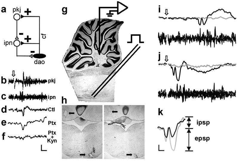
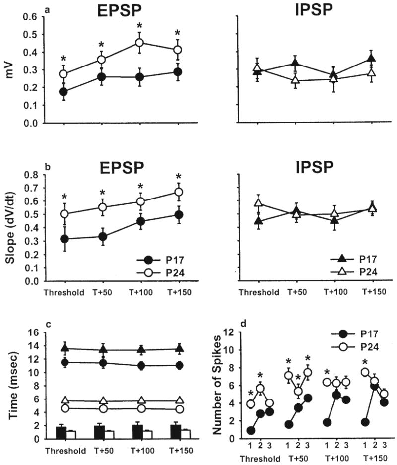
Previous studies have demonstrated that learning-related cerebellar plasticity and stimulus-elicited neuronal activity emerge ontogenetically in parallel with delay eyeblink conditioning in rats. The present study examined cerebellar interpositus field potentials and multiunit neuronal activity evoked by microstimulation of the inferior olive in Postnatal Day 17 and 24 rats. The slope and amplitude of the excitatory postsynaptic potential and the number of evoked multiunit spikes increased with age, whereas the inhibitory postsynaptic potential caused by Purkinje cell input remained stable. These results are consistent with the notion that the postsynaptic depolarization of cerebellar interpositus neurons caused by cerebellar afferents (e.g., the climbing fibers of the inferior olive) is a critical factor contributing to the ontogeny of delay eyeblink conditioning in rats.
Figures
Similar articles
-
Synaptic excitation by climbing fibre collaterals in the cerebellar nuclei of juvenile and adult mice.J Physiol. 2017 Nov 1;595(21):6703-6718. doi: 10.1113/JP274598. Epub 2017 Sep 20. J Physiol. 2017. PMID: 28795396 Free PMC article.
-
Developmental changes in eyeblink conditioning and simple spike activity in the cerebellar cortex.Dev Psychobiol. 2004 Jan;44(1):45-57. doi: 10.1002/dev.10149. Dev Psychobiol. 2004. PMID: 14704989
-
Perineuronal Nets in the Deep Cerebellar Nuclei Regulate GABAergic Transmission and Delay Eyeblink Conditioning.J Neurosci. 2018 Jul 4;38(27):6130-6144. doi: 10.1523/JNEUROSCI.3238-17.2018. Epub 2018 Jun 1. J Neurosci. 2018. PMID: 29858484 Free PMC article.
-
Ontogenetic changes in the neural mechanisms of eyeblink conditioning.Integr Physiol Behav Sci. 2001 Jan-Mar;36(1):15-35. doi: 10.1007/BF02733945. Integr Physiol Behav Sci. 2001. PMID: 11484994 Review.
-
The ontogeny of associative cerebellar learning.Int Rev Neurobiol. 2014;117:53-72. doi: 10.1016/B978-0-12-420247-4.00004-X. Int Rev Neurobiol. 2014. PMID: 25172629 Review.
Cited by
-
Long-term depression at the mossy fiber-deep cerebellar nucleus synapse.J Neurosci. 2006 Jun 28;26(26):6935-44. doi: 10.1523/JNEUROSCI.0784-06.2006. J Neurosci. 2006. PMID: 16807323 Free PMC article.
-
Cerebellum Lecture: the Cerebellar Nuclei-Core of the Cerebellum.Cerebellum. 2024 Apr;23(2):620-677. doi: 10.1007/s12311-022-01506-0. Epub 2023 Feb 13. Cerebellum. 2024. PMID: 36781689 Free PMC article. Review.
-
Oscillations, Timing, Plasticity, and Learning in the Cerebellum.Cerebellum. 2016 Apr;15(2):122-38. doi: 10.1007/s12311-015-0665-9. Cerebellum. 2016. PMID: 25808751 Review.
-
Trigeminal high-frequency stimulation produces short- and long-term modification of reflex blink gain.J Neurophysiol. 2014 Feb;111(4):888-95. doi: 10.1152/jn.00667.2013. Epub 2013 Nov 27. J Neurophysiol. 2014. PMID: 24285868 Free PMC article.
-
Synaptic excitation by climbing fibre collaterals in the cerebellar nuclei of juvenile and adult mice.J Physiol. 2017 Nov 1;595(21):6703-6718. doi: 10.1113/JP274598. Epub 2017 Sep 20. J Physiol. 2017. PMID: 28795396 Free PMC article.
References
-
- Aizenman CD, Huang EJ, Linden DJ. Morphological correlates of intrinsic electrical excitability in neurons of the deep cerebellar nuclei. Journal of Neurophysiology. 2003;89:1738–1747. - PubMed
-
- Aizenman CD, Huang EJ, Manis PB, Linden DJ. Use-dependent changes in synaptic strength at the Purkinje cell to deep nuclear synapse. Progress in Brain Research. 2000;124:247–273. - PubMed
-
- Aizenman CD, Linden DJ. Regulation of the rebound depolarization and spontaneous firing patterns of deep nuclear neurons in slice of rat cerebellum. Journal of Neurophysiology. 1999;82:1697–1709. - PubMed
-
- Carew TJ. Developmental assembly of learning in Aplysia. Trends in Neurosciences. 1989;12:389–394. - PubMed