. 2004 Sep 17:1:4.
doi: 10.1186/1743-422X-1-4.

Divergence of the mRNA targets for the Ssb proteins of bacteriophages T4 and RB69

Affiliations

Divergence of the mRNA targets for the Ssb proteins of bacteriophages T4 and RB69

Jamilah M Borjac-Natour et al. Virol J. .

Abstract

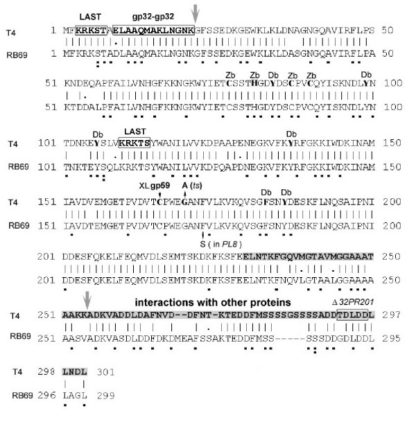
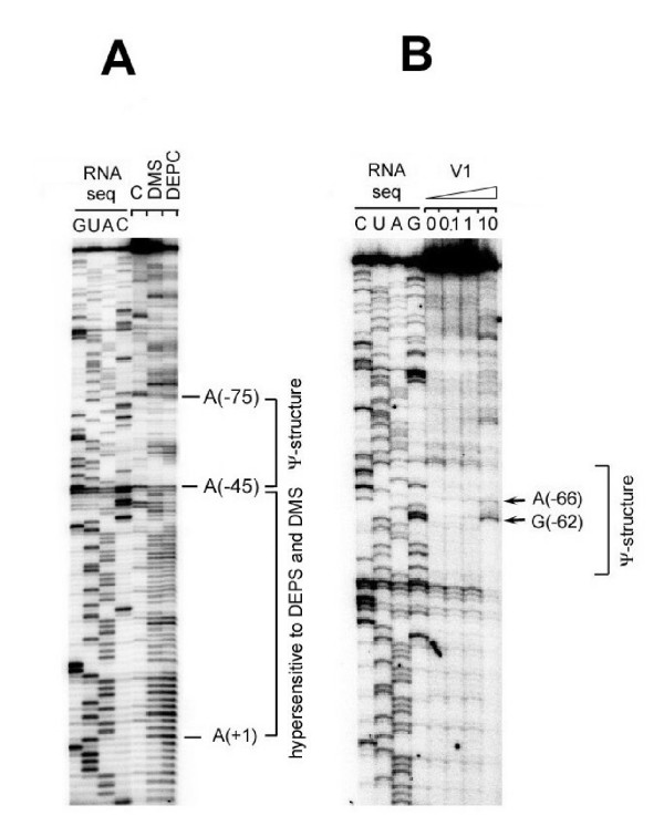
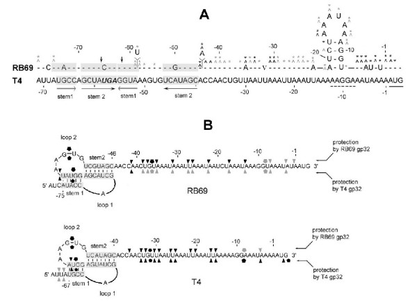
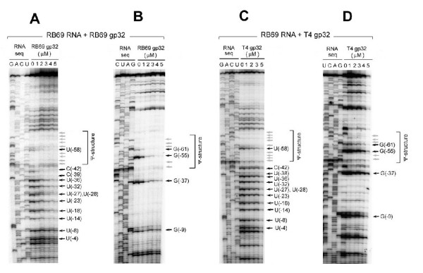
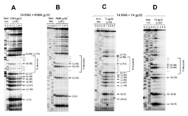
The single-strand binding (Ssb) protein of phage T4 (T4 gp32, product of gene 32) is a mRNA-specific autogenous translational repressor, in addition to being a sequence-independent ssDNA-binding protein that participates in phage DNA replication, repair and recombination. It is not clear how this physiologically essential protein distinguishes between specific RNA and nonspecific nucleic acid targets. Here, we present phylogenetic evidence suggesting that ssDNA and specific RNA bind the same gp32 domain and that plasticity of this domain underlies its ability to configure certain RNA structures for specific binding. We have cloned and characterized gene 32 of phage RB69, a relative of T4 We observed that RB69 gp32 and T4 gp32 have nearly identical ssDNA binding domains, but diverge in their C-terminal domains. In T4 gp32, it is known that the C-terminal domain interacts with the ssDNA-binding domain and with other phage-induced proteins. In translation assays, we show that RB69 gp32 is, like T4 gp32, an autogenous translational repressor. We also show that the natural mRNA targets (translational operators) for the 2 proteins are diverged in sequence from each other and yet can be repressed by either gp32. Results of chemical and RNase sensitivity assays indicate that the gp32 mRNA targets from the 2 related phages have similar structures, but differ in their patterns of contact with the 2 repressors. These and other observations suggest that a range of gp32-RNA binding specificities may evolve in nature due to plasticity of the protein-nucleic acid interaction and its response to modulation by the C-terminal domain of this translational repressor.


Figures

Figure 1
Figure 1
A comparison between the genetic maps of the Ssb protein (gp32) encoding regions of phages T4 and RB69. Note the presence of an open-reading frame (ORF) for a homing endonuclease (SegG protein; [45]) between T4 genes 59 (gp59; primase-helicase loader) and 32 (gp32; Ssb protein). The restriction sites we used for cloning RB69 gene 32 are marked, and compared to the locations of analogous sites in T4. GenBank Accession numbers for the genetic regions of interest are also noted.
Figure 4
Figure 4
Results of experiments showing that RB69 gp32 is an autogenous translational repressor. For Panel A, λCI857PLN-bearing plasmid clones of the diagrammed DNA segments were heat-induced (42°C) and assayed for gp32 synthesis as described in other work [24,27]. RBG32 is a DNA segment that carries the wild-type sequence from -120 through +900 relative to the first base of the initiator AUG of RB69 gene 32. RBG32Δop is a truncated derivative of RBG32 that lacks elements of the putative RNA pseudoknot of RB69 gene 32 (Figs 3 & 6). PL8 is identical to RBG32 except that it carries a single-base substitution (marked with an asterisk) in codon 173, leading to a F173S substitution in RB69 gp32. PL2 is similar to RBG32 and PL8, except that it carries several point mutations (map positions marked with asterisks). Panel B shows results of an experiment in which purified RB69 gp32 was shown to inhibit in vitro translation of purified mRNA from the cloned RBG32 fragment, as well as mRNA from in vitro expressed plasmid clone (coupled transcription/translation). Conditions for these assays are described in METHODS.
Figure 2
Figure 2
Amino-acid sequence alignments between the Ssb proteins (gp32s) of T4 and RB69. Residues and segments of the T4 gp32 sequence that have been implicated in specific biological functions of the protein are marked as follows: Db [DNA binding residue]; Zb (residues that coordinate Zn++ in the zinc-binding domain; [20,46]); gp32-gp32 [residues involved in cooperative gp32 binding to ssDNA]; XLgp59 (residue that cross-links to gp59; [42]); LAST (sequence motifs, (Lys/Arg)3 (Ser/Thr)2, that have been proposed to directly bind nucleic-acids or mediate gp32-gp32 interactions [31]). The shaded C-terminal portion of T4 gp32 has been implicated in interactions with other phage induced proteins [38]. The small deletion (Δ32PR201) alters specificity of T4 gp32 in phage replication without affecting autogenous translational repression [39]. The largest vertical arrows denote trypsin-hypersensitive sites (19) The G-to-A mutation marked "(ts)" was isolated in this laboratory as a missense (temperature-sensitive) suppressor of a defective gp43 function (unpublished). In the RB69 gp32 sequence, residues whose codons differ from their conserved T4 counterpart at the third nucleotide are underscored with a single dot; those differing by 2 nucleotides are marked by 2 dots.
Figure 3
Figure 3
A comparison between the nucleotide sequences of the T4 IC32.1-32 and RB69 IC59-32 regions. These 2 regions contain determinants for translation initiation of the respective phage-induced mRNAs for gp32. The chart emphasizes sequence differences (entered as lettered residues in the RB69 sequence) between the 2 regions. The dashes indicate identity between RB69 and T4 residues. Sequence elements contributing to RNA pseudoknot formation in the T4 gene 32-specific mRNA are marked by horizontal arrows. Note the sequence overlap between elements of the pseudoknot and ORF32.1 (segG) of the T4 sequence. Also, see Fig 6 for a summary of properties of the RB69 sequence.
Figure 5
Figure 5
Portions of autoradiograms from RNA sequencing gels showing sites of cleavage in RB69 gene 32-derived RNA following treatments with DMS and DEPC (Panel A) and RNase V1 (Panel B). These experiments probed the RB69 RNA for secondary and higher-order structure. The lanes marked "RNA seq" show results from sequencing untreated RNA by the RVT-catalyzed chain termination method [23,35]. In Panel A the lane marked with a "minus" sign shows the positions of RVT chain termination caused by RNA structure in the untreated RNA. The DMS and DEPC lanes show sites of hypersensitivity (cleavage) of the same RNA to treatment with these chemical agents. In Panel B, the V1 lanes denote the amount of RNase V1 (×10-5 units) used to digest the RNA substrate.
Figure 6
Figure 6
Summaries of results from the chemical and RNase sensitivity and RNA footprinting studies reported here. Panel A shows our interpretation of experiments that probed the existence of RNA structure in RB69 gene 32-specific RNA (Fig 5). The T4-derived RNA counterpart is shown for comparison The "caret" symbol denotes sensitivity to cleavage after DMS treatment; asterisks denote sensitivity to cleavage after DEPC treatment. The darker symbols denote greater sensitivity. Positions that are not marked by any symbols were resistant to the modifying agents under the conditions used. Vertical arrows mark positions that were sensitive to RNase V1. Panel B shows our interpretation of the RNA footprinting studies described in Figs 7 and 8. Positions of protection from RNaseA1 by gp32 are marked by the triangles and protection from RNase T1 by the pentagonal symbols. The darker symbols denote stronger protection. Unmarked positions were not protected by either gp32 from phage source under the experimental conditions used.
Figure 7
Figure 7
In vitro footprinting of RB69 gene 32-specific RNA with purified RB69 gp32 (Panels A and B) and T4 gp32 (Panels C and D). Preparation of RNA and proteins and experimental conditions for footprinting are described in METHODS. Horizontal arrows mark nucleotide positions (Fig 6B) that exhibited gp32-mediated protection from RNaseA (panels A and C) and RNase T1 (panels B and D). Darker arrows denote stronger protection. The results are summarized in Fig 6B.
Figure 8
Figure 8
In vitro footprinting of T4 gene32-specific RNA with purified RB69 gp32 (Panels A and C; RNase A) and T4 gp32 (Panels B and D; RNase T1). Conditions for these experiments were identical to those described in Fig 7, except that the RNA substrate used for footprinting was derived from clones of T4 gene 32 rather than RB69 gene 32. See also Fig 6C for a summary.

References

    1. Alberts BM, Frey L. T4 bacteriophage gene 32: a structural protein in the replication and recombination of DNA. Nature. 1970;227:1313–1318. - PubMed
    1. Kreuzer KN, Morrical SW. in Initiation of DNA Replication. Molecular Biology of Bacteriophage T4. 1994. pp. 28–42.
    1. Mosig G. in Recombination Models and Pathways. Molecular Biology of Bacteriophage T4. 1994. pp. 54–82.
    1. Bleuit JS, Xu H, Ma Y, Wang T, Liu J, Morrical SW. Mediator proteins orchestrate enzyme-ssDNA assembly during T4 recombination-dependent DNA replication and repair. Proc Natl Acad Sci USA. 2001;98:8298–8305. doi: 10.1073/pnas.131007498. - DOI - PMC - PubMed
    1. Lefebvre SD, Wong ML, Morrical SW. Simultaneous interactions of bacteriophage T4 DNA replication proteins gp59 and gp32 with single-stranded (ss) DNA. Co-modulation of ssDNA binding activities in a DNA helicase assembly intermediate. J Biol Chem. 1999;274:22830–22838. doi: 10.1074/jbc.274.32.22830. - DOI - PubMed

Publication types

LinkOut - more resources