

The combination of PRRS virus and bacterial endotoxin as a model for multifactorial respiratory disease in pigs
- PMID: 15507303
- PMCID: PMC7112634
- DOI: 10.1016/j.vetimm.2004.09.006
The combination of PRRS virus and bacterial endotoxin as a model for multifactorial respiratory disease in pigs
Abstract
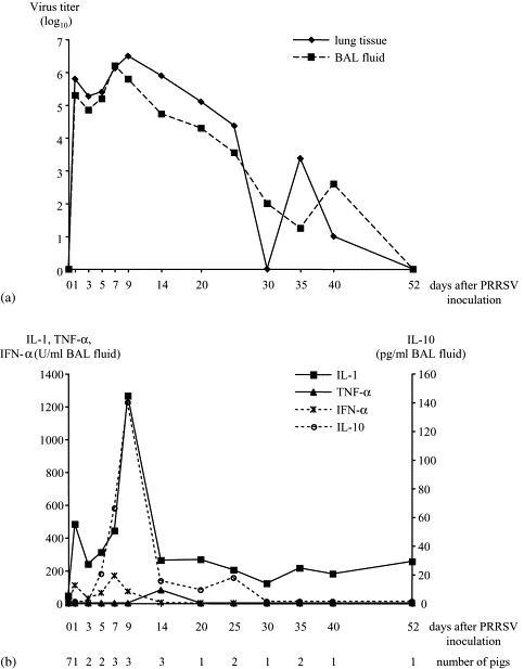
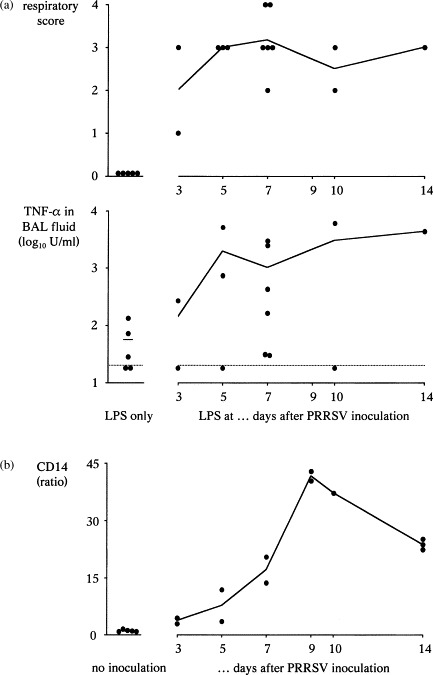
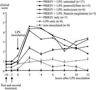
This paper reviews in vivo studies on the interaction between porcine reproductive and respiratory syndrome virus (PRRSV) and LPS performed in the authors' laboratory. The main aim was to develop a reproducible model to study the pathogenesis of PRRSV-induced multifactorial respiratory disease. The central hypothesis was that respiratory disease results from an overproduction of proinflammatory cytokines in the lungs. In a first series of studies, PRRSV was shown to be a poor inducer of TNF-alpha and IFN-alpha in the lungs, whereas IL-1 and the anti-inflammatory cytokine IL-10 were produced consistently during infection. We then set up a dual inoculation model in which pigs were inoculated intratracheally with PRRSV and 3-14 days later with LPS. PRRSV-infected pigs developed acute respiratory signs for 12-24h upon intratracheal LPS inoculation, in contrast to pigs inoculated with PRRSV or LPS only. Moreover, peak TNF-alpha, IL-1 and IL-6 titers were 10-100 times higher in PRRSV-LPS inoculated pigs than in the singly inoculated pigs and the cytokine overproduction was associated with disease. To further prove the role of proinflammatory cytokines, we studied the effect of pentoxifylline, a known inhibitor of TNF-alpha and IL-1, on PRRSV-LPS induced cytokine production and disease. The clinical effects of two non-steroidal anti-inflammatory drugs (NSAIDs), meloxicam and flunixin meglumine, were also examined. Pentoxifylline, but not the NSAIDs, significantly reduced fever and respiratory signs from 2 to 6h after LPS. The levels of TNF-alpha and IL-1 in the lungs of pentoxifylline-treated pigs were moderately reduced, but were still 26 and 3.5-fold higher than in pigs inoculated with PRRSV or LPS only. This indicates that pathways other than inhibition of cytokine production contributed to the clinical improvement. Finally, we studied a mechanism by which PRRSV may sensitize the lungs for LPS. We hypothesized that PRRSV would increase the amount of LPS receptor complex in the lungs leading to LPS sensitisation. Both CD14 and LPS-binding protein, two components of this complex, increased significantly during infection and the amount of CD14 in particular was correlated with LPS sensitisation. The increase of CD14 was mainly due to infiltration of strongly CD14-positive monocytes in the lungs. The PRRSV-LPS combination proved to be a simple and reproducible experimental model for multifactorial respiratory disease in pigs. To what extent the interaction between PRRSV and LPS contributes to the development of complex respiratory disease is still a matter of debate.
Figures
Similar articles
-
Interaction between porcine reproductive-respiratory syndrome virus and bacterial endotoxin in the lungs of pigs: potentiation of cytokine production and respiratory disease.J Clin Microbiol. 2003 Mar;41(3):960-6. doi: 10.1128/JCM.41.3.960-966.2003. J Clin Microbiol. 2003. PMID: 12624016 Free PMC article.
-
Porcine reproductive and respiratory syndrome virus infection increases CD14 expression and lipopolysaccharide-binding protein in the lungs of pigs.Viral Immunol. 2005;18(1):116-26. doi: 10.1089/vim.2005.18.116. Viral Immunol. 2005. PMID: 15802956
-
Porcine reproductive-respiratory syndrome virus infection predisposes pigs for respiratory signs upon exposure to bacterial lipopolysaccharide.Vet Microbiol. 2002 Aug 2;88(1):1-12. doi: 10.1016/s0378-1135(02)00104-9. Vet Microbiol. 2002. PMID: 12119134 Free PMC article.
-
In vivo studies on cytokine involvement during acute viral respiratory disease of swine: troublesome but rewarding.Vet Immunol Immunopathol. 2002 Sep 10;87(3-4):161-8. doi: 10.1016/s0165-2427(02)00047-8. Vet Immunol Immunopathol. 2002. PMID: 12072230 Free PMC article. Review.
-
Proinflammatory cytokines and viral respiratory disease in pigs.Vet Res. 2000 Mar-Apr;31(2):187-213. doi: 10.1051/vetres:2000113. Vet Res. 2000. PMID: 10779199 Review.
Cited by
-
Pig Lung Immune Cytokine Response to the Swine Influenza Virus and the Actinobacillus Pleuropneumoniae Infection.J Vet Res. 2017 Sep 19;61(3):259-265. doi: 10.1515/jvetres-2017-0036. eCollection 2017 Sep. J Vet Res. 2017. PMID: 29978082 Free PMC article.
-
Recombinant Antigen of Type 2 Porcine Reproductive and Respiratory Syndrome Virus (PRRSV-2) Promotes M1 Repolarization of Porcine Alveolar Macrophages and Th1 Type Response.Vaccines (Basel). 2021 Sep 10;9(9):1009. doi: 10.3390/vaccines9091009. Vaccines (Basel). 2021. PMID: 34579246 Free PMC article.
-
Effect of LPS and LTA stimulation on the expression of TLR-pathway genes in PBMCs of Akkaraman lambs in vivo.Trop Anim Health Prod. 2021 Jan 3;53(1):65. doi: 10.1007/s11250-020-02491-4. Trop Anim Health Prod. 2021. PMID: 33392825 Free PMC article.
-
Reactomes of porcine alveolar macrophages infected with porcine reproductive and respiratory syndrome virus.PLoS One. 2013;8(3):e59229. doi: 10.1371/journal.pone.0059229. Epub 2013 Mar 19. PLoS One. 2013. PMID: 23527143 Free PMC article.
-
Describing antimicrobial use and reported treatment efficacy in Ontario swine using the Ontario Swine Veterinary-based Surveillance program.BMC Vet Res. 2013 Dec 1;9:238. doi: 10.1186/1746-6148-9-238. BMC Vet Res. 2013. PMID: 24289212 Free PMC article.
References
-
- Albina E., Kobisch M., Cariolet R., Morvan P., Kéranflec’h A., Beaurepaire B., Hutet E., Labbé A. Le syndrome dysgénésique et respiratoire du porc (SDRP): Etude expérimentale des effets de l’infection sur la réponse immunitaire et la résistance aux infections Aujeszky et Mycoplasma hyopneumoniae chez le porc en croissance. Journées Rech. Porcine en France. 1995;27:107–116.
-
- Albina E., Carrat C., Charley B. Interferon-alpha response to swine arterivirus (PoAV), the porcine reproductive and respiratory syndrome virus. J. Interf. Cytok. Res. 1998;18(7):485–490. - PubMed
-
- Antal-Szalmas P., Strijp J.A., Weersink A.J., Verhoef J., Van Kessel K.P. Quantitation of surface CD14 on human monocytes and neutrophils. J. Leukoc. Biol. 1997;61(6):721–728. - PubMed
-
- Antal-Szalmas P. Evaluation of CD14 in host defence. Eur. J. Clin. Invest. 2000;30(2):167–179. - PubMed
-
- Bielefeldt-Ohmann H. Role of cytokines in the pathogenesis and treatment of respiratory disease. In: Myers M.J., Murtaugh M.P., editors. Cytokines in Animal Health and Disease. Marcel Dekker; New York: 1995. pp. 291–332.
Publication types
MeSH terms
Substances
LinkOut - more resources
Full Text Sources
Research Materials
