. 2005 Jan 7;280(1):164-73.
doi: 10.1074/jbc.M408211200. Epub 2004 Oct 26.

Severe acute respiratory syndrome coronavirus 3C-like proteinase N terminus is indispensable for proteolytic activity but not for enzyme dimerization. Biochemical and thermodynamic investigation in conjunction with molecular dynamics simulations

Affiliations

Severe acute respiratory syndrome coronavirus 3C-like proteinase N terminus is indispensable for proteolytic activity but not for enzyme dimerization. Biochemical and thermodynamic investigation in conjunction with molecular dynamics simulations

Shuai Chen et al. J Biol Chem. .

Abstract

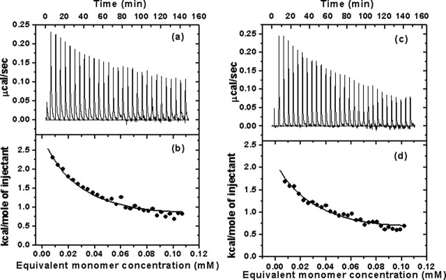
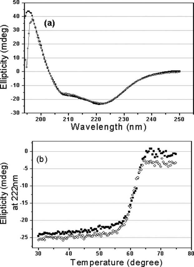
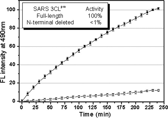
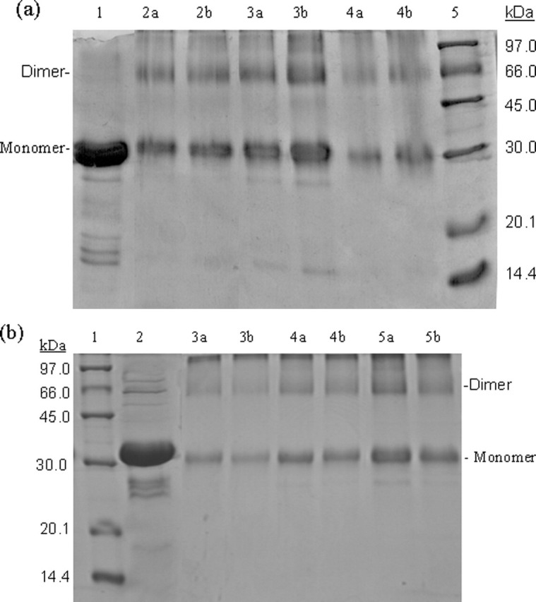
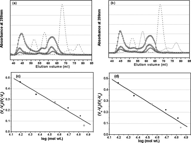
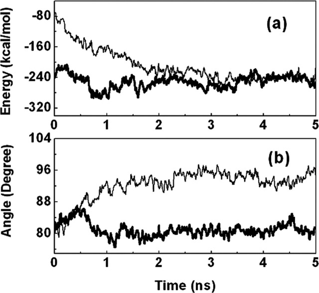
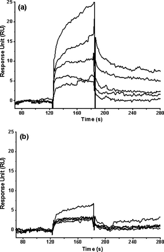
Severe acute respiratory syndrome (SARS) coronavirus is a novel human coronavirus and is responsible for SARS infection. SARS coronavirus 3C-like proteinase (SARS 3CL(pro)) plays key roles in viral replication and transcription and is an attractive target for anti-SARS drug discovery. In this report, we quantitatively characterized the dimerization features of the full-length and N-terminal residues 1-7 deleted SARS 3CL(pro)s by using glutaraldehyde cross-linking SDS-PAGE, size-exclusion chromatography, and isothermal titration calorimeter techniques. Glutaraldehyde cross-linking SDS-PAGE and size-exclusion chromatography results show that, similar to the full-length SARS 3CL(pro), the N-terminal deleted SARS 3CL(pro) still remains a dimer/monomer mixture within a wide range of protein concentrations. Isothermal titration calorimeter determinations indicate that the equilibrium dissociation constant (K(d)) of the N-terminal deleted proteinase dimer (262 microm) is very similar to that of the full-length proteinase dimer (227 microm). Enzymatic activity assay using the fluorescence resonance energy transfer method reveals that N-terminal deletion results in almost complete loss of enzymatic activity for SARS 3CL(pro). Molecular dynamics and docking simulations demonstrate the N-terminal deleted proteinase dimer adopts a state different from that of the full-length proteinase dimer, which increases the angle between the two protomers and reduces the binding pocket that is not beneficial to the substrate binding. This conclusion is verified by the surface plasmon resonance biosensor determination, indicating that the model substrate cannot bind to the N-terminal deleted proteinase. These results suggest the N terminus is not indispensable for the proteinase dimerization but may fix the dimer at the active state and is therefore vital to enzymatic activity.


Figures

F<sc>ig</sc>. 5
Fig. 5
Typical calorimetric dilution data for the dissociation of SARS 3CLpro dimers into monomers measured by VP-ITC.a, the calorimetric dilution profile for the dissociation of the full-length SARS 3CLpro dimers. Consecutive 10 μl volumes of 572.5 μm 3CLpro were diluted into 1.43 ml of 20 mm sodium phosphate, pH 7.5, 100 mm NaCl, 1 mm EDTA, at 25 °C (except an initial injectant of a 3-μl volume). b, integrated dilution heat effects of the dissociation of the full-length SARS 3CLpro dimers, with theoretical fits by a simple dimer-monomer dissociation model to yield a dimer dissociation constant (Kd) of 227 ± 34 μm and a dimerization enthalpy change (ΔHdim0) of -8.283 ± 0.103 kcal/mol. c, the calorimetric dilution profile for the dissociation of the N-terminal deleted SARS 3CLpro dimers. Endothermic responses for sequential 10-μl injections of 544.6 μm 3CLpro into buffer were measured under the same conditions, except a 3-μl initial injection. d, integrated dilution heat effects of the dissociation of the N-terminal deleted SARS 3CLpro dimers, with theoretical fits to yield a Kd of 262 ± 15 μm and a ΔHdim0 of -6.893 ± 0.25 kcal/mol.
F<sc>ig</sc>. 1
Fig. 1
CD spectra of the full-length and N-terminal deleted SARS 3CLpros.a, far-UV CD spectra of the full-length (▪) and N-terminal deleted (○) SARS 3CLpros at 25 °C. b, thermal induced unfolding monitored at 222 nm with a temperature range from 30 to 75 °C for the full-length (▪) and N-terminal deleted (○) SARS 3CLpros. Protein concentrations used in CD experiments were 10 μm, and all protein samples were prepared in 20 mm sodium phosphate, pH 7.5, 100 mm NaCl.
F<sc>ig</sc>. 2
Fig. 2
Representative fluorescence profiles of hydrolysis of the fluorogenic substrate by SARS 3CLpros. The fluorogenic substrate at a concentration of 10 μm was incubated with 1 μm full-length (▪) or N-terminal deleted (○) SARS 3CLpro in 20 mm sodium phosphate, pH 7.5, 100 mm NaCl, 5 mm DTT, 1 mm EDTA, at 25 °C. Increase of emission fluorescence intensity at 490 nm wavelength was recorded at 10-min intervals, λEX = 340 nm. The emission spectrum was recorded for more than 4 h. The initial reaction velocity (ν0) was determined from the linear portion of the progress curve, which corresponded to between 2 and 10% hydrolysis of the substrate. The activity of the full-length SARS 3CLpro was taken as 100%.
F<sc>ig</sc>. 3
Fig. 3
SDS-PAGE (10% acrylamide) profiles of glutaraldehyde cross-linked SARS 3CLpros.a, cross-linking analyses of the full-length SARS 3CLpro. Lane 1, untreated 3CLpro (7 mg/ml); lane 2a, 3CLpro (2 mg/ml) cross-linked by 0.01% glutaraldehyde; lane 2b, 3CLpro (2 mg/ml) cross-linked by 0.1% glutaraldehyde; lanes 3a and 3b, 3CLpro (7 mg/ml) cross-linked by 0.01 and 0.1% glutaraldehyde, respectively; lanes 4a and 4b, 3CLpro (1 mg/ml), 0.01 and 0.1% glutaraldehyde; lane 5, molecular weight SDS calibration kit protein standards are as follows: phosphorylase b (97.0 kDa), albumin (66.0 kDa), ovalbumin (45.0 kDa), carbonic anhydrase (30.0 kDa), trypsin inhibitor (20.1 kDa), α-lactalbumin (14.4 kDa). b, cross-linking analyses of the N-terminal deleted SARS 3CLpro. Lane 1, protein standards; lane 2 untreated 3CLpro (7 mg/ml); lanes 3a and 3b, 3CLpro (1 mg/ml), 0.01 and 0.1% glutaraldehyde, respectively; lanes 4a and 4b, 3CLpro (2 mg/ml), 0.01 and 0.1% glutaraldehyde; lanes 5a and 5b, 3CLpro (7 mg/ml), 0.01 and 0.1% glutaraldehyde.
F<sc>ig</sc>. 4
Fig. 4
Analyses of monomer-dimer equilibria of SARS 3CLpros by SEC.a, elution profiles of the full-length SARS 3CLpro at neutral pH (7.5) and concentrations of 1 (○), 2 mg/ml (▵), and 7 mg/ml (□). Elution profiles of four marker proteins, ribonuclease A (15.6 kDa), chymotrypsinogen A (22.8 kDa), ovalbumin (48.9 kDa), and albumin (65.4 kDa), are also shown as dashed lines. The dimer/monomer ratios for the concentrations of 1, 2, and 7 mg/ml are 0.68, 0.71, and 0.84, respectively. b, elution profiles of the N-terminal deleted SARS 3CLpro at neutral pH (7.5) and concentrations of 1 (○), 2 (▵), and 7 mg/ml (□). Elution profiles of marker proteins are shown as dashed lines. The dimer/monomer ratios for the concentrations of 1, 2, and 7 mg/ml are 0.65, 0.66, and 0.80, respectively. In the SEC-FPLC experiment, each protein sample was loaded to a HiLoad 16/60 Superdex 75 prep grade column pre-equilibrated with the buffer and then eluted at a flow rate of 1 ml/min with a detection of absorbance at 280 nm. The buffer used was 20 mm sodium phosphate, pH 7.5, 100 mm NaCl, 5 mm DTT, 1 mm EDTA. Standard calibration curves were used to determine the molecular masses of monomeric and dimeric form of the full-length (c) and N-terminal deleted (d) SARS 3CLpros. Elution volumes of the full-length and N-terminal deleted proteinases are indicated by open circles (○) and for marker proteins by filled circles (•). Ve and V0 are peak elution volume and column void volume, respectively, and Vt is the total bed volume.
F<sc>ig</sc>. 6
Fig. 6
Interaction energy and dimerization state changes from the full-length SARS 3CLpro to the N-terminal deleted SARS 3CLpro.a, total interaction energies for the full-length (thick line) and N-terminal deleted (thin line) SARS 3CLpros versus MD simulation time (shown as 10 ps average). b, the angles between the two protomers of the full-length (thick line) and N-terminal deleted (thin line) SARS 3CLpros versus MD simulation time (shown as 10 ps average). The angle is defined by two vectors, and each vector points from the center of mass of domain III to the center of mass of domains I and II in one protomer.
F<sc>ig</sc>. 7
Fig. 7
Binding affinity of a 6-amino acid peptide (TSAVLQ) to the full-length and N-terminal deleted SARS 3CLpros determined by SPR. Real time binding affinity measurements of the peptide to the full-length SARS 3CLpro (a) and the N-terminal deleted proteinase (b) using Biacore 3000. Representative sensorgrams were obtained from injections of the peptide at concentrations of 500, 250, 100, 50, and 20 μm (curves from top to bottom). The peptides were injected for 60s, and dissociation was monitored for more than 120s.

References

    1. Kathryn V.H. J. Clin. Investig. 2002;111:1605–1609.
    1. Peiris J.S., Lai S.T., Poon L.L., Guan Y., Yam L.Y., Lim W., Nicholls J., Yee W.K., Yan W.W., Cheung M.T., Cheng V.C., Chan K.H., Tsang D.N., Yung R.W., Ng T.K., Yuen K.Y. Lancet. 2003;361:1319–1325. - PMC - PubMed
    1. Fouchier R.A., Kuiken T., Schutten M., Amerongen VanG., Van Doornum G.J., Van Den Hoogen B.G., Peiris M., Lim W., Stohr K., Osterhaus A.D. Nature. 2003;423:240. - PMC - PubMed
    1. Herold J., Raabe T., Schelle-Prinz B., Siddell S.G. Virology. 1993;195:680–691. - PMC - PubMed
    1. Lee H.J., Shieh C.K., Gorbalenya A.E., Koonin E.V., La Monica N., Tuler J., Bagdzhadzhyan A., Lai M.M. Virology. 1991;180:567–582. - PMC - PubMed

Publication types

LinkOut - more resources