. 2004 Nov 2;101(44):15567-72.
doi: 10.1073/pnas.0406901101. Epub 2004 Oct 26.

Human fatty acid synthase: structure and substrate selectivity of the thioesterase domain

Affiliations

Human fatty acid synthase: structure and substrate selectivity of the thioesterase domain

Bornali Chakravarty et al. Proc Natl Acad Sci U S A. .

Abstract

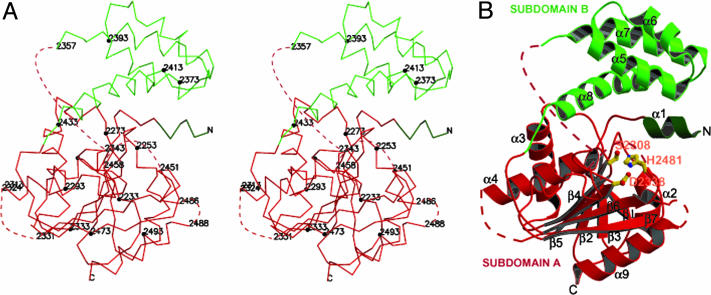
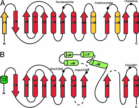
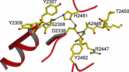
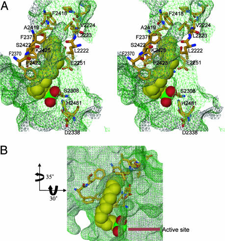
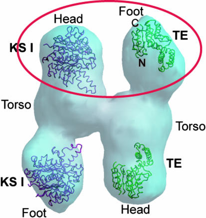
Human fatty acid synthase is a large homodimeric multifunctional enzyme that synthesizes palmitic acid. The unique carboxyl terminal thioesterase domain of fatty acid synthase hydrolyzes the growing fatty acid chain and plays a critical role in regulating the chain length of fatty acid released. Also, the up-regulation of human fatty acid synthase in a variety of cancer makes the thioesterase a candidate target for therapeutic treatment. The 2.6-A resolution structure of human fatty acid synthase thioesterase domain reported here is comprised of two dissimilar subdomains, A and B. The smaller subdomain B is composed entirely of alpha-helices arranged in an atypical fold, whereas the A subdomain is a variation of the alpha/beta hydrolase fold. The structure revealed the presence of a hydrophobic groove with a distal pocket at the interface of the two subdomains, which constitutes the candidate substrate binding site. The length and largely hydrophobic nature of the groove and pocket are consistent with the high selectivity of the thioesterase for palmitoyl acyl substrate. The structure also set the identity of the Asp residue of the catalytic triad of Ser, His, and Asp located in subdomain A at the proximal end of the groove.


Figures

Fig. 1.
Fig. 1.
Crystal structure of the TE domain. (A) Stereoview of the Cα trace. Every 20 residues (filled circles) and residues at ends of disordered loops are labeled. Dotted lines represent segments that are disordered (also shown in Figs. 2B and 3). Subdomain A is in red and dark green. Subdomain B is in light green. (B) Ribbon diagram of the TE monomer. The catalytic residues (Ser-2308, His-2481, and Asp-2338) are in ball-and-stick representation and are labeled in orange.
Fig. 2.
Fig. 2.
Schematic diagram of α/β hydrolase fold. Helices are represented as cylinders, strands as arrows, and random coils as lines. (A) Canonical/core hydrolase fold along with positions of catalytic triad (37). Differences between the core and TE structure are in yellow, whereas similarities are in red. (B) Modified hydrolase fold of TE structure. Subdomain A is in red and dark green. Subdomain B is in light green. Position of the catalytic triad is shown as filled black circles. The core has an extra strand in place of helix α1 in TE structure. Thus, strand β2 in the core corresponds to strand β1 in TE fold, and helix α1 in core corresponds to helix α2 in TE structure. Also helix α4 in the core is replaced by the all-helical subdomain B. Helix α5 in the core also is missing in the corresponding TE structure.
Fig. 3.
Fig. 3.
Sequence alignment of FAS TE domains of different species. The alignment was carried out by using blast (www.ncbi.nlm.nih.gov/blast) and clustalw (www.ebi.ac.uk/clustalW) (44). The domain originally was described in ref. . Conserved residues are boxed. Invariants to partially conserved residues are colored from red to yellow, respectively. The catalytic residues are marked with a star. Yellow circles represent residues forming the candidate palmitoyl binding groove. The elements of the secondary structure from the crystal structure are shown above the amino acid sequence, with helices represented as cylinders, strands as arrows, and random coils as lines. Subdomain A is in red and dark green; subdomain B is in light green.
Fig. 4.
Fig. 4.
Hydrogen bonding network among the various catalytic residues. Dotted lines denote hydrogen bonds connecting the corresponding residues. Hydrogen bond distances range from 2.4 to 3.2 Å.
Fig. 5.
Fig. 5.
Lipid binding site. (A) Stereoview of the candidate palmitoyl binding groove for the first 11–12 carbons and the pocket for the last 5–4 carbons. The hexadecyl sulfonyl inhibitor is represented as a Corey–Pauling–Koltun space-filling model (C, yellow; O, red; S, green). Side chains lining the groove are shown as ball-and-stick figures (C, yellow; O, red; N, blue). The side chains making up the active site are also shown as ball-and-stick figures with the color scheme similar to that of the residues. The groove surface map is shown in green wire mesh. The residues making up the groove are labeled except for Ile-2250, which cannot be seen because it falls below the inhibitor. The initial 11–12 carbon atoms from the sulfonyl group are solvent-exposed with the 11th and 12th carbon atoms at the mouth of the pocket. (B) A different view of the groove and pocket. The arrows denote the rotations in going from the orientation of the view in A to B. This view shows that most of the fatty acyl chain of hexadecyl inhibitor is exposed to the solvent.
Fig. 6.
Fig. 6.
Preliminary fit of TE (green backbone trace) and KSI (maroon trace) in the 20-Å resolution cryo-EM mass density. The substructures seen from the density are designated Head, Torso, and Foot. The TE trace fits best in the substructure designated Foot, whereas the KSI backbone trace fit is best in substructure designated Head. The best fit for both KSI and TE is circled in red. The backbone traces of TE and KSI at the bottom of the mass density show less satisfactory fits in the head and foot, respectively. Based on prior proteolytic digestion experiments of intact FAS, three domains were identified: domain I, which comprises the KS-AT/MT-DH enzyme centers and the linker region; domain II, which comprises the ER-KR-ACP region; and domain III, which is assigned to the TE (1, 2). The Head and Torso substructures, which are essentially coalesced, could correspond to domain I and parts of domain II, and the Foot could represent the rest of domain II and domain III.

References

    1. Wakil, S. J. (1989) Biochemistry 28, 4523-4530. - PubMed
    1. Smith, S. (1994) FASEB J. 8, 1248-1259. - PubMed
    1. Chirala, S. S., Chang, H., Matzuk, M., Abu-Elheiga, L., Mao, J., Mahon, K., Finegold, M. & Wakil, S. J. (2003) Proc. Natl. Acad. Sci. USA 100, 6358-6363. - PMC - PubMed
    1. Must, A., Spadano, J., Coakley, E. H., Field, A. E., Colditz, G. & Dietz, W. H. (1999) J. Am. Med. Assoc. 282, 1523-1529. - PubMed
    1. Alo, P. L., Visca, P., Marci, A., Mangoni, A., Botti, C. & Di Tondo, U. (1996) Cancer 77, 474-482. - PubMed

Publication types

Substances

Associated data

LinkOut - more resources