. 2004 Nov;78(22):12157-68.
doi: 10.1128/JVI.78.22.12157-12168.2004.

A retroviral promoter and a cellular enhancer define a bipartite element which controls env ERVWE1 placental expression

Affiliations

A retroviral promoter and a cellular enhancer define a bipartite element which controls env ERVWE1 placental expression

Sarah Prudhomme et al. J Virol. 2004 Nov.

Abstract

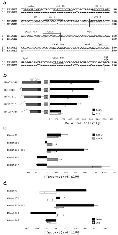
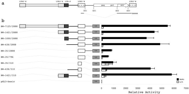
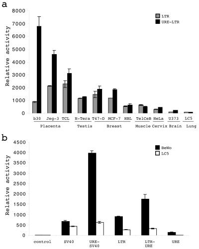
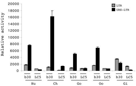
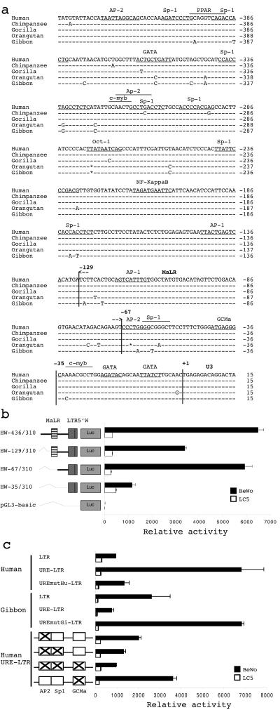
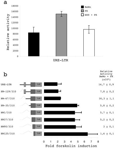
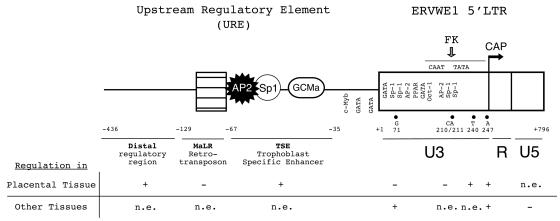
The HERV-W family contains hundreds of loci diversely expressed in several physiological and pathological contexts. A unique locus termed ERVWE1 encodes an envelope glycoprotein (syncytin) involved in hominoid placental physiology. Here we show that syncytin expression is regulated by a bipartite element consisting of a cyclic AMP (cAMP)-inducible long terminal repeat (LTR) retroviral promoter adjacent to a cellular enhancer conferring a high level of expression and placental tropism. Deletion mutant analysis showed that the ERVWE1 5' LTR contains binding sites essential for basal placental activity in the region from positions +1 to +125. The region from positions +125 to +310 represents a cAMP-responsive core HERV-W promoter active in all cell types. Site-directed mutagenesis analysis highlighted the complexity of U3 regulation. ERVWE1 placenta-specific positive (e.g., T240) and negative (e.g., G71) regulatory sites were identified, as were essential sites required for basic activity (e.g., A247). The flanking sequences of the ERVWE1 provirus contain several putative regulatory elements. The upstream HERV-H and HERV-P LTRs were found to be inactive. Conversely, the 436-bp region located between the HERV-P LTR and ERVWE1 was shown to be an upstream regulatory element (URE) which is significantly active in placenta cells. This URE acts as a tissue-specific enhancer. Genetic and functional analyses of hominoid UREs revealed large differences between UREs of members of the Hominidae and the Hylobatidae. These data allowed the identification of a positive regulatory region from positions -436 to -128, a mammalian apparent LTR retrotransposon negative regulatory region from positions -128 to -67, and a trophoblast-specific enhancer (TSE) from positions -67 to -35. Putative AP-2, Sp-1, and GCMa binding sites are essential constituents of the 33-bp TSE.


Figures

FIG. 1.
FIG. 1.
ERVWE1 LTR analysis. (a) ERVWE1 5′ and 3′ LTR alignment. Nucleotide numbering starts from the first position in the U3 region. Putative transcription factor binding sites, the TATA box, and the CAAT box are indicated. The CAP transcription initiation site at the 5′ end of the R region is represented by an arrow. (b) Relative activities of 5′ LTR deletion mutants. Constructs are schematically represented on the left. Their promoter activities were measured in BeWo choriocarcinoma cells (black bars) and in LC5 lung fibroblasts (white bars). Firefly luciferase activities were normalized to the activity of the pRL-TK renilla luciferase plasmid. The mean and standard deviation from at least three independent experiments are shown. (c) Relative activity variations of ERVWE1 5′ and 3′ LTR mutants. Point mutants were generated by PCR-directed mutagenesis, allowing the swap between precise ERVWE1 5′ and 3′ nucleotides: G71A (HWmut71), G151A (HWmut151), C210T and A211G (HWmut210-211), T240C (HWmut240), and A247G (HWmut247). ERVWE1 5′ (black bars) and 3′ (gray bars) LTR mutants were used to transiently transfect BeWo cells. Luciferase activities were normalized and expressed as a percentage of the relative activity increase or decrease compared to the activity of the wild-type reference sequence (shown on the x axis): [(mutant − wild type)/wild type] × 100. The mean and standard deviation from at least three independent experiments are shown. (d) Relative activity variations of ERVWE1 5′ LTR mutants shown in panel c in BeWo (black bars) and LC5 (white bars) cells. The mean and standard deviation from at least three independent experiments are shown.
FIG. 2.
FIG. 2.
Identification of a URE. (a) Schematic representation of the ERVWE1 locus and its integration context. Retroviral sequences are depicted by rectangles, whereas host nonretroviral DNA is represented by a line. The U3, R, and U5 regions of the ERVWE1 5′ and 3′ LTRs are delineated. The subgenomic env transcript is indicated below the ERVWE1 provirus. Donor (DS) and acceptor (AS) splice sites are represented, as is the 2-kb intron (stippled line). The ERVWE1 upstream flanking sequence consists of a complete HERV-H provirus (light gray), a truncated HERV-P LTR (dark gray), and a host nonretroviral region (black line). (b) Relative activities of the 5′ LTR within its environment. HW-7125/2888 and the corresponding 5′ deletion mutants (HW-1421/2888, HW-1000/2888, HW-436/2888, and HW-35/2888) were used to transiently transfect BeWo (black bars) and LC5 (white bars) cells. 3′ Deletion mutants that eliminated the 2-kb intron (HW-35/796) and the U5 region (HW-35/310, HW-436/310, and HW-1421/310) were also tested. The promoterless pGL3-basic vector was used as a negative control. The mean and standard deviation from at least three independent experiments are shown.
FIG. 3.
FIG. 3.
TSE role of the URE. (a) Analysis of URE-LTR relative activities in various cell types. The LTR (gray bars) and the URE-LTR (black bars) were used to transfect 11 human cell types (BeWo [b30], Jeg-3, TCL-1, N-Tera-2, T-47D, MCF-7, HBL-100, TelCeB6, HeLa, U373, and LC5) corresponding to seven organs. Luciferase relative activities from at least three independent experiments (mean and standard deviation) are shown. (b) URE relative activities in heterologous contexts. The URE (positions −436 to +1) was cloned upstream from a heterologous SV40 promoter controlling the firefly luciferase gene (URE-SV40), in a reverse orientation far downstream from the firefly luciferase gene controlled by the ERVWE1 5′ LTR (LTR-URE), and upstream from the firefly luciferase gene (URE). The pGL3-basic (control) and pGL3-SV40 (SV40) vectors were used as negative controls. Promoter activities were measured in BeWo (black bars) and LC5 (white bars) cells. Luciferase relative activities from at least three independent experiments (mean and standard deviation) are shown. LTR means the HW-35/310 construction and corresponds to the ERVWE1 5′ LTR cloned upstream of the luciferase gene from position −35 to position 310.
FIG. 4.
FIG. 4.
Comparison of hominoid UREs. Relative activities of the LTR (gray bars) and the URE-LTR (black bars) from human (Hu), chimpanzee (Ch), gorilla (Go), orangutan (Oo), and gibbon (Gi) are shown. Promoter activities were measured in BeWo (b30) and LC5 (LC5) cells. The mean and standard deviation from at least three independent experiments are shown.
FIG. 5.
FIG. 5.
Characterization of TSE constituents. (a) Alignment of orthologous UREs of human, chimpanzee, gorilla, orangutan, and gibbon. Putative transcription factor binding sites are indicated. The truncated MaLR is delineated between positions −129 and −67. The +1 position corresponds to the 5′ start site of the LTR U3 region. (b) Dissection of the human URE. Sequential 5′ deletions of the URE are schematically represented on the left. Constructs HW-129/310 and HW-67/310 started, respectively, from MaLR (hatched box) and after MaLR. The promoterless pGL3-basic vector was used as a negative control. Promoter activities were measured in BeWo (black bars) and LC5 (white bars) cells. The mean and standard deviation from at least three independent experiments are shown. LTR5′W corresponds to the U3 region and the beginning of the R region of the ERVWE1 5′ LTR from position +1 to positioin 310. (c) Relative activities of URE mutants. Point mutations were generated by PCR-directed mutagenesis with the primers listed in Table 1, allowing the mutation of particular residues. Swapping of nucleotides at positions −63 and −59 between human and gibbon was realized in the context of human (UREmutGi-LTR) and gibbon (UREmutHu-LTR) wild-type URE-LTRs. Mutations that specifically eliminated putative transcription factor binding sites for AP-2, AP-2-Sp-1, AP-2-Sp-1-GCMa, and GCMa were introduced into the human wild-type URE-LTR construct, as shown on the left by crosses inside boxes. Promoter activities were measured in BeWo (black bars) and LC5 (white bars) cells. The mean and standard deviation from at least three independent experiments are shown.
FIG. 6.
FIG. 6.
Identification of cAMP-responsive regions on the syncytin promoter. (a) Effect of cAMP on the syncytin promoter. The activities of the URE-LTR (HW-436/310) in BeWo cells (black bars), BeWo cells treated with 50 μM forskolin (FK) for 9 h after transfection (gray bars), and BeWo cells pretreated with 20 μM H89 for 30 min before the addition of 50 μM forskolin (white bars) are shown. Normalized luciferase activities from at least three experiments (mean and standard deviation) are shown. (b) Effect of cAMP on 5′ URE sequential deletion mutants. The constructs schematically represented on the left were used to transfect BeWo cells incubated with or without 50 μM forskolin for 9 h. The fold increase in the presence of forskolin represented the ratio of the relative luciferase activity with forskolin to the relative luciferase activity without forskolin for each construct. Luciferase relative activities after forskolin treatment are indicated for each construct on the right. Data represent the mean and standard deviation of three independent transfection experiments.
FIG. 7.
FIG. 7.
Schematic representation of regulatory elements controlling ERVWE1 env gene expression. The URE is composed of a distal regulatory region (black line), truncated MaLR (hatched box), and a TSE region containing putative transcription factor AP-2, Sp-1, and GCMa binding sites. The ERVWE1 5′ LTR (white box) is composed of the U3, R, and U5 regions. The U3 region contains particular regulatory nucleotides (71, 210-211, 240, and 247) and the CAAT and TATA boxes. The CAP transcription initiation site (arrow) is located at the 5′ end of the R region. The forskolin (FK)-responsive domain of the U3 region corresponds to the basal promoter region (stippled line). The positive (+) or negative (−) involvement or the absence of an effect (n.e.) of regulatory domains and nucleotides on syncytin promoter activity in placental and other tissues is annotated below the schematic representation. The GCMa site in the TSE region and the Oct-1 site in the LTR were proven to interact with their relative transcription factors (9, 52).

Similar articles

Cited by

References

    1. Anson-Cartwright, L., K. Dawson, D. Holmyard, S. J. Fisher, R. A. Lazzarini, and J. C. Cross. 2000. The glial cells missing-1 protein is essential for branching morphogenesis in the chorioallantoic placenta. Nat. Genet. 25:311-314. - PubMed
    1. Ben Zimra, M., M. Koler, and J. Orly. 2002. Transcription of cholesterol side-chain cleavage cytochrome P450 in the placenta: activating protein-2 assumes the role of steroidogenic factor-1 by binding to an overlapping promoter element. Mol. Endocrinol. 16:1864-1880. - PubMed
    1. Bi, S., O. Gavrilova, D. W. Gong, M. M. Mason, and M. Reitman. 1997. Identification of a placental enhancer for the human leptin gene. J. Biol. Chem. 272:30583-30588. - PubMed
    1. Bieche, I., A. Laurent, I. Laurendeau, L. Duret, Y. Giovangrandi, J. L. Frendo, M. Olivi, J. L. Fausser, D. Evain-Brion, and M. Vidaud. 2003. Placenta-specific INSL4 expression is mediated by a human endogenous retrovirus element. Biol. Reprod. 68:1422-1429. - PubMed
    1. Blond, J. L., F. Beseme, L. Duret, O. Bouton, F. Bedin, H. Perron, B. Mandrand, and F. Mallet. 1999. Molecular characterization and placental expression of HERV-W, a new human endogenous retrovirus family. J. Virol. 73:1175-1185. - PMC - PubMed

Publication types