. 2004 Nov;78(22):12428-37.
doi: 10.1128/JVI.78.22.12428-12437.2004.

Emergence of a drug-dependent human immunodeficiency virus type 1 variant during therapy with the T20 fusion inhibitor

Affiliations

Emergence of a drug-dependent human immunodeficiency virus type 1 variant during therapy with the T20 fusion inhibitor

Chris E Baldwin et al. J Virol. 2004 Nov.

Abstract

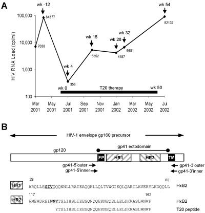
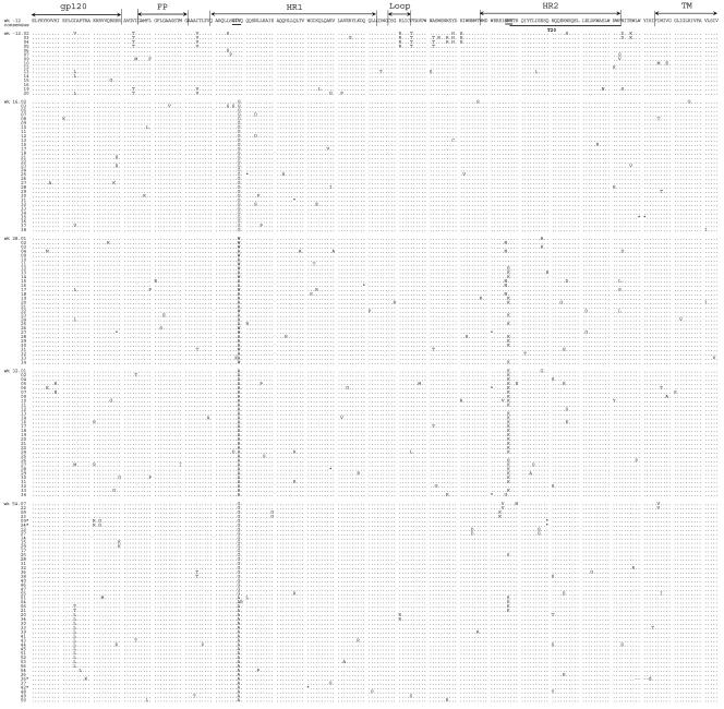
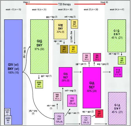
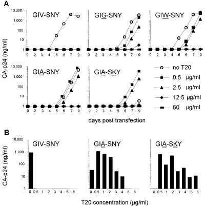
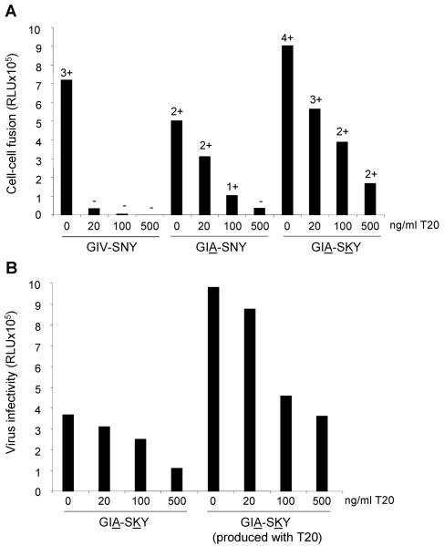
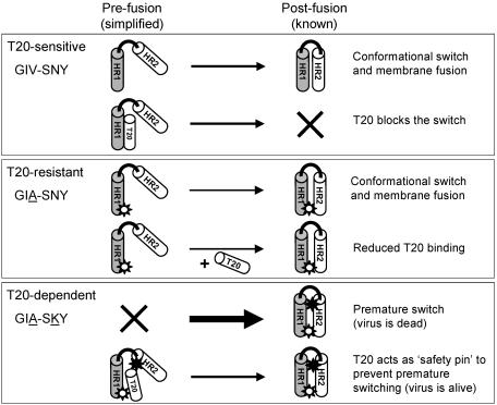
The fusion inhibitor T20 belongs to a new class of anti-human immunodeficiency virus type 1 (HIV-1) drugs designed to block entry of the virus into the host cell. However, the success of T20 has met with the inevitable emergence of drug-resistant HIV-1 variants. We describe an evolutionary pathway taken by HIV-1 to escape from the selective pressure of T20 in a treated patient. Besides the appearance of T20-resistant variants, we report for the first time the emergence of drug-dependent viruses with mutations in both the HR1 and HR2 domains of envelope glycoprotein 41. We propose a mechanistic model for the dependence of HIV-1 entry on the T20 peptide. The T20-dependent mutant is more prone to undergo the conformational switch that results in the formation of the fusogenic six-helix bundle structure in gp41. A premature switch will generate nonfunctional envelope glycoproteins (dead spikes) on the surface of the virion, and T20 prevents this abortive event by acting as a safety pin that preserves an earlier prefusion conformation.


Figures

FIG. 1.
FIG. 1.
(A) Plasma HIV-1 RNA load during T20 therapy. Plasma samples that were analyzed by sequencing are marked in the graph as week -12 (pretherapy), week 16, week 28, week 32, and week 54. T20 therapy was started at week 0 and stopped at week 50. At the initiation of T20 therapy, the patient also received 300 mg of ziduvidine, 150 mg of lamivudine, and three caps of lopinavir twice daily. The patient's treatment regimen was modified at week 16 to 150 mg of lamivudine, 600 mg of amprenavir, 100 mg of ritonavir, 90 mg of T20 twice daily, and 300 mg of tenofovir disoproxil fumarate once daily. (B) PCR amplification of the gp41 ectodomain. The HIV-1 env gene is shown (not to scale) with the location of primers used to amplify the gp41e region. The HXB2 amino acid sequences for HR1 and HR2 are indicated. The GIV sequence in HR1 and SNY sequence in HR2 (NNY for strain HXB2) are underlined and bolded. The T20 peptide sequence within HR2 is indicated.
FIG. 2.
FIG. 2.
Amino acid sequence of gp41e in individual HIV-1 clones from a patient under T20 therapy. The PCR fragments that were sequenced directly were ligated into the TA cloning vector, and 20 to 38 individual clones per time point were sequenced. Only the sequences that passed the quality control are shown (full-length sequence of correct product). All clones were aligned to a consensus sequence based on the 15 sequences of the pretreatment sample (week -12). Dots mark amino acids that are identical to this consensus sequence. Stop codons are indicated with *. The GIV and SNY motifs are underlined and in bold. The HR2 region corresponding to the T20 peptide sequence (amino acids 127 to 162) is underlined.
FIG. 3.
FIG. 3.
Evolution of T20 resistance in the ectodomain of HIV-1 gp41. This schematic of the viral quasispecies in the course of T20 therapy focuses exclusively on the GIV sequence in HR1 and the SNY sequence in HR2. The situation is depicted at week -12 (pretherapy), weeks 16, 28, and 32 (during T20 therapy), and week 54 (posttherapy). The number of clones that were sequenced per time point is indicated by n. The initial virus population present at week -12 consists of the fully wild-type sequence (GIV-SNY). The starting virus population is shown as a blue box, all single mutants are shown as hatched and colored boxes (e.g., GIG-SNY at week 16) and the double mutants in full color boxes (e.g., GIA-SKY at weeks 28 and 32). The sizes of the boxes reflect the abundance of that particular variant within the viral population. The actual percentage of that variant in the viral quasispecies and the number of clones with that sequence are indicated within the boxes. The evolution scheme was derived in part from inspection of the actual codon changes that are indicated alongside the arrows. The mutations are ranked according to their likelihood (2, 17, 18): the easy G-to-A and C-to-T transitions (1), the more difficult A-to-G and T-to-C transitions (2), and all transversions (3).
FIG. 4.
FIG. 4.
Replication of wild-type and mutant HIV-1 LAI molecular clones. (A) Viral replication curves over a 9-day period. The open-circle curve represents replication fitness in the absence of T20, and the other curves show the replication fitness of the mutants in the presence of different T20 concentrations. Similar results were obtained from six separate transfection experiments. (B) We used T20 concentrations that are comparable to that of patient plasma after twice daily subcutaneous 100-mg T20 injections (19). We only show CA-p24 values for day 6 posttransfection, which best represent the relative differences in replication capacity in the absence of T20 and the resistance-dependence phenotypes observed for the GIA-SNY and GIA-SKY mutants.
FIG. 5.
FIG. 5.
(A) T20-dependent Env has increased fusogenic activity. SupT1 cells were transfected with the mutants indicated below the x axis. One day later, transfected cells were mixed with SupT1 cells containing a Tat-responsive long terminal repeat-luciferase reporter gene construct. After 24 h, formation of syncytia was analyzed by light microscopy (−, no syncytia; ++++, all cells involved in syncytia) and quantitated by measurement of luciferase activity in cell extracts. We consistently measured reduced fusion activity (syncytia and luciferase counts) for the GIA mutant and increased activity for the GIA-SKY mutant. This is a representative experiment, which shows 126% fusion activity for the GIA-SKY mutant compared to the wild-type control (set at 100%). Similar results were obtained in independent experiments (134%, 136%, and 150%; results not shown). (B) T20 prevents the loss of infectivity of GIA-SKY virions. SupT1 cells containing a Tat-responsive long terminal repeat-luciferase reporter gene construct were infected with equal amounts of the GIA-SKY double mutant virus, which was produced in the presence (100 ng/ml) or absence of T20 and scored for infectivity in a single-cycle infection assay. Virus infectivity is expressed in relative light units (RLU) from a luciferase assay performed on the cell lysate.
FIG. 6.
FIG. 6.
Biophysical characterization of the wild-type and mutant gp41 ectodomain cores. (A) Schematic representation of HIV-1 gp41. The important functional features of gp41 and the sequences of the N36 and C34 segments are shown. Recombinant N36(L6)C34 protein consists of the N36 and C34 peptides plus a six-residue linker. The disulfide bond and four potential N-glycosylation sites are depicted. The residues are numbered according to their position in gp160 of the HIV-1 HXB2 strain. (B) Thermal melts of the wild-type peptide N36(L6)C34 (solid fill) and the GIA (open) and GIA-SKY (dotted fill) mutants monitored by circular dichroism at 222 nm at 10 μM protein concentration in phosphate-buffered saline. We performed thermal unfolding experiments four times for the wild-type and mutant peptides. The error of Tm determination is ±0.5°C. It should be emphasized that melting temperature is useful only as a qualitative guide to stability. (C) Representative sedimentation equilibrium data for the GIA-SKY mutant (≈30 μM) were collected at 20°C and 18,000 rpm in phosphate-buffered saline. The data fit closely to a trimer model. The deviation in the data from the linear fit for a trimer model is plotted (upper).
FIG. 7.
FIG. 7.
Proposed model for T20-dependent viral entry. Each box depicts one of three scenarios: T20-sensitive (GIV-SNY), T20-resistant (GIA-SNY), and T20-dependent (GIA-SKY). A simplified gp41 ectodomain comprising only one subunit of HR1 (shaded cylinder) and HR2 (white cylinder) joined by a loop region (black line) is used to depict the prefusion and postfusion states of the peptide. The thickness of the arrows represents the speed of the conformational switch between pre- and postfusion conformations. A white star represents the GIA mutation in HR1, and a black star represents the SKY mutation in HR2. Explanations for each reaction are provided on the right hand side.

References

    1. Baldwin, C. E., R. W. Sanders, and B. Berkhout. 2003. Inhibiting HIV-1 entry with fusion inhibitors. Curr. Med. Chem. 10:1633-1642. - PubMed
    1. Berkhout, B., A. T. Das, and N. Beerens. 2001. HIV-1 RNA editing, hypermutation and error-prone reverse transcription. Science 292:7. - PubMed
    1. Boom, R., C. J. A. Sol, M. M. M. Salimans, C. L. Jansen, P. M. E. Wertheim-van Dillen, and J. Van der Noordaa. 1990. A rapid and simple method for purification of nucleic acids. J. Clin. Microbiol. 28:495-503. - PMC - PubMed
    1. Cantor, C., and P. Schimmel. 1980. Biophysical chemistry, part III. W. H. Freeman and Company, New York, N.Y.
    1. Chan, D. C., D. Fass, J. M. Berger, and P. S. Kim. 1997. Core structure of gp41 from the HIV envelope glycoprotein. Cell 89:263-273. - PubMed

Publication types