. 2004 Nov;78(22):12446-54.
doi: 10.1128/JVI.78.22.12446-12454.2004.

Structural basis for coevolution of a human immunodeficiency virus type 1 nucleocapsid-p1 cleavage site with a V82A drug-resistant mutation in viral protease

Affiliations

Structural basis for coevolution of a human immunodeficiency virus type 1 nucleocapsid-p1 cleavage site with a V82A drug-resistant mutation in viral protease

Moses Prabu-Jeyabalan et al. J Virol. 2004 Nov.

Abstract

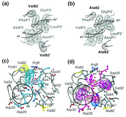
Maturation of human immunodeficiency virus (HIV) depends on the processing of Gag and Pol polyproteins by the viral protease, making this enzyme a prime target for anti-HIV therapy. Among the protease substrates, the nucleocapsid-p1 (NC-p1) sequence is the least homologous, and its cleavage is the rate-determining step in viral maturation. In the other substrates of HIV-1 protease, P1 is usually either a hydrophobic or an aromatic residue, and P2 is usually a branched residue. NC-p1, however, contains Asn at P1 and Ala at P2. In response to the V82A drug-resistant protease mutation, the P2 alanine of NC-p1 mutates to valine (AP2V). To provide a structural rationale for HIV-1 protease binding to the NC-p1 cleavage site, we solved the crystal structures of inactive (D25N) WT and V82A HIV-1 proteases in complex with their respective WT and AP2V mutant NC-p1 substrates. Overall, the WT NC-p1 peptide binds HIV-1 protease less optimally than the AP2V mutant, as indicated by the presence of fewer hydrogen bonds and fewer van der Waals contacts. AlaP2 does not fill the P2 pocket completely; PheP1' makes van der Waals interactions with Val82 that are lost with the V82A protease mutation. This loss is compensated by the AP2V mutation, which reorients the peptide to a conformation more similar to that observed in other substrate-protease complexes. Thus, the mutant substrate not only binds the mutant protease more optimally but also reveals the interdependency between the P1' and P2 substrate sites. This structural interdependency results from coevolution of the substrate with the viral protease.


Figures

FIG. 1.
FIG. 1.
Conformation of the NC-p1 substrates. Stereo diagram of the superposition of the WT (cyan) and AP2V (magenta) variants of NC-p1 peptides. An arrow indicates the difference in conformation of the scissile bonds between the two structures. The superposition was based on structurally conserved parts of the respective protease structures. Disordered side chains, whose conformation could not be resolved in the electron density, have their names listed in parentheses.
FIG. 2.
FIG. 2.
Double difference plots. Relative shifts in the V82A complexes in reference to the corresponding WT complexes with NC-p1 (a), CA-p2 (b), and p1-p6 (c). Double difference plots contour differences in internal Cα-Cα distances between two complexes. Each contour line represents a deviation by 0.25 Å. Black, green, blue, and red distinguish the contour ranges −1.0 Å and below, −1.0 to −0.5 Å, 0.5 to 1.0 Å, and 1 Å and above, respectively. In panel a, box A corresponds to conformational changes between the P1 loops (Gly78-Asn83) from each of the two monomers, and boxes B1 and B2 correspond to the conformational changes between the P1 loop with respect to the flap (Ile50) within each of the two monomers. All of the boxed regions emphasize internal regions of the protease whose relative conformation has changed by more than 1.0 Å between the two complexes.
FIG. 3.
FIG. 3.
Superposition of WT and V82A complexes. (a) Stereo diagram of the superimposed active site residues in the WTNC-p1WT (in gray and cyan) and AP2VNC-p1V82A (in magenta and purple) complexes. Protease residues within 4.2Å of the substrate are shown in ball-and-stick representation, and residue 82 is highlighted in yellow. The substrate mutation site (P2) is circled, and regions exhibiting large structural changes are indicated with arrows. Similarly, panels b and c show the superposition of CA-p2 and p1-p6, respectively, in complex with the WT and V82A protease variants. Note that there are virtually no conformational changes seen between the WT and V82A complexes in these two complexes.
FIG. 4.
FIG. 4.
Variations in the substrate-protease hydrogen bonds between the WTNC-p1WT and AP2VNC-p1V82A complexes. In the superimposed active site regions of the WTNC-p1WT (gray and cyan) and the AP2VNC-p1V82A (magenta and purple) complexes, only those side chains that form protease-substrate hydrogen bonds are explicitly shown (Asn 25, 25′; Asp29, 29′; Asp30, 30′; and AsnP1). Water molecules are shown with van der Waals surfaces. The AP2VNC-p1V82A complex forms additional hydrogen bonds compared with the WTNC-p1WT complex. These are highlighted with dotted yellow lines (Tables 2 and 3).
FIG. 5.
FIG. 5.
Variations in the substrate-protease van der Waals interactions between the WTNC-p1WT and AP2VNC-p1V82A complexes. van der Waals surfaces of protease residue 82, viewed down the dimer twofold axis, and neighboring atoms of the corresponding NC-p1 substrate: (a) WTNC-p1WT complex (note that Val82 contacts PheP1′) and (b) AP2VNC-p1V82A complex (note that no contact is made between Ala82 and the substrate). Substrates and proteases are distinguished in gray and black, respectively. A Cα trace of the overlying flaps is also shown. (c) Active site residues in the WTNC-p1WT. The protease is shown in gray and the substrate in cyan. Protease residues within 4.2Å of the substrate are shown in ball-and-stick representation, and residue 82 is highlighted in yellow. van der Waals contacts that are lost compared with the AP2VNC-p1V82A complex are highlighted with dotted surfaces and the P2 site is circled. (d) Active site residues in the AP2VNC-p1V82A. The protease is shown in gray, and the substrate is shown in magenta; protease residues within 4.2Å of the substrate are shown in ball-and-stick representation, and residue 82 is highlighted in yellow. van der Waals contacts that are lost compared with the WTNC-p1WT complex are highlighted with dotted surfaces, and the P2 site is circled.

References

    1. Bally, F., R. Martinez, S. Peters, P. Sudre, and A. Telenti. 2000. Polymorphism of HIV-1 type 1 Gag p7/p1 and p1/p6 cleavage sites: clinical significance and implications for resistance to protease inhibitors. AIDS Res. Hum. Retrovir. 16:1209-1213. - PubMed
    1. Chapman, M. S. 1995. Restrained real-space macromolecular refinement using a new resolution-dependent electron density function. Acta Crystallogr. Sect. D 51:69-80. - PubMed
    1. Chou, K.-C., A. G. Tomasselli, I. M. Reardon, and R. L. Heinrikson. 1996. Predicting human immunodeficiency virus protease cleavage sites in proteins by a discriminant function method. Proteins 24:51-72. - PubMed
    1. Collaborative Computational Project, Number 4. 1994. The CCP4 suite: programs for protein crystallography. Acta Crystallogr. Sect. D 50:760-763. - PubMed
    1. Condra, J. H., W. A. Schleif, O. M. Blahy, L. J. Gabryelski, D. J. Graham, J. C. Quintero, A. Rhodes, H. L. Robbins, E. Roth, M. Shivaprakash, D. Titus, T. Yang, H. Teppler, K. E. Squires, P. J. Deutsch, and E. Emini. 1995. In vivo emergence of HIV-1 variants resistant to multiple protease inhibitors. Nature 374:569-571. - PubMed

Publication types

LinkOut - more resources