

doi: 10.1128/JVI.78.22.12683-12688.2004.
The nucleoprotein is required for efficient coronavirus genome replication
Affiliations
- PMID: 15507657
- PMCID: PMC525053
- DOI: 10.1128/JVI.78.22.12683-12688.2004
Item in Clipboard
The nucleoprotein is required for efficient coronavirus genome replication
J Virol.
2004 Nov.
Abstract
The construction of a set of transmissible gastroenteritis coronavirus (TGEV)-derived replicons as bacterial artificial chromosomes is reported. These replicons were generated by sequential deletion of nonessential genes for virus replication, using a modified TGEV full-length cDNA clone containing unique restriction sites between each pair of consecutive genes. Efficient activity of TGEV replicons was associated with the presence of the nucleoprotein provided either in cis or in trans. TGEV replicons were functional in several cell lines, including the human cell line 293T, in which no or very low cytopathic effect was observed, and expressed high amounts of heterologous protein.
Figures
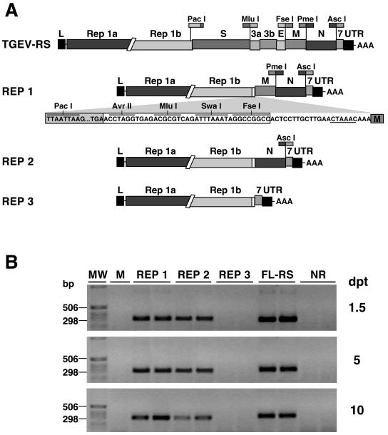
Functional analysis of TGEV-derived replicons. (A) Genetic structures of TGEV-derived replicons. The genetic structures of the TGEV cDNA clone (TGEV-RS) and the replicons generated from this cDNA (REP 1, REP 2, and REP 3) are illustrated. To construct REP 1, a 125-bp fragment containing the restriction sites PacI, AvrII, MluI, SwaI, and FseI, generated by PCR using two overlapping oligonucleotides, was cloned in TGEV-RS digested with PacI and FseI. REP 2 and REP 3 were generated from REP 1 by deletion of fragments SwaI-PmeI and SwaI-AscI, respectively. To avoid possible interferences with the expression of heterologous genes, the TRS of the S gene, located at the 3′ end of the replicase gene, was eliminated by introduction of three silent point mutations in its conserved core sequence. Letters and numbers above the bars indicate the viral genes. L, leader sequence; UTR, untranslated region. Relevant restriction sites are indicated. The core sequence is underlined. (B) Functional analysis of TGEV-derived replicons by RT-PCR. Human 293T cells were mock transfected (M) or transfected with TGEV replicons (REP 1, 2, and 3), the full-length cDNA clone (FL-RS), or a nonreplicative cDNA clone (NR), using Lipofectamine 2000 (Invitrogen) according to the manufacturer's specifications. Total intracellular RNA was isolated at 1.5, 5, and 10 dpt and analyzed by RT-PCR with specific oligonucleotides to detect gene 7 mRNA. Duplicate RT-PCR products amplified in parallel were resolved by electrophoresis in 1% agarose gels. MW, molecular weight markers.
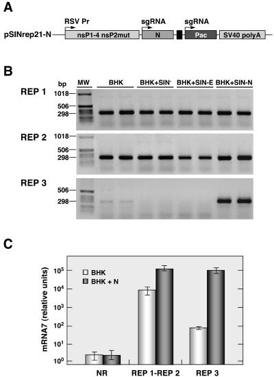
Effect of N protein on TGEV-derived replicon activity. (A) Scheme of the Sindbis virus replicon construct encoding the TGEV N gene. RSV Pr, Rous sarcoma virus promoter; sgRNA, subgenomic RNA; nsP1-4, nonstructural proteins 1 to 4; nsP2mut, mutant of nonstructural protein 2; Pac, puromycin resistance gene; SV40 poly A, transcription-termination polyadenylation signal from simian virus 40. (B) RT-PCR analysis of gene 7 mRNA. BHK-pAPN cells (BHK) or BHK-pAPN cells transformed with either the Sindbis virus replicon pSINrep21 alone (BHK + SIN−) or pSINrep21 expressing TGEV E protein (BHK + SIN-E) or TGEV N protein (BHK + SIN-N) were transfected with TGEV replicons (REP 1, 2, and 3), using Lipofectamine 2000 (Invitrogen) following the manufacturer's specifications. At 36 hpt total intracellular RNA was extracted and used as the template for RT-PCR analysis with specific primers to detect gene 7 mRNA. Duplicate RT-PCR products amplified in parallel were resolved by electrophoresis in 1% agarose gels. MW, molecular weight markers. (C) Real-time RT-PCR quantification of gene 7 mRNA. The amount of mRNA7, expressed as relative units, was determined by real-time RT-PCR with specific oligonucleotides to detect gene 7 mRNA in RNA samples isolated at 36 hpt from BHK-pAPN cells (BHK) or BHK-pAPN cells expressing N protein (BHK + N) transfected with either a nonreplicative cDNA clone (NR), REP 1, REP 2, or REP 3. REP 1-REP 2 indicates mean values from REP 1 and 2, both encoding the N gene. Mean values from three experiments are represented, with standard deviations shown as error bars.
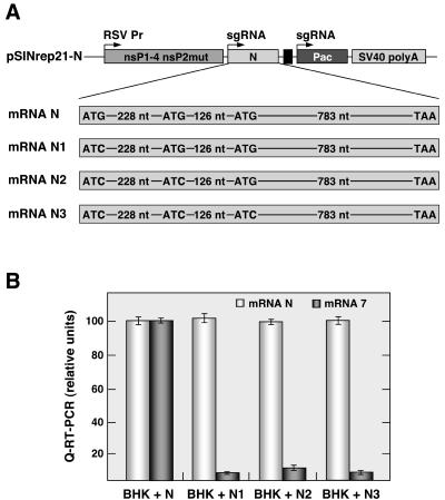
Effect of N protein and its mRNA on TGEV-derived replicon activity. (A) Schematic representation of the Sindbis virus replicon encoding the different gene N mRNA mutants. The positions of the three ATGs in the N gene coding sequence, as well as the point mutations introduced, are indicated. The designations above the top bar are the same that those described for Fig. 2A. (B) Real-time RT-PCR quantification of genes 7 and N mRNAs. The amounts of genes 7 and N mRNAs, expressed as relative units, were determined by real-time RT-PCR with specific oligonucleotides in RNA samples isolates at 36 hpt from BHK cells expressing the wild-type gene N mRNA (BHK + N) or gene N mRNA with the first ATG mutated (BHK + N1), with the two first ATGs mutated (BHK + N2), or with the three ATGs mutated (BHK + N3) transfected with REP 3. Mean values from two experiments are represented, with standard deviations shown as error bars.
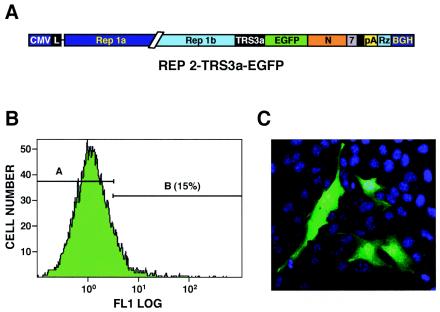
Analysis of EGFP expression by TGEV-derived REP 2. (A) Schematic structure of the cDNA encoding the REP2-TRS3a-EGFP RNA with the EGFP gene under the control of gene 3a TRS. The numbers and letters inside the boxes indicate the viral genes. L, leader sequence; CMV, cytomegalovirus immediate-early promoter; pA, poly(A); Rz, hepatitis delta virus ribozyme; BGH, bovine growth hormone termination and polyadenylation sequences. (B) Flow cytometry analysis of EGFP expression in BHK-pAPN cells transfected with REP2-TRS3a-EGFP. A and B represent the EGFP-negative and -positive cell populations, respectively. Green fluorescence intensity is revealed in logarithmic units on the x axis. (C) EGFP expression analyzed by confocal microscopy of BHK-pAPN cells transfected with REP2-TRS3a-EGFP. Cell nuclei were stained with TOPRO 3 (Molecular Probes).
References
-
- Delmas, B., J. Gelfi, H. Sjöström, O. Noren, and H. Laude. 1994. Further characterization of aminopeptidase-N as a receptor for coronaviruses. Adv. Exp. Med. Biol. 342:293-298. - PubMed
MeSH terms
Substances
LinkOut - more resources
Full Text Sources
Other Literature Sources
