

Intratumoral patterns of clonal evolution in meningiomas as defined by multicolor interphase fluorescence in situ hybridization (FISH): is there a relationship between histopathologically benign and atypical/anaplastic lesions?
- PMID: 15507670
- PMCID: PMC1867491
- DOI: 10.1016/S1525-1578(10)60527-2
Intratumoral patterns of clonal evolution in meningiomas as defined by multicolor interphase fluorescence in situ hybridization (FISH): is there a relationship between histopathologically benign and atypical/anaplastic lesions?
Abstract
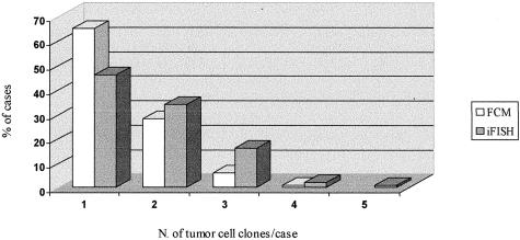
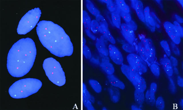
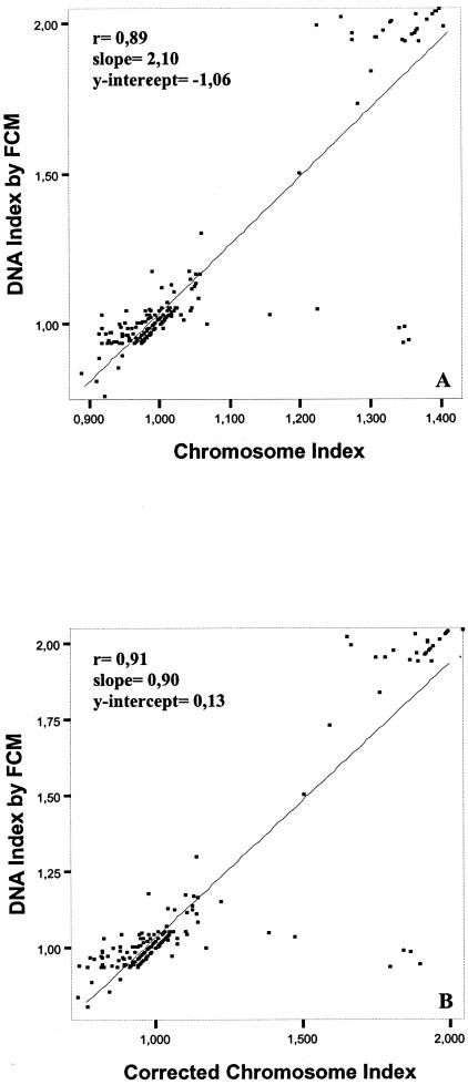
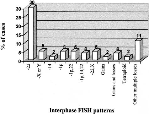
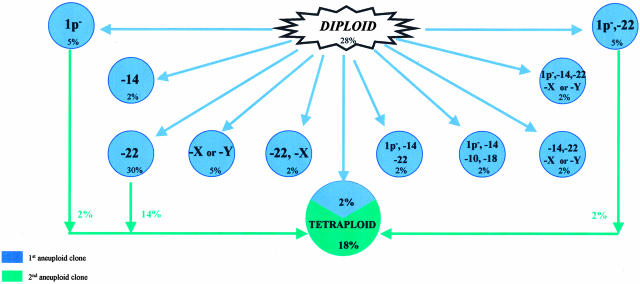
Meningiomas are cytogenetically heterogeneous tumors in which chromosome gains and losses frequently occur. Based on the intertumoral cytogenetic heterogeneity of meningiomas, hypothetical models of clonal evolution have been proposed in these tumors which have never been confirmed at the intratumoral cell level. The aim of this study was to establish the intratumoral patterns of clonal evolution associated with chromosomal instability in individual patients as a way to establish tumor progression pathways in meningiomas and their relationship with tumor histopathology and behavior. A total of 125 meningioma patients were analyzed at diagnosis. In all cases, multicolor interphase fluorescence in situ hybridization (iFISH) studies were performed on fresh tumor samples for the detection of quantitative abnormalities for 11 different chromosomes. In addition, overall tumor cell DNA content was measured in parallel by flow cytometry. iFISH studies were also performed in parallel on tissue sections in a subset of 30 patients. FISH studies showed that 56 (45%) of the 125 cases analyzed had a single tumor cell clone, all these cases corresponding to histologically benign grade I tumors. In the remaining cases (55%) more than one tumor cell clone was identified: two in 45 cases (36%), three in 19 (15%), and four or more clones in five cases (4%). Overall, flow cytometric analysis of cell DNA contents showed the presence of DNA aneuploidy in 44 of these cases (35%), 30% corresponding to DNA hyperdiploid and 5% to hypodiploid cases; from the DNA aneuploid cases, 35 (28%) showed two clones and 9 (7%) had three or more clones. A high degree of correlation (r >/= 0.89; P < 0.001) was found between FISH and flow cytometry as regards the overall quantitative DNA changes detected with both techniques, the former being more sensitive. Among the cases with chromosome abnormalities, the earliest tumor cell clone observed was frequently characterized by the loss of one or more chromosomes (64% of all meningiomas); loss of either a single chromosome 22 or, less frequently, of a sex chromosome (X or Y) and del (1p) was commonly found as the single initial cytogenetic aberration (30%, 5%, and 5% of the cases, respectively). Interestingly, an isolated loss of chromosome 22 was only found as the initial abnormality in one out of 14 atypical/anaplastic meningiomas, while the same cytogenetic pattern was present in the ancestral tumor cell clone of 32% of the benign tumors. Cytogenetic patterns based on chromosome gains were found in the ancestral tumor cell clone in 4% of the patients, 2% corresponding to tetraploid tumors. Overall, cytogenetic evolution of the earliest tumor cell clones was frequently associated with tetraploidization (31%). Our results show that meningiomas are genetically heterogeneous tumors that display different patterns of numerical chromosome changes, with the presence of more than one tumor cell clone detected in almost half of the cases including all atypical/anaplastic cases. Interestingly, the pathways of intratumoral clonal evolution observed in the benign tumors were different from those observed in atypical/anaplastic meningiomas, suggesting that the latter tumors might not always represent a more advanced stage of histologically benign meningiomas.
Figures
References
-
- Zattara-Cannoni H, Gambarelli D, Dufour H, Figarella D, Vollot F, Grisoli F, Vagner-Capodano AM. Contribution of cytogenetics and FISH in the diagnosis of meningiomas: a study of 189 tumors. Ann Genet. 1998;41:164–175. - PubMed
-
- Ketter R, Henn W, Niedermayer I, Steilen-Gimbel H, Konig J, Zang KD, Steudel WI. Predictive value of progression-associated chromosomal aberrations for the prognosis of meningiomas: a retrospective study of 198 cases. J Neurosurg. 2001;95:601–607. - PubMed
-
- Zang KD. Meningioma: a cytogenetic model of a complex benign human tumor, including data on 394 karyotyped cases. Cytogenet Cell Genet. 2001;93:207–220. - PubMed
-
- Cai DX, Banerjee R, Scheithauer BW, Lohse CM, Kleinschmidt-Demasters BK, Perry A. Chromosome 1p and 14q FISH analysis in clinicopathologic subsets of meningioma: diagnostic and prognostic implications. J Neuropathol Exp Neurol. 2001;60:628–636. - PubMed
Publication types
MeSH terms
Substances
LinkOut - more resources
Full Text Sources
