. 2004 Nov;165(5):1519-33.
doi: 10.1016/S0002-9440(10)63410-4.

T-cell properties determine disease site, clinical presentation, and cellular pathology of experimental autoimmune encephalomyelitis

Affiliations

T-cell properties determine disease site, clinical presentation, and cellular pathology of experimental autoimmune encephalomyelitis

Sara Abromson-Leeman et al. Am J Pathol. 2004 Nov.

Abstract

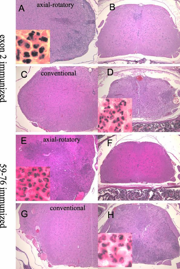
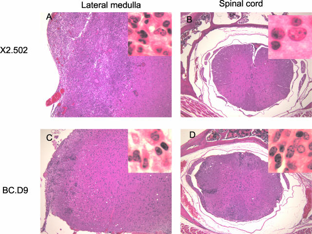
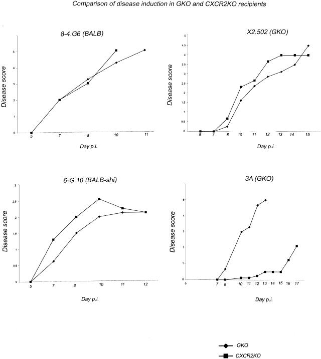
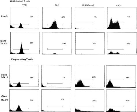
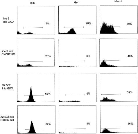
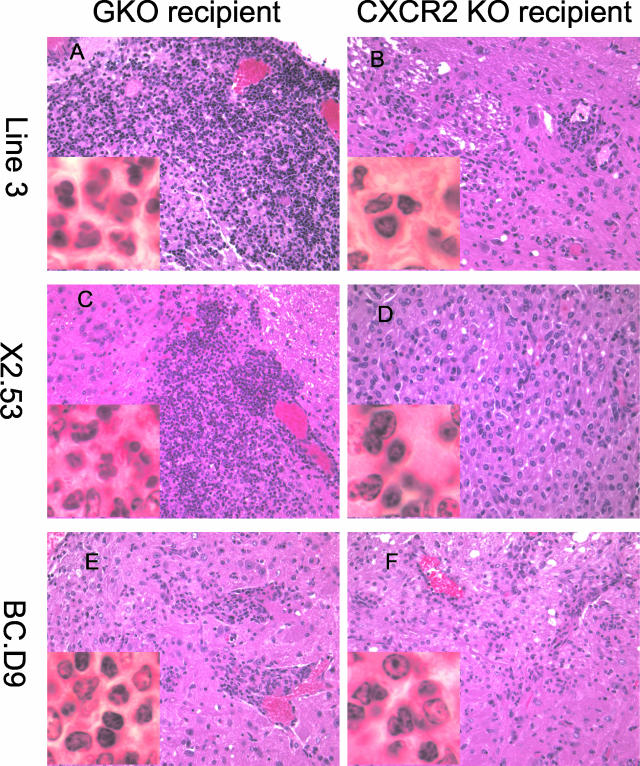
Two distinct clinical phenotypes of experimental autoimmune encephalomyelitis are observed in BALB interferon-gamma knockout mice immunized with encephalitogenic peptides of myelin basic protein. Conventional disease, characterized by ascending weakness and paralysis, occurs with greater frequency after immunizing with a peptide comprising residues 59 to 76. Axial-rotatory disease, characterized by uncontrolled axial rotation, occurs with greater frequency in mice immunized with a peptide corresponding to exon 2 of the full length 21.5-kd protein. The two clinical phenotypes are histologically distinguishable. Conventional disease is characterized by inflammation and demyelination primarily in spinal cord, whereas axial-rotatory disease involves inflammation and demyelination of lateral medullary areas of brain. Both types have infiltrates in which neutrophils are a predominating component. By isolating T cells and transferring disease to naive recipients, we show here that the type of disease is determined entirely by the inducing T cell. Furthermore, studies using CXCR2 knockout recipients, unable to recruit neutrophils to inflammatory sites, show that although neutrophils are critical for some of these T cells to effect disease, there are also interferon-gamma-deficient T cells that induce disease in the absence of both interferon-gamma and neutrophils. These results highlight the multiplicity of T-cell-initiated effector pathways available for inflammation and demyelination.


Figures

Figure 1
Figure 1
Comparison of histopathology of actively induced axial-rotatory and conventional EAE in BALB-GKO mice. H&E-stained sections from mice immunized with either MBP exon 2 peptide (A–D) or MBP 59-76 peptide (E–H). A, C, E, G: Lateral medullary regions of brain. B, D, F, H: Spinal cord. A and B are from an exon 2-immunized mouse with axial-rotatory disease. C and D are from an exon 2-immunized mouse with conventional ascending paralysis. E and F are from a peptide 59-76-immunized mouse with axial-rotatory disease. G and H are from a peptide 59-76-immunized mouse with conventional ascending disease. Original magnifications: ×100; ×1000 (insets).
Figure 2
Figure 2
Sections of lateral medulla and spinal cord from BALB-GKO recipients of GKO-derived clones X2.502 (A and B) and IFN-γ-secreting clone BC.D9 (C and D). A and C show lateral medulla. B and D show corresponding spinal cord sections. All sections are H&E stained. Original magnifications: ×100; ×1000 (insets).
Figure 3
Figure 3
Analysis of intraexon 2 peptide epitopes recognized by exon 2-specific T cells. Proliferative responses of line 3 and clone 3a.56 (from line 3), clones X2.51 and X2.502 (both from line 1), and two IFN-γ-secreting clones 6-G.10 and BC.D9, to the full length exon 2 peptide (residues 1 to 26), or peptides of exon 2 residues 1 to 15, 6 to 20, or 11 to 26. Results show mean cpm of 3H-thymidine incorporated in triplicate wells. Results shown are for 10 μg/ml of each peptide.
Figure 4
Figure 4
A representative experiment showing clinical outcomes after injecting two IFN-γ-secreting T-cell lines (8-4.G6 and 6-G.10) and two GKO lines (X2.502 and line 3) into either BALB-GKO or BALB-CXCR2 KO recipients. Each T-cell line was activated in vitro, then injected in parallel into recipient groups of five mice each. Clinical scores were assigned as described in Materials and Methods. Both IFN-γ-secreting clones, shown at left, induced essentially identical disease with similar kinetics in either GKO or CXCR2 KO recipient mice. GKO clone X2.502 (derived from line 1), was also able to induce EAE with essentially the same kinetics and severity whether or not recipients expressed CXCR2. In contrast, GKO-derived line 3 is critically dependent on neutrophil recruitment to effect clinical disease.
Figure 5
Figure 5
Flow cytometric profiles of mononuclear cells isolated from CNS of diseased GKO recipient mice 10 days after T-cell injections. T cells injected were line 3 or X2.502, from GKO mice (top) or IFN-γ-secreting clones 6-G.10 and BC.D9 (bottom). Mononuclear cells were isolated on Percoll gradients, as described in Materials and Methods. Monoclonal antibodies used were H57-597, to detect TCR+ cells, anti-Gr-1 (RB6-8C5) to detect neutrophils, anti-MHC class II I-Ad/I-Ed (2G9) and anti-Mac-1 (CD11b) antibody M1/70, which binds to macrophages (lower intensity peak) and neutrophils (higher intensity peak). Analysis was done on three to five mice per group; representative examples from individual mice are shown.
Figure 6
Figure 6
Comparative quantitation of mononuclear cell composition from CNS of GKO versus CXCR2 KO recipients of line 3 (top rows) and clone X2.502 (bottom rows). Cells from diseased recipients were isolated by Percoll gradient 12 days after injection. Monoclonal antibodies were used to detect TCR+ cells (H57-597), neutrophils (anti-Gr-1), or Mac-1+ cells (anti-CD11b). Profiles shown are from individual representative mice; three to five mice per group were injected and analyzed.
Figure 7
Figure 7
H&E-stained photographs of lateral medulla from either GKO (A, C, E) or CXCR2 KO (B, D, F) recipients of T cells. A and B: Day 14 after injection of line 3 into GKO (A) or CXCR2 KO (B) recipients. C and D: Day 11 after injection of clone X2.53 into GKO (C) or CXCR2 KO (D) recipients. E and F: Day 17 after injection of clone BC.D9 into GKO (E) or CXCR2 KO (F) recipients. Original magnifications: ×400; ×1000 (insets).
Figure 8
Figure 8
Comparison of RPA results for a panel of chemokines is shown. A: mRNA expressed in brain (B, lanes 1 and 3) or spinal cord (SC, lanes 2 and 4) of GKO recipients of either 6-G.10 (IFN-γ-producing, lanes 1 and 2) or line 3 (non-IFN-γ-producing, lanes 3 and 4) T cells. B: Decreased MIP-2 in a CXCR2KO recipient of X2.502 (lane 2), as compared with MIP-2 in a CXCR2-expressing (GKO) recipient of this clone (lane 1). C: RPA of brain tissue from recipients of a C57BL/6-GRKO (B6-IFN-γR knockout)-derived myelin oligodendrocyte glycoprotein-specific clone, clone G. The activated clone was transferred in parallel into two wild-type C57BL/6 recipients (lanes 1 and 2) or two B6-GRKO recipients (lanes 3 and 4).

References

    1. Abromson-Leeman S, Hayashi M, Martin C, Sobel R, al-Sabbagh A, Weiner H, Dorf ME. T cell responses to myelin basic protein in experimental autoimmune encephalomyelitis-resistant BALB/c mice. J Neuroimmunol. 1993;45:89–101. - PubMed
    1. Abromson-Leeman S, Alexander J, Bronson R, Carroll J, Southwood S, Dorf M. Experimental autoimmune encephalomyelitis-resistant mice have highly encephalitogenic myelin basic protein (MBP)-specific T cell clones that recognize a MBP peptide with high affinity for MHC class II. J Immunol. 1995;154:388–398. - PubMed
    1. Abromson-Leeman S, Bronson R, Dorf ME. Experimental autoimmune peripheral neuritis induced in BALB/c mice by myelin basic protein-specific T cell clones. J Exp Med. 1995;182:587–592. - PMC - PubMed
    1. Yoshizawa I, Bronson R, Ben-Nun A, Richert JR, Dorf ME, Abromson-Leeman S. Differential recognition of MBP epitopes in BALB/c mice determines the site of inflammatory disease induction. J Neuroimmunol. 1998;89:73–82. - PubMed
    1. Yoshizawa I, Bronson R, Dorf ME, Abromson-Leeman S. T-cell responses to myelin basic protein in normal and MBP-deficient mice. J Neuroimmunol. 1998;84:131–138. - PubMed

Publication types

MeSH terms

LinkOut - more resources