

Rheumatic heart disease: proinflammatory cytokines play a role in the progression and maintenance of valvular lesions
- PMID: 15509528
- PMCID: PMC1618676
- DOI: 10.1016/S0002-9440(10)63415-3
Rheumatic heart disease: proinflammatory cytokines play a role in the progression and maintenance of valvular lesions
Abstract
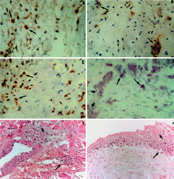
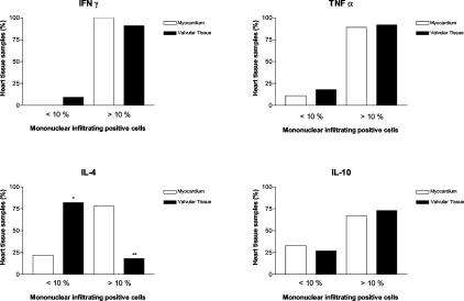
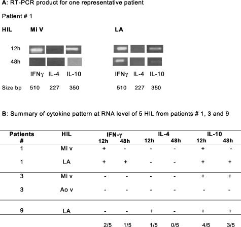
Heart lesions of rheumatic heart disease (RHD) patients contain T-cell clones that recognize heart proteins and streptococcal M peptides. To functionally characterize heart-infiltrating T lymphocytes, we evaluated their cytokine profile, both directly in situ and in T-cell lines derived from the heart (HIL). Interferon (IFN)-gamma, tumor necrosis factor (TNF)-alpha, interleukin (IL)-4, and IL-10 expressions were characterized in 20 heart tissue infiltrates from 14 RHD patients by immunohistochemistry. IFN-gamma-, TNF-alpha-, and IL-10-positive cells were consistently predominant, whereas IL-4 was scarce in the valves. In agreement with these data, the in vitro experiments, in which 13 HILs derived from heart samples of eight patients were stimulated with M5 protein and the immunodominant M5 (81-96) peptide, IL-4 was detected in HIL derived from the atrium (three of six) but not from the valve (zero of seven). IFN-gamma and IL-10 production were detected in culture supernatants in 11 of 13 and 6 of 12 HILs, respectively. The predominant IFN-gamma and TNF-alpha expression in the heart suggests that Th1-type cytokines could mediate RHD. Unlike in reversible myocardium inflammation, the significantly lower IL-4 expression in the valvular tissue (P = 0.02) may contribute to the progression of the RHD leading to permanent valvular damage (relative risk, 4.3; odds ratio, 15.8). The lack of IL-4 in vitro production by valve-derived HIL also emphasizes the more severe tissue destruction in valves observed in RHD.
Figures
Similar articles
-
Rheumatic fever: how S. pyogenes-primed peripheral T cells trigger heart valve lesions.Ann N Y Acad Sci. 2005 Jun;1051:132-40. doi: 10.1196/annals.1361.054. Ann N Y Acad Sci. 2005. PMID: 16126952 Review.
-
Pathology and pathogenesis of rheumatic heart disease.Indian J Pathol Microbiol. 2007 Oct;50(4):685-97. Indian J Pathol Microbiol. 2007. PMID: 18306530 Review.
-
Heart-directed autoimmunity: the case of rheumatic fever.J Autoimmun. 2001 May;16(3):363-7. doi: 10.1006/jaut.2000.0487. J Autoimmun. 2001. PMID: 11334505
-
How an autoimmune reaction triggered by molecular mimicry between streptococcal M protein and cardiac tissue proteins leads to heart lesions in rheumatic heart disease.J Autoimmun. 2005 Mar;24(2):101-9. doi: 10.1016/j.jaut.2005.01.007. J Autoimmun. 2005. PMID: 15829402
-
Rheumatic fever and rheumatic heart disease: genetics and pathogenesis.Scand J Immunol. 2007 Aug-Sep;66(2-3):199-207. doi: 10.1111/j.1365-3083.2007.01974.x. Scand J Immunol. 2007. PMID: 17635797 Review.
Cited by
-
Cardiac valves: Another "Disaster-hit area" of COVID-19 patients?Heart Lung. 2020 Nov-Dec;49(6):890-891. doi: 10.1016/j.hrtlng.2020.05.004. Epub 2020 May 14. Heart Lung. 2020. PMID: 32410761 Free PMC article. No abstract available.
-
Rheumatic heart valve disease: navigating the challenges of an overlooked autoimmune disorder.Front Cardiovasc Med. 2025 Mar 13;12:1537104. doi: 10.3389/fcvm.2025.1537104. eCollection 2025. Front Cardiovasc Med. 2025. PMID: 40182432 Free PMC article. Review.
-
Effect of hydroxymethylglutaryl coenzyme-a reductase inhibitors on the long-term progression of rheumatic mitral valve disease.Circulation. 2010 May 18;121(19):2130-6. doi: 10.1161/CIRCULATIONAHA.109.891598. Epub 2010 May 3. Circulation. 2010. PMID: 20439789 Free PMC article.
-
Transfusion of fresh murine red blood cells reverses adverse effects of older stored red blood cells.Transfusion. 2011 Dec;51(12):2695-702. doi: 10.1111/j.1537-2995.2011.03197.x. Epub 2011 Jun 3. Transfusion. 2011. PMID: 21645005 Free PMC article.
-
Strategies in the development of vaccines to prevent infections with group A streptococcus.Hum Vaccin Immunother. 2013 Nov;9(11):2393-7. doi: 10.4161/hv.25506. Epub 2013 Jun 28. Hum Vaccin Immunother. 2013. PMID: 23863455 Free PMC article. Review.
References
-
- Stollerman GH. Rheumatogenic streptococci and autoimmunity. Clin Immunol Immunopathol. 1991;61:113–142. - PubMed
-
- Raizada V, Williams RC, Jr, Chopra P, Gopinath N, Prakash K, Sharma KB, Cherian KM, Panday S, Arora R, Nigam M, Zabriskie JB, Husby G. Tissue distribution of lymphocytes in rheumatic heart valves as defined by monoclonal anti-T cells antibodies. Am J Med. 1983;74:90–96. - PubMed
-
- Kemeny E, Grieve T, Marcus R, Sareli P, Zabriskie JB. Identification of mononuclear cells and T cell subsets in rheumatic valvulitis. Clin Immunol Immunopathol. 1989;52:225–237. - PubMed
-
- Roberts S, Kosanke S, Dunn ST, Jankelow D, Duran CMG, Cunningham MW. Pathogenic mechanisms in rheumatic carditis: focus on valvular endothelium. J Infect Dis. 2001;183:507–511. - PubMed
-
- Guilherme L, Cunha-Neto E, Coelho V, Snitcowsky R, Pomerantzeff PMA, Assis RV, Pedra F, Neumann J, Goldberg A, Patarroyo ME, Pillegi F, Kalil J. Human-infiltrating T cell clones from rheumatic heart disease patients recognize both streptococcal and cardiac proteins. Circulation. 1995;92:415–420. - PubMed
Publication types
MeSH terms
Substances
LinkOut - more resources
Full Text Sources
Other Literature Sources
