. 2004 Nov;165(5):1633-41.
doi: 10.1016/S0002-9440(10)63420-7.

PITX2 gain-of-function in Rieger syndrome eye model

Affiliations

PITX2 gain-of-function in Rieger syndrome eye model

Johan Holmberg et al. Am J Pathol. 2004 Nov.

Abstract

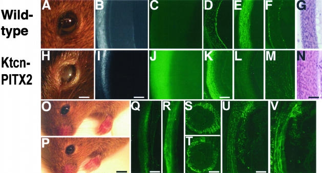
The human autosomal-dominant disorder Axenfeld-Rieger syndrome presents with defects in development of the eyes, teeth, and umbilicus. The eye manifests with iris ruptures, irido-corneal adhesions, cloudy corneas, and glaucoma. Transcription factors such as PITX2 and FOXC1 have been found to carry point mutations, causing the disorder. However, for approximately 40% of the cases, the pathogenesis is unknown. It has been reported that some mutations in PITX2 increase transactivation, whereas most mutations cause defects in DNA binding or transactivation. It is not known whether up-regulation of PITX2 activity can cause the disorder as well. Here we test this hypothesis directly by overexpressing PITX2A as a transgene in mouse corneal mesenchyme and iris, using keratocan-flanking sequences. The mice presented with corneal opacification, corneal hypertrophy, irido-corneal adhesions, and severely degenerated retina, resembling glaucoma. The corneal hypertrophy also resembles the corneal hypertrophy of Pitx2-/- mice. Control transgenic mice carrying point mutations T68P or K88E in PITX2A were normal. These findings indicate a novel pathogenetic mechanism in which excess corneal and iridal PITX2A cause glaucoma and anterior defects that closely resemble Axenfeld-Rieger syndrome.


Figures

Figure 1
Figure 1
A: Ktcn-PITX2 transgenic construct. The mouse keratocan (Ktcn) 5′ flank, promoter, noncoding exon 1, intron 1, and 6 bp of noncoding exon 2 are depicted together in the white box. The human PITX2A reading frame (gray box) and rabbit poly-A signals (black box) were fused to the Ktcn sequences. Key restriction enzyme sites are marked above the construct. Start and stop codons for PITX2A are marked below. The horizontal arrows depict location of PCR primers used for genotyping. The map is not to scale. The Ktcn-T68P and Ktcn-K88E constructs are shown below, with sites for point mutations introduced marked with vertical arrows. B: Genotyping and RT-PCR. Typical results from genotyping Ktcn-PITX2 transgenic (t) and wild-type (w) mouse tail DNA, using PCR (left), or Southern blotting (middle). Right: Results from RT-PCR of 1-month-old Ktcn-PITX2 mouse eyes.
Figure 2
Figure 2
Transgenic mice corneas become thick and cloudy. A–C: Wild-type 1-month-old mouse eyes; H–J: Ktcn-PITX2 1-month-old mouse eyes; D–G: wild-type 5-day-old corneas; K–N: Ktcn-PITX2 5-day-old corneas. A and H, Digital photographs; B and I, light microscopy photographs; C and J, whole-mount immunofluorescence of PITX2; D and K, immunofluorescence of sections with PITX2 antibody; E and L, immunofluorescence of sections with Ktcn antibody; F and M, immunofluorescence of sections with Ki-67 antibody; G and N, H&E-stained sections. O: Two-month-old Ktcn-K88E mouse; P, 2-month-old Ktcn-T68P mouse. Q and R: PITX2 immunostainings of 1-month-old cornea sections. Q, Ktcn-K88E; R, Ktcn-T68P. S and T: PITX2 immunostainings of E11.5 eyes. S, wild-type; T, Ktcn-PITX2. U and V: PITX2 immunostainings of E17.5 corneas and iris. U, wild-type; V, Ktcn-PITX2. Scale bars: 2 mm (H); 100 μm (I); 50 μm (K); 60 μm (N); 5 mm (P); 300 μm (Q); 150 μm (T); 200 μm (U).
Figure 3
Figure 3
Dramatic retinal degeneration in Ktcn-PITX2 retinas. Wild-type 3-month-old (A), Ktcn-PITX2 3-month-old (B), and Ktcn-PITX2 8-month-old (C) mice retinas were sectioned and stained with H&E. Arrows indicate retinal ganglion cells. D: Wild-type retina TUNEL assay; E: Ktcn-PITX2 retina TUNEL assay. F and G: H&E-stained sections of irido-corneal angles. F, Wild type, 9 days old, arrow indicates angle; G, Ktcn-PITX2, 9 days old. Scale bars: 20 μm (A); 150 μm (G).
Figure 4
Figure 4
Severe irido-corneal phenotype of newborn Ktcn-PITX2 mice. A–F: Wild-type 5-day-old littermate; G–M: Transgenic Ktcn-PITX2 mouse. A, G, M: H&E staining; B and H: immunofluorescence of PITX2; C, D, I, J: immunofluorescence of MKi-67; E and K: immunofluorescence of Ktcn; F and L: TUNEL assay of retinas. Scale bar, 40 μm.
Figure 5
Figure 5
Ktcn-PITX2 corneas display disrupted collagen morphology. A–D: Electron microscopy. A, D: Wild-type corneas. B, C: Ktcn-PITX2 corneas. Scale bars: 10 μm (A, C, D); 2 μm (B).
Figure 6
Figure 6
Model for gain-or loss-of-function in Rieger syndrome. Known heterozygous defects in the PITX2 gene can cause eye disorders. The human and mouse homozygotes are early embryonic lethal. These mice develop hypertrophic corneas. Heterozygous mice develop phenotypes resembling human patients, even the hypomorphic variants, with PITX2 expression levels between heterozygous and normal. The Rieger-syndrome-causing mutation V45L elevates PITX2 transactivating activity. The Ktcn-PITX2 mice overexpress PITX2 in the cornea and iris, resulting in hypertrophic corneas like the mouse Pitx2 homozygotes, and Rieger syndrome-like effects including irido-corneal adhesions and retinal degeneration/glaucoma.

Similar articles

Cited by

References

    1. Quigley HA. Number of people with glaucoma worldwide. Br J Ophthalmol. 1996;80:389–393. - PMC - PubMed
    1. Clark AF, Yorio T. Ophthalmic drug discovery. Nat Rev Drug Disc. 2003;2:448–459. - PubMed
    1. Amendt BA, Semina EV, Alward WLM. Rieger syndrome: a clinical, molecular, and biochemical analysis. Cell Mol Life Sci. 2000;57:1652–1666. - PMC - PubMed
    1. Gage PJ, Suh H, Camper SA. The bicoid-related Pitx gene family in development. Mamm Genome. 1999;10:197–200. - PubMed
    1. Lines MA, Kozlowski K, Walter MA. Molecular genetics of Axenfeld-Rieger malformations. Hum Mol Genet. 2002;11:1177–1184. - PubMed

Publication types

MeSH terms

LinkOut - more resources