. 2004 Nov;165(5):1653-62.
doi: 10.1016/S0002-9440(10)63422-0.

Effects of angiotensin II receptor signaling during skin wound healing

Affiliations

Effects of angiotensin II receptor signaling during skin wound healing

Hikaru Takeda et al. Am J Pathol. 2004 Nov.

Abstract

The tissue angiotensin (Ang) system, which acts independently of the circulating renin Ang system, is supposed to play an important role in tissue repair in the heart and kidney. In the skin, the role of the system for wound healing has remained to be ascertained. Our study demonstrated that oral administration of selective AngII type-1 receptor (AT(1)) blocker suppressed keratinocyte re-epithelization and angiogenesis during skin wound healing in rats. Immunoprecipitation and Western blot analysis indicated the existence of AT(1) and AngII type-2 receptor (AT(2)) in cultured keratinocytes and myofibroblasts. In a bromodeoxyuridine incorporation study, induction of AT(1) signaling enhanced the incorporation into keratinocytes and myofibroblasts. Wound healing migration assays revealed that induction of AT(1) signaling accelerated keratinocyte re-epithelization and myofibroblasts recovering. In these experiments, induction of AT(2) signaling acted vice versa. Taken together, our study suggests that skin wound healing is regulated by balance of opposing signals between AT(1) and AT(2).


Figures
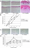
Figure 1
Figure 1
Effects of AT1 blocker on re-epithelization and angiogenesis during skin wound healing. A: Histological findings of the skin wounds on day 12 after wounding (H&E staining). The control group and the group with TCV-116, 1 mg/kg, showed 100% re-epithelization. The group with TCV-116, 10 mg/kg, showed delayed re-epithelization and delayed dermal repair. The crust still attached on the surface. Arrows indicate the re-epithelized edges. B: Time course of re-epithelization. The re-epithelization index (percentage of the wound site with new epithelization) was evaluated microscopically using tissue sections stained with H&E. Rats were orally administered with vehicle or TCV-116, 1 or 10 mg/kg/day. Data are represented as mean ± SD (n = 5). *, Significantly different from controls (P < 0.05). Administration of TCV-116 suppressed the re-epithelization dose dependently and postponed the day of 100% re-epithelization. C: Immunohistochemical staining for von Willebrand factor of the wound bed on day 9. The control wound showed many blood vessels. Administration of TCV-116 suppressed vascular growth dose dependently. D: Time course of angiogenesis. Number of blood vessels was evaluated microscopically using tissue sections immunohistochemically stained for von Willebrand factor. Each time point was analyzed separately. Data are represented as mean ± SD (n = 5). *, Significantly different from controls (P ≤ 0.05). Administration of TCV-116, 1 mg/kg/day, slightly (on days 3, 6, 12, and 15) or significantly (day 9) suppressed vascular growth. At all time points, administration of TCV-116, 10 mg/kg/day, significantly inhibited vascular growth. Original magnifications: ×10 [A (top layer)]; ×20 [A (bottom layer), C].
Figure 2
Figure 2
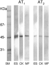
Immunoprecipitation and Western blot analysis of AngII receptors. In the epidermal strips, cultured keratinocytes, and myofibroblasts, the signals of both AT1 and AT2 were detected. The signal of AT1 is prominent compared to that of AT2. SM, size marker; ES, epidermal strips; CK, cultured keratinocytes; MF, myofibroblasts.
Figure 3
Figure 3
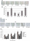
BrdU incorporation and wound healing migration assays of cultured keratinocytes. A: Detection of BrdU incorporation by immunohistochemical staining. The BrdU-labeled nuclei stained red. CV, CV-11974; PD, PD-123319. B: Effects of stimulation or blockade of AngII receptor signaling on BrdU incorporation. Data are represented as mean ± SD (n = 3). *, Significantly different from controls (P ≤ 0.05). , Significantly different from AngII-treated group (P ≤ 0.05). AngII or PD-123319 with AngII enhanced BrdU incorporation. CV-11974 with AngII suppressed BrdU incorporation. C: Wound healing migration assays. The phase contrast microscopic findings at 40 hours after scraping. D: Results of wound healing migration assays. Data are represented as mean ± SD (n = 3). *, Significantly different from controls (P ≤ 0.05). , Significantly different from AngII-treated group (P ≤ 0.05). AngII or PD-123319 with AngII accelerated re-epithelization. CV-11974 with AngII inhibited keratinocyte re-epithelization. Original magnifications: ×20 (A); ×10 (C).
Figure 4
Figure 4
BrdU incorporation and wound healing migration assays of cultured myofibroblasts. A: Detection of BrdU incorporation by immunohistochemical staining. The BrdU-labeled nuclei stained red. CV, CV-11974; PD, PD-123319. B: Effects of stimulation or blockade of AngII receptor signaling on BrdU incorporation. Data are represented as mean ± SD (n = 3). *, Significantly different from controls (P ≤ 0.05). , Significantly different from AngII-treated group (P ≤ 0.05). AngII or PD-123319 with AngII enhanced BrdU incorporation. CV-11974 with AngII suppressed BrdU incorporation. C: Wound healing migration assays. The phase contrast microscopic findings at 36 hours after scraping. D: Results of wound healing migration assays. Data are represented as mean ± SD (n = 3). *, Significantly different from controls (P ≤ 0.05). , Significantly different from AngII-treated group (P ≤ 0.05). AngII or PD-123319 with AngII accelerated myofibroblast recovering. CV-11974 with AngII inhibited recovering. Original magnifications: ×20 (A); ×10 (C).

References

    1. Gyurko R, Kimura B, Kurian P, Crews FT, Philips MI. Angiotensin II receptor subtypes play opposite roles in regulating phosphatidylinositol hydrolysis in rat skin slices. Biochem Biophys Res Commun. 1992;186:285–292. - PubMed
    1. Fujiyama S, Matsubara H, Nozawa Y, Maruyama K, Mori Y, Tsutsumi Y, Masaki H, Uchiyama Y, Koyama Y, Nose A, Iba O, Tateishi E, Ogata N, Jyo N, Higashiyama S, Iwasaka T. Angiotensin AT1 and AT2 receptors differentially regulate angiopoietin-2 and vascular endothelial growth factor expression and angiogenesis by modulating heparin binding-epidermal growth factor (EGF)-mediated EGF receptor transactivation. Circ Res. 2001;88:22–29. - PubMed
    1. Shibasaki Y, Matsubara H, Nozawa Y, Mori Y, Masaki H, Kosaki A, Tsutsumi Y, Uchiyama Y, Fujiyama S, Nose A, Iba O, Tateishi E, Hasegawa T, Horiuchi M, Nahmias C, Iwasaka T. Angiotensin II type 2 receptor inhibits epidermal growth factor receptor transactivation by increasing association of SHP-1 tyrosine phosphatase. Hypertension. 2001;38:367–372. - PubMed
    1. Murasawa S, Mori Y, Nozawa Y, Gotoh N, Shibuya M, Masaki H, Maruyama K, Tsutsumi Y, Moriguchi Y, Shibasaki Y, Tanaka Y, Iwasaka T, Inada M, Matsubara H. Angiotensin II type 1 receptor-induced extracellular signal-related protein kinase activation is mediated by Ca2+/calmodulin-dependent transactivation of epidermal growth factor receptor. Circ Res. 1998;82:1338–1348. - PubMed
    1. Eguchi S, Numaguchi K, Iwasaki H, Matsumoto T, Yamakawa T, Utsumonoa H, Motley ED, Kawakatsu H, Owada KM, Hirata Y, Marumo F, Inagami T. Calcium-dependent epidermal growth factor receptor transactivation mediates the angiotensin II-induced mitogen-activated protein kinase activation in vascular smooth muscle cells. J Biol Chem. 1998;273:8890–8896. - PubMed

MeSH terms