. 2004 Nov;165(5):1719-30.
doi: 10.1016/S0002-9440(10)63427-X.

Biliary dysgenesis in the PCK rat, an orthologous model of autosomal recessive polycystic kidney disease

Affiliations

Biliary dysgenesis in the PCK rat, an orthologous model of autosomal recessive polycystic kidney disease

Tatyana V Masyuk et al. Am J Pathol. 2004 Nov.

Abstract

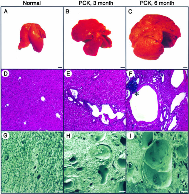
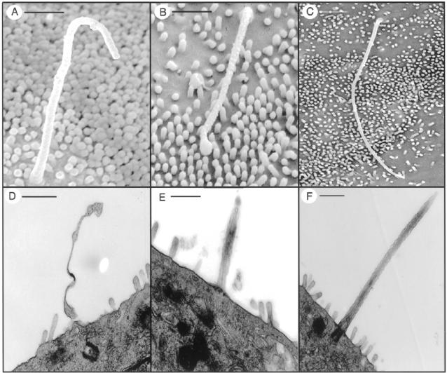
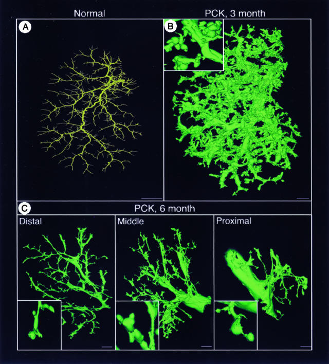
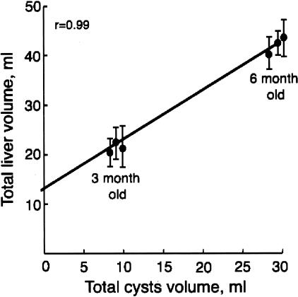
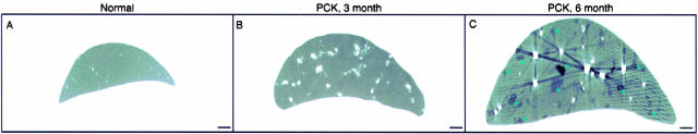
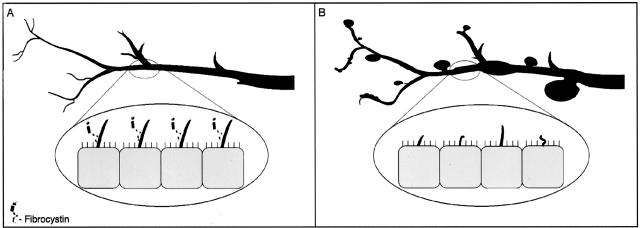
Hepatic polycystic disease occurs alone or in combination with polycystic kidney disease (PKD). In autosomal recessive PKD (ARPKD), liver lesions are the major cause of morbidity and mortality in older patients. ARPKD is caused by a mutation to PKHD1 and the PCK rat is an orthologous model of disease. Recently, we showed that fibrocystin, Pkhd1 protein, is localized to primary cilia in rat cholangiocytes and that disruption of its ciliary expression results in biliary cystogenesis. This study describes biliary phenotype in the PCK rat using micro-computed tomography scanning and three-dimensional reconstruction, and light, scanning, and transmission microscopy. Our results show that the biliary tree undergoes extensive remodeling resulting in bile duct dilatation, focal budding, and formation of cysts that are initially connected to bile ducts, but throughout time separate from them. Progressive liver enlargement results from massive cyst formation while liver parenchymal volume remains unchanged. Cilia in cystic cells are abnormal consistent with the notion that the primary defect in ARPKD resulting in cystogenesis may be linked to ciliary dysfunction. Our results suggest that the PCK rat is a useful model for studies of biliary cystogenesis and treatment options of inherited cystic liver disease.


Figures

Figure 1
Figure 1
Gross anatomy and liver morphology in normal and PCK rats. A–C: At the gross level, livers in the PKC rats progressively enlarged with age. D–F: Light microscopy (H&E) and G–I: Scanning electron microscopy shows that some cysts are present in PCK rats at 3 month of age; by 6 months cysts replace most of the liver parenchyma. Portal inflammation was found occasionally in both groups of the PCK rats (F, asterisk). Scale bars: 5 mm (A–C); 200 μm (G–I). Original magnifications, ×40 (D–F).
Figure 2
Figure 2
Representative images of the liver from 3-month-old (A) and 6-month-old (B) PCK rats stained with Masson’s trichrome. Mild fibrosis without septa formation was found in 6-month-old PCK rats. Original magnifications, ×40.
Figure 3
Figure 3
Morphology of the liver cyst. Single cyst (A) and cluster of cysts (B) isolated from the liver of the PCK rat by microdissection. C and D: Scanning electron microscopy of liver cysts. E: Cystic epithelia are positive for ductal epithelial cell marker, CK-19. Transmission electron microscopy of cells lining liver cysts in PCK rats (F) and bile duct in normal rats (G). Asterisks indicate mitochondria. Scale bars: 100 μm (A, B); 50 μm (C, D). Original magnifications: ×200 (E); ×6000 (F); ×15,000 (G).
Figure 4
Figure 4
Scanning electron microscopy of bisected liver cyst from 6-month-old PCK rats. A: Single liver cyst embedded in liver parenchyma. B and C: Complementary hemispheres of the bisected liver cyst showed that cyst has no openings and thus, no connections to the bile duct. Scale bars: 20 μm (A); 200 μm (B, C).
Figure 5
Figure 5
The diversity in ciliary length and the structural abnormalities in the PCK rat by scanning (top) and transmission (bottom) electron microscopy. A and B: Cilia from cystic epithelial cells are variable in length compared to cilia of normal cholangiocytes (C). D and E: Malformed cilia with bulbous extension of the axonemal membrane were seen in the PCK rat but not in normal rat (F). Scale bars: 500 nm (A, B); 1 μm (C–F).
Figure 6
Figure 6
The brightest voxel projection of the intrahepatic biliary tree scanned with resolutions of 20 μm of the left lateral lobe from 3-month-old normal (A) and 3-month-old (B) and 6-month-old (C) PCK rats. The liver lobe in 6-month-old PCK rat was cut into three parts: distal, middle, and proximal. Insets in B and C show a high-magnification image. Scale bars: 2 mm (A); 1 mm (B, C).
Figure 7
Figure 7
Micro-CT images of the intact liver lobes of normal (A) and 3-month-old (B) and 6-month-old (C) PCK rats reconstructed in three-dimension. These images were used to measure total liver, parenchymal, and cystic volumes. Scale bars, 2 mm.
Figure 8
Figure 8
Relationship between total liver and total cystic volume in 3- and 6-month-old PCK rats shows that cyst volume correlates strongly with total liver volume.
Figure 9
Figure 9
Micro-CT images of the single liver slice (21 μm) of 3-month-old normal (A) and 3-month-old (B) and 6-month-old (C) PCK rats. White dots represent cross-sections of the bile ducts filled with contrast agent. Black spots are liver cysts. Note that cysts do not fill with contrast agent suggesting that they are disconnected from the biliary tree. Asterisks indicate liver cysts that do not fill with contrast agent. Scale bars, 2 mm.
Figure 10
Figure 10
Model of fibrocystin-dependent terminal differentiation of the biliary tee. A: In normal rats, fibrocystin is localized to cholangiocyte cilia and functions as a bile duct size sensor controlling maturation of the biliary tree. B: In PCK rats, cilia are shortened, malformed, and do not express fibrocystin leading to abnormalities in cell proliferation and differentiation, ultimately resulting in significant bile duct dilatation and cyst formation.

References

    1. Iglesias DM, Palmitano J, Arrizurieta E, Kornblihtt AR, Herrera M, Bernath V, Matrin RS. Isolated polycystic liver disease not linked to polycystic kidney disease 1 and 2. Dig Dis Sci. 1999;44:385–388. - PubMed
    1. Pirson Y, Lannoy N, Peters D, Guubel A, Gigot J-F, Breuning M, Verellen-Dumoulin C. Isolated polycystic liver disease as a distinct genetic disease, unlinked to polycystic kidney disease 1 and polycystic kidney disease 2. Hepatology. 1996;23:249–252. - PubMed
    1. Li A, Davila S, Furu L, Qian Q, Tian X, Kamath PS, King BF, Torres VE, Somlo S. Mutations in PRKCSH cause isolated autosomal dominant polycystic liver disease. Am J Hum Genet. 2003;72:691–703. - PMC - PubMed
    1. Drenth JPH, teMorsche RHM, Smink R, Bonifacino JS, Jansen JBMJ. Germline mutations in PRKCSH are associated with autosomal dominant polycystic liver disease. Nat Genet. 2003;33:345–347. - PubMed
    1. Calvet JP, Grantham JJ. The genetic and physiology of polycystic kidney disease. Semin Nephrol. 2001;21:107–123. - PubMed

Publication types

MeSH terms