. 2004 Nov;165(5):1767-72.
doi: 10.1016/S0002-9440(10)63431-1.

Bone marrow-derived cells contribute to epithelial engraftment during wound healing

Affiliations

Bone marrow-derived cells contribute to epithelial engraftment during wound healing

Xenia Borue et al. Am J Pathol. 2004 Nov.

Abstract

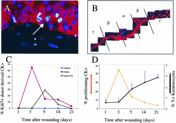
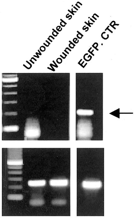
Recent findings suggest that bone marrow-derived cells (BMDC) may contribute to tissue maintenance throughout the body. However, it is not yet known whether marrow-derived epithelial cells are capable of undergoing proliferation. Our laboratory has shown that BMDC engraft as keratinocytes in the skin at low levels (</= 1%) in the absence of injury. Here we show that skin damage affects the degree of engraftment of BMDC as keratinocytes and that the keratinocytes are actively cycling. Female mice reconstituted with sex-mismatched BM were wounded by punch biopsy and incision. At the wound site, engraftment of BMDC as epidermal cells increased within 1 day, and continued to increase to approximately 4% by 3 weeks after injury. Using a Cre-lox system, fusion of BMDC with epithelial cells was ruled out. BMDC-derived epithelial cells at the wound edges expressed Ki67, a marker for actively cycling cells, and this proliferation correlated with an increase in the number of donor-derived cells within the wound. Donor-derived cytokeratin 5-expressing cells were rare, suggesting that BMDC do not engraft as epidermal stem cells, and the level of engraftment peaked and then decreased over time, further suggesting that BMDC may assist in early wound healing by engrafting as transit-amplifying cells, which then differentiate into keratinocytes.


Figures

Figure 1
Figure 1
A: Sample from day 3 showing an ulcer defect (left) filled by crust, with a mixed underlying inflammatory infiltrate. The epidermis adjacent to the ulcer exhibits acanthosis and reactive enlargement of keratinocytes. B: Sample from day 5 exhibiting greater organization of fibrin at the surface of the ulcer, diminished crust, and growth of epidermis over the edge of the ulcer. C: Sample from day 14 showing complete re-epithelialization. The epidermis over the scar (right) exhibits acanthosis, hypergranulosis, and hyperkeratosis. Fibrin has been replaced by horizontally oriented collagen bundles, interspersed by plump fibroblasts.
Figure 2
Figure 2
Shown are representative images from two different mice, 5 days (top) and 3 weeks (bottom) after wounding. Images were taken of the same slide after immunohistochemical staining for cytokeratin (left) and Y-chromosome FISH (right). After development of the immunohistochemical staining with DAB, the slides were lightly counter-stained with hematoxylin to allow for visualization of nuclei. In the fluorescence panels (B and D), the red signal derived from the autofluorescence of the DAB in the Cy5 channel, the white signal is from the Y-chromosome FISH, the blue represent DAPI-stained nuclei and the green signal is autofluorescence. The white arrows indicate Y+ positive cytokeratin-positive cells. The green arrows indicate Y chromosome-positive cells that do not appear to stain for cytokeratin. Original magnification, ×60.
Figure 3
Figure 3
Shown in this graph is the mean ± SD of the percentage of Y chromosome-positive keratinocytes at different times after wounding with either a cut (black bars) or punch biopsy (gray bars). Clear bars indicate the engraftment of marrow-derived keratinocytes in an unwounded area of skin of the same animals that had received wounds at each time point. Note that no Y+ cytokeratin+ cells were found in unwounded skin on d1 or d14. Uncut refers to the control animals that were transplanted but did not receive wounds.
Figure 4
Figure 4
A: Fluorescent staining for Ki67 (light blue nuclei), Y chromosome (yellow), cytokeratin (red), and DAPI for the nuclei (dark blue). The arrow indicates Y chromosome+, Ki67+, and cytokeratin+ cells in the basal layer of the skin in the β region 5 days after wounding. B: Shown is a composite of a skin wound 21 days after wounding. The α region is the center of the wounded area, and the β region is located at the periphery of the wound. Just outside the highly proliferative β region is the γ α region. C: Graph shows the percentage of donor-derived epithelial cells that are Ki67+ in each of the regions delineated in B at different times after wounding (α, circles; β, squares; γ, triangles). D: Graph shows the percentage of all donor-derived epithelial cells that are proliferating over time (squares) on the left axis and the percentage of keratinocytes that are donor-derived (diamonds) on the right axis at different times after wounding.
Figure 5
Figure 5
RT-PCR for GFP (top) in unwounded and wounded skin as indicated. At right, the GFP-positive control is shown. The band present in the lane labeled unwounded skin is significantly smaller than the expected size of the GFP product and represents the primers. β-actin controls are shown on the bottom.

References

    1. Herzog EL, Chai L, Krause DS. Plasticity of marrow-derived stem cells. Blood. 2003;102:3483–3493. - PubMed
    1. Orlic D, Kajstura J, Chimenti S, Jakoniuk I, Anderson SM, Li B, Pickel J, McKay R, Nadal-Ginard B, Bodine DM, Leri A, Anversa P. Bone marrow cells regenerate infarcted myocardium. Nature. 2001;410:701–705. - PubMed
    1. Quaini F, Urbanek K, Beltrami AP, Finato N, Beltrami CA, Nadal-Ginard B, Kajstura J, Leri A, Anversa P. Chimerism of the transplanted heart. N Engl J Med. 2002;346:5–15. - PubMed
    1. Lagasse E, Connors H, Al-Dhalimy M, Reitsma M, Dohse M, Osborne L, Wang X, Finegold M, Weissman IL, Grompe M. Purified hematopoietic stem cells can differentiate into hepatocytes in vivo. Nat Med. 2000;6:1229–1234. - PubMed
    1. Krause DS, Ito T, Fackler MJ, Collector MI, Sharkis SJ, May WS. Characterization of murine CD34, a marker for hematopoietic progenitor and stem cells. Blood. 1994;84:691–701. - PubMed

Publication types

MeSH terms