. 2004 Nov;165(5):1781-7.
doi: 10.1016/S0002-9440(10)63433-5.

CEACAM1 enhances invasion and migration of melanocytic and melanoma cells

Affiliations

CEACAM1 enhances invasion and migration of melanocytic and melanoma cells

Alireza Ebrahimnejad et al. Am J Pathol. 2004 Nov.

Abstract

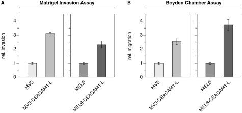
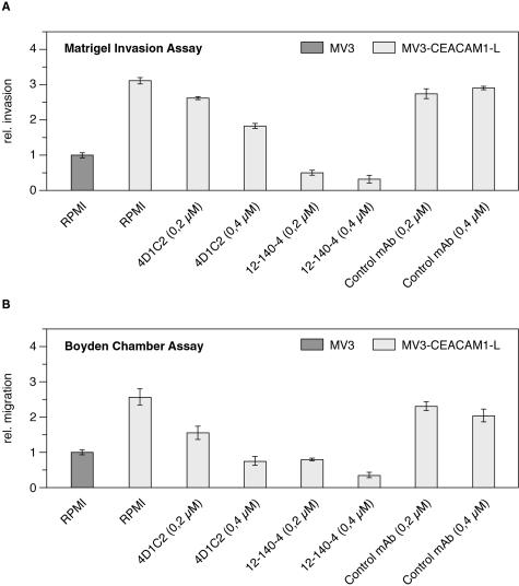
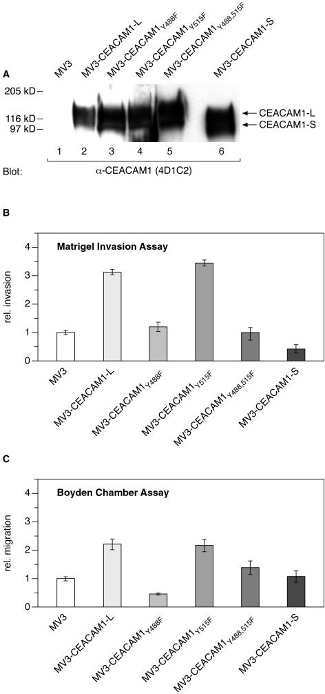
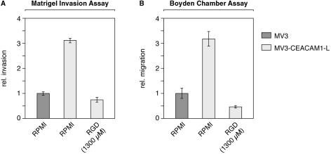
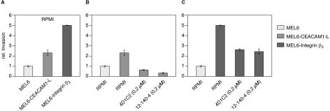
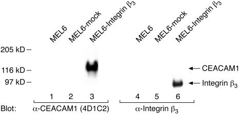
Expression of the cell adhesion molecule CEACAM1 in melanomas is an independent factor for the risk of metastasis with a predictive value superior to that of tumor thickness. We have previously shown that CEACAM1 co-localizes at the tumor-stroma interface of invading melanoma masses with integrin beta(3) and that these two adhesion molecules interact via the CEACAM1 cytoplasmic domain. To address the functional consequences of CEACAM1 expression, we investigated invasion and migration of melanocytic and melanoma cells that stably express CEACAM1 using two different in vitro systems. Here, we demonstrate that CEACAM1 expression markedly enhances cell invasion and migration. The enhanced invasion and migration of CEACAM1-transfected cells was dependent on the presence of Tyr-488 within the full-length cytoplasmic CEACAM1 domain. Treatment with anti-CEACAM monoclonal antibodies blocked CEACAM1-enhanced cell invasion and cell migration in a dose-dependent manner. Furthermore, the enhanced invasion and migration of CEACAM1-transfected melanoma cells was blocked by integrin-antagonizing RGD peptides. Expression of integrin beta(3) induces the up-regulation of CEACAM1 in melanocytic MEL6 cells. These results strengthen the view that CEACAM1 and alpha(v)beta(3) integrin are functionally interconnected with respect to the invasive growth of melanomas.


Figures

Figure 1
Figure 1
CEACAM1 expression enhances cell invasion and cell migration. A: Matrigel invasion assay. B: Boyden chamber assay. Cells (2.5 × 10⁴) within passages 6 to 10 were seeded in the upper compartment of transwell chambers containing PET filters precoated with Matrigel (A) or polyester filters precoated with collagen I (B). After 24 hours (invasion assay) or 5 hours (migration assay), under serum-free conditions (RPMI), cells that invaded through the 8-μm pores were stained. The data are means ± SD of five separate experiments. The mean invasion of untransfected MV3 or untransfected MEL6 cells was assigned a value of 1.
Figure 2
Figure 2
Anti-CEACAM mAb inhibit enhanced CEACAM1-mediated cell invasion and migration. A: Matrigel invasion assay and B: Boyden chamber assay performed as described in Figure 1. Cells that invaded, under serum-free conditions in the absence (RPMI in A and B) or presence of inhibitors, were stained. The data are means ± SD of five separate experiments. The mean invasion of untransfected MV3 cells was assigned a value of 1. Inhibitors were used as follows: 0.2 μmol/L and 0.4 μmol/L 4D1C2 mAb and 0.2 μmol/L and 0.4 μmol/L 12-140-4 mAb as anti-CEACAM1 blocking agents. Control bars are invasion (A) or migration (B) in the presence of 0.2 μmol/L and 0.4 μmol/L nonimmune IgG.
Figure 3
Figure 3
CEACAM1-enhanced invasion or migration requires CEACAM1-Tyr-488. A: Western blot analysis of MV3 cells (lane 1) and MV3 cells transfected with wild-type (lane 2) and mutant (lanes 3 to 6) CEACAM1. Cell lysates containing 50 μg of protein each were resolved by sodium dodecyl sulfate-polyacrylamide gel electrophoresis, transferred to nitrocellulose, and immunoblotted with anti-CEACAM1 mAb 4D1C2. Positions of CEACAM1-L and mutant CEACAM1, containing the short cytoplasmic isoform (CEACAM1-S), are indicated on the right. Matrigel invasion assay (B) and Boyden chamber assay (C) performed as described in Figure 1. The data are means ± SD of five separate experiments. The mean invasion of untransfected MV3 cells was assigned a value of 1.
Figure 4
Figure 4
CEACAM1-enhanced invasion and migration is blocked by inhibitor of integrin β3 function. A: Matrigel invasion assay and B: Boyden chamber assay performed as described in Figure 1. Cells that invaded, under serum-free conditions in the absence (RPMI) or presence of RGD peptides (1300 μmol/L), were stained. The data are means ± SD of five separate experiments. The mean invasion of untransfected MV3 cells was assigned a value of 1.
Figure 5
Figure 5
Expression of CEACAM1 and integrin β3 in MEL6 cells enhances invasion. A: MEL6 cells or MEL6 cells transfected either with cDNA for CEACAM1 or for integrin β3 were studied using the Matrigel invasion assay as described in Figure 1. B: Invasion of CEACAM1 transfected MEL6 cells in the absence (RPMI) or presence of anti-CEACAM1 inhibitors (0.2 μmol/L 4D1C2 and 0.2 μmol/L 12-140-4). C: Invasion of integrin β3 transfected MEL6 cells in the absence (RPMI) and presence of anti-CEACAM1 inhibitors (0.2 μmol/L 4D1C2 and 0.2 μmol/L 12-140-4). The data are means ± SD of five separate experiments. The mean invasion of untransfected MEL6 cells was assigned a value of 1.
Figure 6
Figure 6
Expression of integrin β3 induces the up-regulation of CEACAM1. Eighty-μg aliquots of cell lysates of untransfected (lanes 1 and 4), and mock (lanes 2 and 5)- and integrin β3 (lanes 3 and 6)-transfected MEL6 cells were resolved by 7.5% sodium dodecyl sulfate-polyacrylamide gel electrophoresis and immunoblotted with anti-CEACAM1 mAb 4D1C2 (lanes 1 to 3) or anti-integrin β3 mAb (lanes 4 to 6). The positions of CEACAM1 and integrin β3 are indicated on the right.

Similar articles

Cited by

References

    1. Edward M. Integrins and other adhesion molecules involved in melanocytic tumor progression. Curr Opin Oncol. 1995;7:185–191. - PubMed
    1. Buttner P, Garbe C, Bertz J, Burg G, d’Hoedt B, Drepper H, Guggenmoos-Holzmann I, Lechner W, Lippold A, Orfanos CE, Peters A, Rassner G, Stadler R, Stroebel W. Primary cutaneous melanoma. Optimized cutoff points of tumor thickness and importance of Clark’s level for prognostic classification. Cancer. 1995;75:2499–2506. - PubMed
    1. Johnson JP. Cell adhesion molecules in the development and progression of malignant melanoma. Cancer Metastasis Rev. 1999;18:345–357. - PubMed
    1. Natali PG, Hamby CV, Felding-Habermann B, Liang B, Nicotra MR, Di Filippo F, Giannarelli D, Temponi M, Ferrone S. Clinical significance of alpha(v)beta3 integrin and intercellular adhesion molecule-1 expression in cutaneous malignant melanoma lesions. Cancer Res. 1997;57:1554–1560. - PubMed
    1. Hsu MY, Shih DT, Meier FE, Van Belle P, Hsu JY, Elder DE, Buck CA, Herlyn M. Adenoviral gene transfer of beta3 integrin subunit induces conversion from radial to vertical growth phase in primary human melanoma. Am J Pathol. 1998;153:1435–1442. - PMC - PubMed

Publication types

MeSH terms