. 2004 Oct 27;24(43):9531-40.
doi: 10.1523/JNEUROSCI.3085-04.2004.

Human immunodeficiency virus type 1 gp120 induces apoptosis in human primary neurons through redox-regulated activation of neutral sphingomyelinase

Affiliations

Human immunodeficiency virus type 1 gp120 induces apoptosis in human primary neurons through redox-regulated activation of neutral sphingomyelinase

Arundhati Jana et al. J Neurosci. .

Abstract

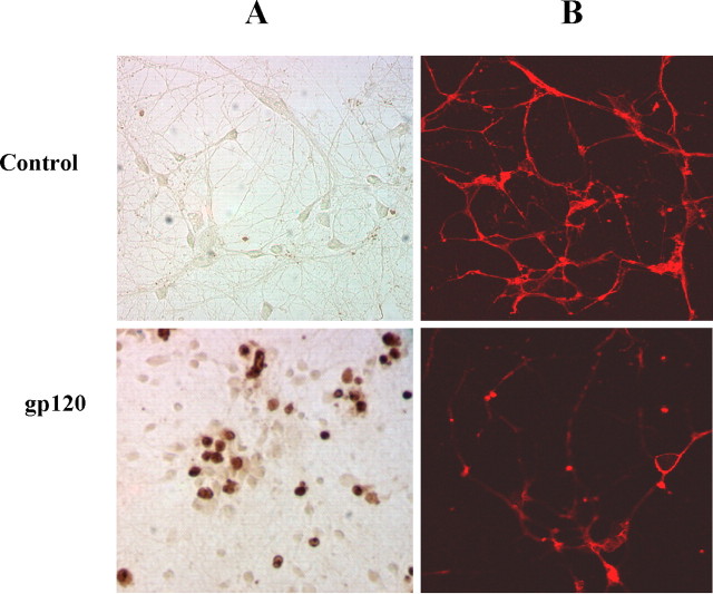
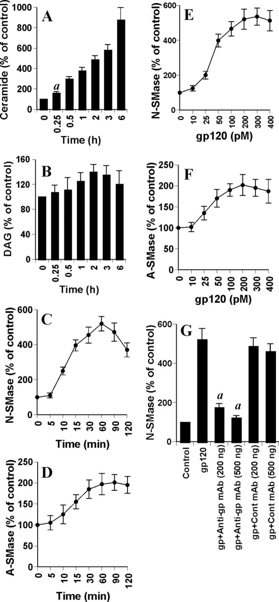
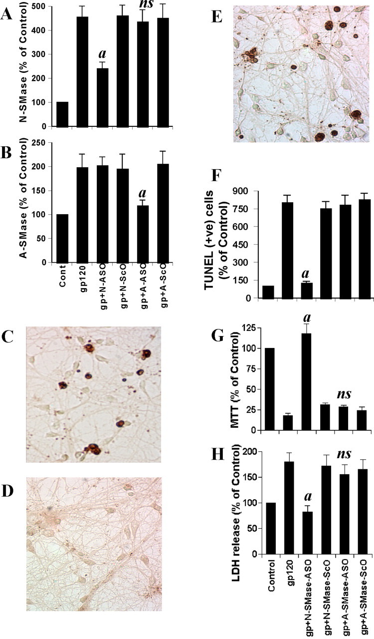
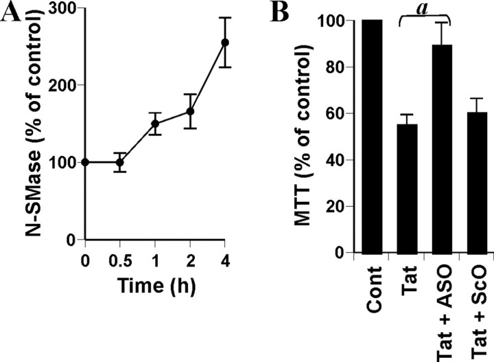
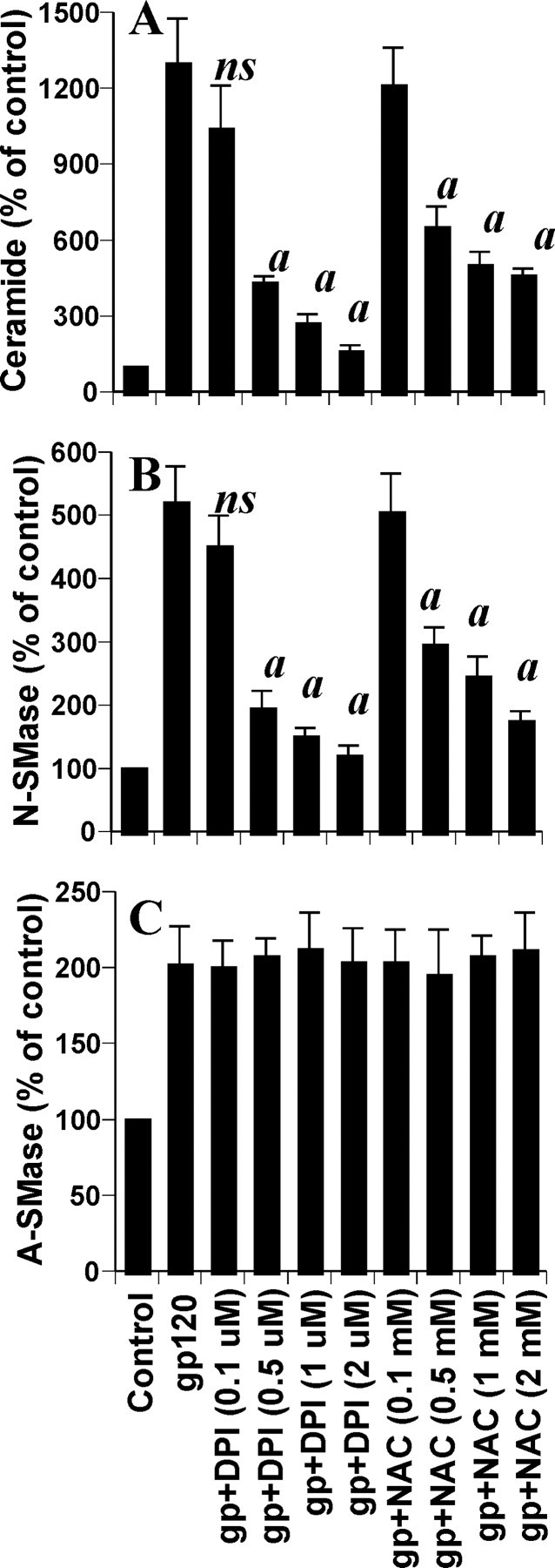
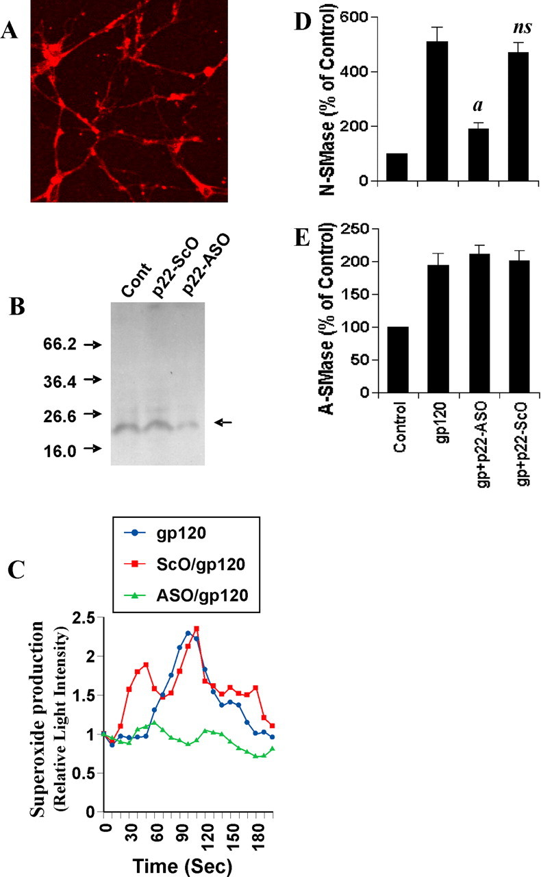
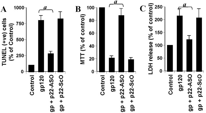
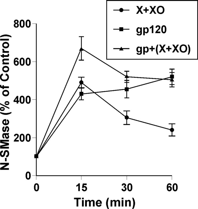
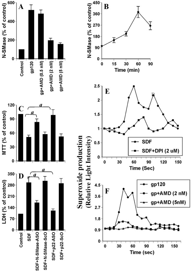
Human immunodeficiency virus type 1 (HIV-1) infection is known to cause disorders of the CNS, including HIV-associated dementia (HAD). HIV-1 coat protein gp120 (glycoprotein 120) induces neuronal apoptosis and has been implicated in the pathogenesis of HAD. However, the mechanism by which gp120 causes neuronal apoptosis is poorly understood. The present study underlines the importance of gp120 in inducing the production of ceramide, an important inducer of apoptosis, in human primary neurons. gp120 induced the activation of sphingomyelinases (primarily the neutral one) and the production of ceramide in primary neurons. Antisense knockdown of neutral (NSMase) but not acidic (ASMase) sphingomyelinase markedly inhibited gp120-mediated apoptosis and cell death of primary neurons, suggesting that the activation of NSMase but not ASMase plays an important role in gp120-mediated neuronal apoptosis. Similarly, the HIV-1 regulatory protein Tat also induced neuronal cell death via NSMase. Furthermore, gp120-induced production of ceramide was redox sensitive, because reactive oxygen species were involved in the activation of NSMase but not ASMase. gp120 coupled CXCR4 (CXC chemokine receptor 4) to induce NADPH oxidase-mediated production of superoxide radicals in neurons, which was involved in the activation of NSMase but not ASMase. These studies suggest that gp120 may induce neuronal apoptosis in the CNS of HAD patients through the CXCR4-NADPH oxidase-superoxide-NSMase-ceramide pathway.


Figures

Figure 1.
Figure 1.
gp120 induces apoptosis and cell death in human primary neurons. A, Coverslips containing human primary neurons were incubated with 200 pm gp120 for 6 hr, followed by TdT-mediated end labeling of 3′-OH ends of DNA fragments. B, After 18 hr of treatment with 200 pm gp120, cells were immunostained with MAP-2.
Figure 2.
Figure 2.
Effect of gp120 on the induction of ceramide production and the activation of NSMase and ASMase in human primary neurons. Cells were incubated with 200 pm gp120 for different time intervals. Lipids were extracted, and ceramide (A) and DAG (B) contents were determined. Activities of NSMase (C) and ASMase (D) were assayed in total-cell extract. Cells were treated with different concentrations of gp120 for 1 hr. Activities of NSMase (E) and ASMase (F) were assayed. G, Cells preincubated with anti-gp120 mAb (2G12) and control IgG1 for 30 min were treated with 200 pm gp120. Activity of NSMase was assayed after 1 hr of treatment. Control group served as 100%, and data from other groups were expressed as percentage of control. Results are mean ± SD of three different experiments. ap < 0.001 versus 0 hr.
Figure 3.
Figure 3.
Effect of antisense oligonucleotides against NSMase and ASMase on gp120-mediated cell death in human primary neurons. Cells received 1 μm of either ASO or ScO against NSMase and ASMase. After 48 hr of incubation, cells were treated with 200 pm gp120, and, after 1 hr of stimulation, activities of NSMase (A) and ASMase (B) were measured. Coverslips containing primary neurons were preincubated with 1 μm ASO or ScO against NSMase and ASMase for 40 hr, followed by stimulation with 200 pm gp120. Control cells received only vehicle (PBS). After 6 hr of stimulation, TUNEL (C, control; D, ASO; E, ScO) was performed. TUNEL-positive (+ve) cells were counted manually in four different images of each of three coverslips by three blinded individuals (F). Cells preincubated with 1 μm ASO or ScO against NSMase and ASMase for 40 hr received 200 pm gp120. After 18 hr of stimulation, cells were subjected to MTT reduction activity assay (G), and culture medium was collected for LDH release assay (H). Values obtained from the control group served as 100%, and data obtained in other groups were calculated as percentage of control accordingly. Results are mean ± SD of three different experiments. ap < 0.001 versus gp120. ns, Not significant.
Figure 4.
Figure 4.
Role of NSMase in Tat-mediated cell death in human primary neurons. A, Cells were incubated with 100 ng/ml Tat for different time intervals. Activity of NSMase was assayed in total-cell extract. B, Cells received 1 μm of either ASO or ScO against NSMase. After 40 hr of incubation, cells were treated with 100 ng/ml Tat, and, after 18 hr of treatment, cells were subjected to MTT reduction activity assay. Values obtained from the control group (Cont) served as 100%, and data obtained in other groups were calculated as percentage of control accordingly. Results are mean ± SD of three different experiments. ap < 0.001 versus Tat.
Figure 5.
Figure 5.
Effects of NAC and DPI on gp120-induced production of ceramide and activation of sphingomyelinases in human primary neurons. Cells preincubated with different concentrations of either NAC or DPI for 1 hr received 200 pm gp120. A, After 12 hr of incubation, level of ceramide was measured. After 1 hr of incubation, activities of NSMase (B) and ASMase (C) were assayed. Values obtained from the control group served as 100%, and data obtained in other groups were calculated as percentage of control accordingly. Results are mean ± SD of three different experiments. ap < 0.001 versus gp120. ns, Not significant.
Figure 6.
Figure 6.
Role of NADPH oxidase in gp120-induced activation of NSMase but not ASMase in human primary neurons. A, Cells were immunostained with antibodies against p22phox. B, Cells were incubated with 1 μm of either ASO or ScO against p22phox. Control cells (Cont) received only vehicle (PBS). After 48 hr, the expression of p22phox was examined by Western blot. Briefly, total-cell homogenates containing protease inhibitor mixture (Sigma) were electrophoresed in 14% SDS-polyacrylamide gel, followed by transfer onto a nitrocellulose membrane and immunoblotting with polyclonal antibodies against human p22phox. C, Cells were incubated with 1 μm either ASO or ScO against p22phox. Control cells received only vehicle (PBS). After 40 hr, cells were stimulated with 200 pm gp120. At different second intervals, superoxide production was assayed in whole cells using the LumiMax Superoxide Anion Detection kit (Stratagene). Results are the mean of two separate experiments. After 1 hr of stimulation of gp120, activities of NSMase (D) and ASMase (E) were assayed. Values obtained from the control group served as 100%, and data obtained in other groups were calculated as percentage of control accordingly. Results are mean ± SD of three different experiments. ap < 0.001 versus gp120. ns, Not significant.
Figure 7.
Figure 7.
Role of NADPH oxidase in gp120-induced apoptosis and cell death in human primary neurons. Cells were incubated with 1 μm of either ASO or ScO against p22phox. Control cells received only vehicle (PBS). After 40 hr, cells were stimulated with 200 pm gp120. After 6 hr of stimulation, TUNEL-positive (+ve) cells were counted manually in four different images of each of three coverslips by three blinded individuals (A). After 18 hr of stimulation, cells were subjected to MTT reduction activity assay (B), and culture medium was collected for LDH release assay (C). Values obtained from the control group served as 100%, and data obtained in other groups were calculated as percentage of control accordingly. Results are mean ± SD of three different experiments. ap < 0.001 versus gp120.
Figure 8.
Figure 8.
Superoxide radicals alone induce the activation of NSMase in human primary neurons. Cells were treated with 200 pm gp120 in the presence or absence of hypoxanthine (5 μm) and xanthine oxidase (0.5 mU/ml) (X plus XO). At different minute intervals, activity of NSMase was assayed. Values obtained from the control group served as 100%, and data obtained in other groups were calculated as percentage of control accordingly. Results are mean ± SD of three different experiments.
Figure 9.
Figure 9.
Role of CXCR4 in gp120-induced activation of NSMase in human primary neurons. A, Cells incubated with different concentrations of AMD3100 for 1 hr were treated with 200 pm gp120. After 1 hr, activity of NSMase was assayed. B, Cells were incubated with 100 ng/ml SDF-1α for different time intervals, followed by assay of NSMase. C, D, Cells preincubated with 1 μm ASO or ScO against NSMase and p22phox for 40 hr received 100 ng/ml SDF-1α. After 18 hr of stimulation, cells were subjected to MTT reduction activity assay (C), and culture medium was collected for LDH release assay (D). Values obtained from the control group served as 100%, and data obtained in other groups were calculated as percentage of control accordingly. Results are mean ± SD of three different experiments. ap < 0.001 versus SDF-1α. E, Cells preincubated with 2 μm DPI for 1 hr were stimulated with 100 ng/ml SDF-1α. F, Cells preincubated with different concentrations of AMD3100 were stimulated with 200 pm gp120. At different second intervals, superoxide production was assayed in whole cells. Values obtained from the control group served as 100%, and data obtained in other groups were calculated as percentage of control accordingly. Results are the mean of two separate experiments.

References

    1. Adam-Klages S, Adam D, Wiegmann K, Struve S, Kolanus W, Schneider-Mergener J, Kronke M (1996) FAN, a novel WD-repeat protein, couples the p55 TNF-receptor to neutral sphingomyelinase. Cell 86: 937-947. - PubMed
    1. Bachis A, Major EO, Mocchetti I (2003) Brain-derived neurotrophic factor inhibits human immunodeficiency virus-1/gp120-mediated cerebellar granule cell death by preventing gp120 internalization. J Neurosci 23: 5715-5722. - PMC - PubMed
    1. Bagasra O, Lavi E, Bobroski L, Khalili K, Pestaner JB, Pomerantz RJ (1996) Cellular reservoirs of HIV-1 in the central nervous system of infected individuals: identification by the combination of in situ polymerase chain reaction and immunohistochemistry. AIDS 10: 573-585. - PubMed
    1. Bansal AK, Mactutus CF, Nath A, Maragos W, Hauser KF, Booze RM (2000) Neurotoxicity of HIV-1 proteins gp120 and Tat in the rat striatum. Brain Res 879: 42-49. - PubMed
    1. Bennett BA, Rusyniak DE, Hollingsworth CK (1995) HIV-1 gp120-induced neurotoxicity to midbrain dopamine cultures. Brain Res 705: 168-176. - PubMed

Publication types

MeSH terms

LinkOut - more resources