. 2004 Oct 27;24(43):9658-68.
doi: 10.1523/JNEUROSCI.2973-04.2004.

Distinct roles for the kainate receptor subunits GluR5 and GluR6 in kainate-induced hippocampal gamma oscillations

Affiliations

Distinct roles for the kainate receptor subunits GluR5 and GluR6 in kainate-induced hippocampal gamma oscillations

André Fisahn et al. J Neurosci. .

Abstract

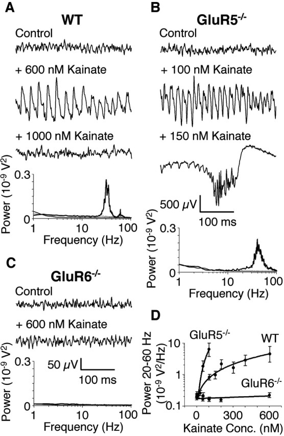
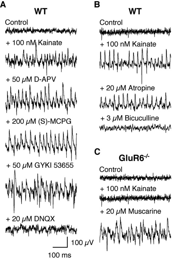
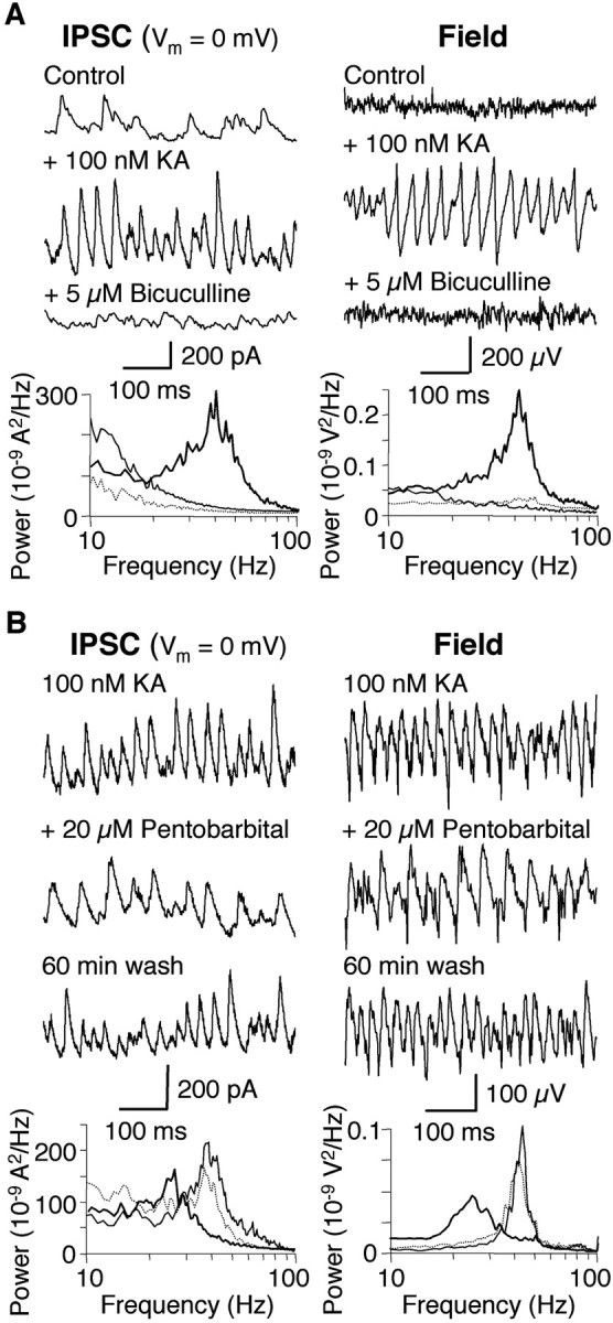
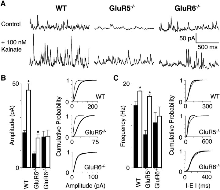
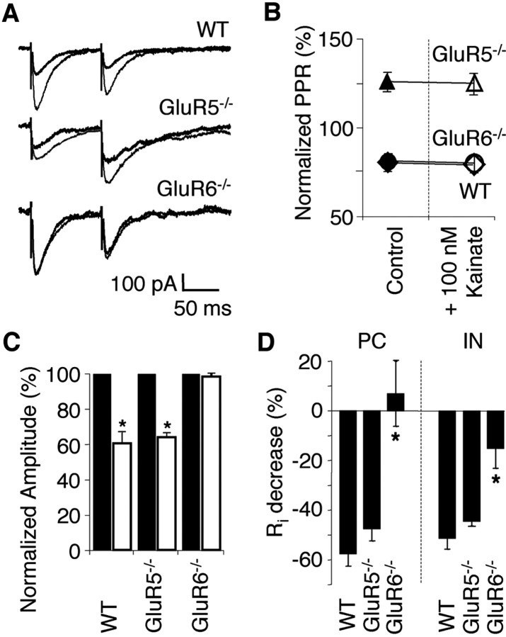
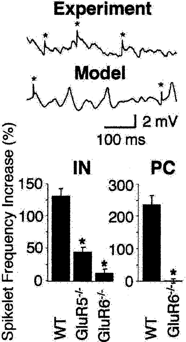
Kainate receptors (KARs) play an important role in synaptic physiology, plasticity, and pathological phenomena such as epilepsy. However, the physiological implications for neuronal networks of the distinct expression patterns of KAR subunits are unknown. Using KAR knock-out mice, we show that subunits glutamate receptor (GluR) 5 and GluR6 play distinct roles in kainate-induced gamma oscillations and epileptiform burst activity. Ablation of GluR5 leads to a higher susceptibility of the network to the oscillogenic and epileptogenic effects of kainate, whereas lack of GluR6 prevents kainate-induced gamma oscillations or epileptiform bursts. Based on experimental and simulated neuronal network data as well as the consequences of GluR5 and GluR6 expression for cellular and synaptic physiology, we propose that the functional interplay of GluR5-containing KARs on axons of interneurons and GluR6-containing KARs in the somatodendritic region of both interneurons and pyramidal cells underlie the oscillogenic and epileptogenic effects of kainate.


Figures

Figure 1.
Figure 1.
Kainate-induced gamma oscillations are disrupted in GluR6-/- but not GluR5-/- hippocampal slices. A, Example traces of extracellular field recordings in the CA3 area. In WT slices, no rhythmic network activity is seen in control conditions (no drug; thin line). Bath application of kainate induces gamma oscillations (kainate concentration that resulted in the maximal amplitude oscillations is shown; thick line). Increasing the kainate concentration leads to a breakdown of gamma oscillations (dotted line). Power spectra of the recorded traces are shown below. B, In GluR5-/- slices, maximal amplitude gamma oscillations are induced by much lower concentrations of kainate compared with WT (thick line). Increasing the kainate concentration leads to the occurrence of epileptiform burst activity (dotted line; 500 μV scale bar applies only to 150 nm kainate trace). Power spectra of the recorded traces are shown below (the power spectrum of the trace showing an epileptiform burst is too large to be displayed). C, In GluR6-/- slices, kainate fails to induce either gamma oscillations or epileptiform bursts (thick line). Power spectra of the recorded traces are shown below. D, Summary diagram showing the dependence of gamma oscillation power (integrated between 20 and 60 Hz) on kainate concentration. WT and GluR5-/- data points are shown only for kainate concentrations that resulted in gamma oscillations (n = 6 for WT, GluR5-/-, and GluR6-/-).
Figure 2.
Figure 2.
Pharmacology of kainate-induced gamma oscillations in hippocampal slices. A, Example traces of extracellular field recordings in the CA3 area. Kainate-induced gamma oscillations do not depend on NMDAR, mGluR, or AMPAR activation but on KARs (n = 6). B, Kainate-induced gamma oscillations do not depend on cholinergic receptors but do require intact inhibitory GABAergic neurotransmission (n = 6). C, In GluR6-/- slices, kainate-induced gamma oscillations are absent but can still be induced by muscarine (n = 4).
Figure 3.
Figure 3.
Kainate-induced gamma oscillations depend on inhibitory synaptic currents. Concomitantly recorded intracellular IPSCs (whole-cell patch-clamp mode) and extracellular field activity in WT hippocampal slices show rhythmicity in the gamma frequency range after superfusion with 100 nm kainate (thick line). A, Blocking GABAA receptors with 5 μm bicuculline terminates extracellular and intracellular gamma oscillations (n = 4; dotted line). Power spectra are shown below the example traces. B, Lengthening the time course of IPSCs by application of the benzodiazepine pentobarbital (20 μm) reversibly decreases the frequency of the intracellular and extracellular gamma oscillation (n = 3; thick line; wash, dotted line). Power spectra are shown below the example traces.
Figure 4.
Figure 4.
Kainate-induced inward current and depolarization are absent in GluR6-/- pyramidal cells and interneurons. A, Concomitant recording of IPSCs and EPSCs (whole-cell patch clamp) and extracellular gamma oscillations. The corresponding cross-correlation analyses show IPSCs are in antiphase and EPSCs are in phase with the extracellular gamma oscillation (n = 4). B, In the absence of AMPAR antagonists, kainate (arrows) induces inward currents in CA3 pyramidal cells of WT but fails to induce any inward current at even the highest kainate concentration tested in pyramidal cells of GluR6-/- hippocampal slices (voltage clamp; Vh = -60 mV). In current clamp (pyramidal cells held below firing threshold), bath application of 100 nm kainate (arrows) leads to a depolarization and increase in firing frequency in WT (Vm = -62.2 ± 2.0 mV) but not GluR6-/- pyramidal cells (Vm = -58.8 ± 1.7 mV). Action potentials are truncated for figure clarity. C, In voltage clamp (Vh = -60 mV), bath application of 100 nm kainate (arrows) induces an inward current in WT (-50.8 ± 9.5 pA) but not GluR6-/- interneurons (-1.9 ± 1.3 pA). In current clamp (interneurons held below firing threshold), bath application of 100 nm kainate (arrows) leads to a depolarization and increase in firing frequency in WT (Vm = -61.7 ± 0.9 mV) but not GluR6-/- interneurons (Vm = -58.0 ± 2.0 mV). Action potentials are truncated for figure clarity. D, Summary histograms of the experiments in C. The kainate-induced inward current is absent in GluR6-/- interneurons (n = 5; GluR5-/-, n = 5; WT, n = 8; p = 0.002). Kainate-induced depolarization and increase in firing frequency are significantly bigger in GluR5-/- (Vm = -63.3 ± 2.0 mV) than in WT interneurons. Partly compromising inhibitory neurotransmission by application of 10 μm picrotoxin results in increased depolarization and firing frequency in WT interneurons (n = 11 for WT; n = 9 for GluR5-/-; n = 5 for GluR6-/-; n = 4 for WT plus picrotoxin; *p < 0.02 for depolarization; p < 0.0001 for frequency).
Figure 5.
Figure 5.
Kainate-induced changes in sIPSC are absent in GluR6-/- but not WT and GluR5-/- hippocampal pyramidal cells. A, In voltage clamp (Vh = 0 mV), bath application of kainate induces an increase in sIPSC amplitude and frequency in both WT and GluR5-/- but not GluR6-/- pyramidal cells. sIPSCs are observed as upward deflections at 0 mV. B, Summary histogram for sIPSC amplitude analysis and representative cumulative probability plots of the experiment in A (control, black bars and thin lines; kainate, white bars and thick lines). Kainate increases sIPSC amplitude in WT. In GluR5-/-, sIPSC control amplitude is less than half of WT control amplitude. Kainate increases sIPSC amplitude in GluR5-/- to WT control levels. In GluR6-/-, sIPSC amplitude remains unchanged by kainate at WT control levels (n = 6 for WT and GluR5-/-; n = 5 for GluR6-/-; *p = 0.02 for WT; p < 0.09 for GluR5-/-). C, Summary histogram for sIPSC frequency analysis and representative cumulative probability plots of the experiment in A (control, black bars and thin lines; kainate, white bars and thick lines). Kainate increases sIPSC frequency in WT as well as GluR5-/-. However, sIPSC control frequency in GluR5-/- is approximately half of WT sIPSC control frequency. In GluR6-/-, sIPSC frequency remains unchanged by kainate near WT control levels (n = 6 for WT and GluR5-/-; n = 5 for GluR6-/-; *p < 0.1 for WT; p < 0.02 for GluR5-/-).
Figure 6.
Figure 6.
Kainate-induced changes in eIPSC are absent in GluR6-/- but not WT and GluR5-/- hippocampal pyramidal cells. A, Paired eIPSCs show paired-pulse depression in WT and GluR6-/- but facilitation in GluR5-/- pyramidal cells in control conditions (thin line) as well as after application of 100 nm kainate (thick line). Concomitantly, kainate depresses eIPSC amplitude in WT and GluR5-/- but has no effect in GluR6-/-. B, PPRs remain unchanged after the application of kainate. C, Summary histogram of eIPSC amplitude changes in the experiment in A (n = 5 for WT and GluR6-/-; n = 4 for GluR5-/-; *p < 0.0001; control, black bars; kainate, white bars). D, Kainate (100 nm) decreases the input resistance of WT and GluR5-/- pyramidal cells and interneurons by indirect action via GABAA receptors (postsynaptic shunting; GABAB receptors are absent at this developmental stage). The kainate-induced change in input resistance in GluR6-/- is significantly smaller (pyramidal cells: n = 9 for WT; n = 6 for GluR5-/-; n = 7 for GluR6-/-; interneurons: n = 8 for WT; n = 7 for GluR5-/-; n = 5 for GluR6-/-; *p < 0.0002 for pyramidal cells; p = 0.001 for interneurons).
Figure 7.
Figure 7.
Kainate-induced gamma oscillations are PKC independent. A, In voltage clamp (Vh = -50 mV), IsAHP charge transfer in control conditions (thin line) was decreased by 100 nm kainate (thick line) in WT pyramidal cells (n = 7). Preincubation (2 hr) in the PKC antagonist calphostin C (2 μm) significantly reduced this effect (n = 6; p = 0.005). B, In voltage clamp (Vh = -60 mV) after 2 hr preincubation in 2 μm calphostin C, bath application of 100 nm kainate (arrows) leads to an inward current in WT pyramidal cells that is similar to the inward current induced in naive WT pyramidal cells (n = 6 for WT; n = 4 for WT plus calphostin C). C, Kainate induces extracellular gamma oscillations in both naive and preincubated (2 hr in 2 μm PKC antagonist calphostin C) hippocampal slices. Power spectra are shown below the example traces (n = 6 for WT and WT plus calphostin C).
Figure 8.
Figure 8.
Ectopic action potentials and kainate-induced gamma oscillations. The two example traces show ectopic action potentials recorded as spikelets (*) in pyramidal cells during an ongoing gamma oscillation. The top trace is a physiological recording during kainate-induced gamma oscillations in WT; the bottom trace is computer generated. The summary histograms show the kainate-induced increase in spikelet frequency recorded in interneurons (IN) and pyramidal cells (PC). The increase in spikelet frequency seen in WT interneurons is significantly reduced in GluR5-/- (*p < 0.0002) and GluR6-/- interneurons (p < 0.0001). Note that the increase in spikelet frequency in GluR5-/- is significantly larger than in GluR6-/- (p < 0.012). Likewise, the increase in spikelet frequency seen in WT pyramidal cells is significantly reduced in GluR6-/- (*p < 0.0001) (interneurons: n = 5 for WT, n = 6 for GluR5-/-, n = 5 for GluR6-/-; pyramidal cells: n = 6 for WT, n = 5 for GluR6-/-).

References

    1. Ben-Ari Y (1985) Limbic seizure and brain damage produced by kainic acid: mechanisms and relevance to human temporal lobe epilepsy. Neuroscience 14: 375-403. - PubMed
    1. Ben-Ari Y, Cossart R (2000) Kainate, a double agent that generates seizures: two decades of progress. Trends Neurosci 23: 580-587. - PubMed
    1. Bortolotto ZA, Clarke VR, Delany CM, Parry MC, Smolders I, Vignes M, Ho KH, Miu P, Brinton BT, Fantaske R, Ogden A, Gates M, Ornstein PL, Lodge D, Bleakman D, Collingridge GL (1999) Kainate receptors are involved in synaptic plasticity. Nature 402: 297-301. - PubMed
    1. Bragin A, Jando G, Nadasdy Z, Hetke J, Wise K, Buzsaki G (1995) Gamma (40-100 Hz) oscillations in the hippocampus of the behaving rat. J Neurosci 15: 47-60. - PMC - PubMed
    1. Bureau I, Bischoff S, Heinemann SF, Mulle C (1999) Kainate receptor-mediated responses in the CA1 field of wild-type and GluR6-deficient mice. J Neurosci 19: 653-663. - PMC - PubMed

Publication types