. 2004 Nov;24(22):9695-704.
doi: 10.1128/MCB.24.22.9695-9704.2004.

Proteasome inhibitor PS-341 induces apoptosis through induction of endoplasmic reticulum stress-reactive oxygen species in head and neck squamous cell carcinoma cells

Affiliations

Proteasome inhibitor PS-341 induces apoptosis through induction of endoplasmic reticulum stress-reactive oxygen species in head and neck squamous cell carcinoma cells

Andrew Fribley et al. Mol Cell Biol. 2004 Nov.

Abstract

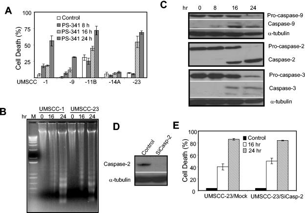
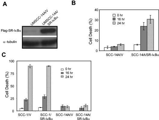
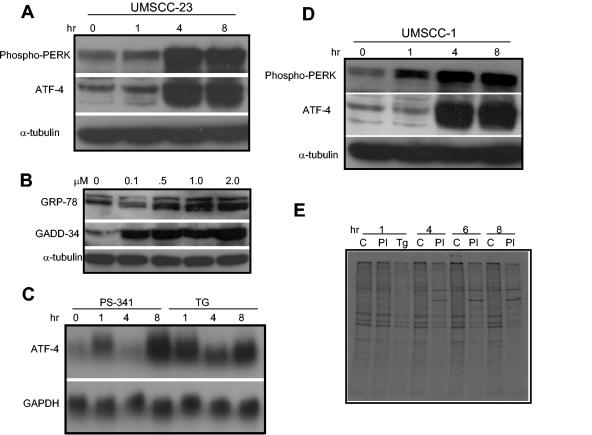
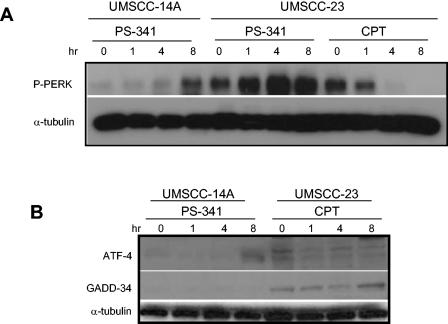
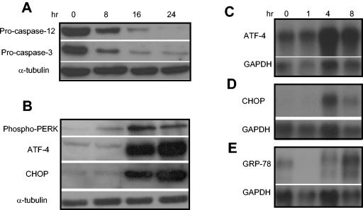
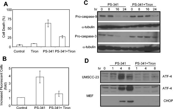
PS-341, also known as Velcade or Bortezomib, represents a new class of anticancer drugs which has been shown to potently inhibit the growth and/or progression of human cancers, including head and neck squamous cell carcinoma (HNSCC). Although it has been logically hypothesized that NF-kappaB is a major target of PS-341, the underlying mechanism by which PS-341 inhibits tumor cell growth is unclear. Here we found that PS-341 potently activated the caspase cascade and induced apoptosis in human HNSCC cell lines. Although PS-341 could inhibit NF-kappaB activation, the inhibition of NF-kappaB was not sufficient to initiate apoptosis in HNSCC cells. Using biochemical and microarray approaches, we found that proteasome inhibition by PS-341 induced endoplasmic reticulum (ER) stress and reactive oxygen species (ROS) in HNSCC cells. The inhibition of ROS significantly suppressed caspase activation and apoptosis induced by PS-341. Consistently, PS-341 could not induce the ER stress-ROS in PS-341-resistant HNSCC cells. Taken together, our results suggest that in addition to the abolishment of the prosurvival NF-kappaB, PS-341 might directly induce apoptosis by activating proapoptotic ER stress-ROS signaling cascades in HNSCC cells, providing novel insights into the PS-341-mediated antitumor activity.


Figures

FIG. 1.
FIG. 1.
PS-341 induces caspase activation and apoptosis in HNSCC cells. (A) HNSCC cell lines UMSCC-1, -9, -11B, -14A, and -23 were treated with PS-341 (0.5 μM) for 0, 8, 16, or 24 h. Cell viability was determined by trypan blue exclusion assay. The assays were performed with triplicate samples, and the results are representative of three independent experiments. Error bars depict standard deviations. (B) UMSCC-1 and UMSCC-23 cells were treated with PS-341 for 0, 16 or 24 h. The detached and attached cells were collected, and genomic DNA was extracted with phenol-chloroform. Genomic DNA was separated on a 1.2% agarose gel. (C) UMSCC-23 cells were treated with PS-341 for 0, 8, 16 or 24 h. The whole-cell extracts were prepared with RIPA buffer, and 50-μg aliquots of protein extracts were resolved on a SDS-12% PAGE. The membrane was probed with antibodies against caspases 9, 2, and 3 (1:500). For loading control, the membrane was stripped and reprobed with anti-α-tubulin monoclonal antibodies (1:5,000). (D) UMSCC-23 cells were transfected with caspase 2 siRNA (SiCasp-2), using oligofectamine. Twenty-four hours after transfection, cells were harvested and probed with monoclonal antibodies against caspase 2. For loading control, the membrane was stripped and reprobed with α-tubulin. (E) UMSCC-23/SiCasp-2 cells and control cells were treated with PS-341 for the indicated times, and cell death was determined.
FIG. 2.
FIG. 2.
The inhibition of NF-κB is not sufficient to induce apoptosis in HNSCC cells. (A) UMSCC-14A cells were transduced with retroviruses expressing SR-IκBα (UMSCC-14A/SR-IκBα) or control vector (UMSCC-14A/V) and selected with neomycin (600 μg/ml) for 1 week. Cells expressing SR-IκBα were confirmed with monoclonal antibodies against the Flag epitope (1:1,000) by Western blot analysis. For loading control, the membrane was stripped and reprobed with monoclonal antibodies against α-tubulin. (B) Both UMSCC-14A/SR-IκBα cells (SCC-14A/SR-IκBα) and control cells (SCC-14A/V) were treated with TNF (20 ng/ml) for 0, 16 or 24 h. Cell viability was determined with a trypan blue exclusion assay. (C) Cells were treated with PS-341 for 0, 16, or 24 h. Cell viability was determined with a trypan blue exclusion assay. The assay was performed in triplicate, and the results represent average values from three independent experiments.
FIG. 3.
FIG. 3.
PS-341 induces ER stress in HNSCC cells. (A and B) UMSCC-23 cells were treated with PS-341 for the indicated time periods and concentrations. Whole-cell extracts were prepared, and 50-μg aliquots of proteins were probed with polyclonal antibodies against phospho-PERK, ATF-4, GRP-78, and GADD-34. For loading control, the membranes were stripped and reprobed with monoclonal antibodies against α-tubulin. (C) UMSCC-23 cells were treated with PS-341 and thapsigargin (TG) for the indicated time periods. The total RNAs were extracted, and 10-μg aliquots of RNAs were resolved on 1.5% agarose formaldehyde gels. The membrane was probed with 32P-labeled ATF cDNA probes. For loading control, the membrane was stripped and reprobed with 32P-labeled glyceraldehyde-3-phosphate dehydrogenase (GAPDH) probe. (D) UMSCC-1 cells were treated with PS-341 for the indicated time periods, and Western blot analysis was performed as described in (A). (E) UMSCC-23 cells were treated with PS-341 or TG as a positive control for the indicated time periods. Cells were labeled with 35S for 15 min. Ten micrograms of 35S-labeled proteins were resolved by SDS-10% PAGE and exposed to autoradiographic film. C, vehicle control; PI, PS-341; Tg, TG.
FIG. 4.
FIG. 4.
The DNA-damaging agent CPT does not induce ER stress in HNSCC cells. (A) UMSCC-23 or UMSCC-14A cells were treated with CPT (10 μM) or PS-341 for the indicated time periods. The phosphorylation of PERK was detected by Western blot analysis as described in the legend to Fig. 3A. (B) Cells were treated with PS-341 or CPT for the indicated time periods. The expression of ATF-4 and GADD-34 was examined by Western blot analysis as described in the legend to Fig. 3A.
FIG. 5.
FIG. 5.
PS-341 induces ER stress and activates caspase 12 in MEFs. (A) MEFs were treated with PS-341 for the indicated time periods, and the whole-cell extracts were prepared. Fifty-microgram aliquots of proteins were probed with polyclonal antibodies against caspase 12 and caspase 3. For loading control, the membrane was stripped and reprobed with monoclonal antibodies against α-tubulin. (B) MEFs were treated with PS-341 for the indicated time periods, and the whole-cell extracts were prepared. Fifty-microgram aliquots of proteins were probed with polyclonal antibodies against ATF-4 and CHOP. For loading control, the membrane was stripped and reprobed with monoclonal antibodies against α-tubulin. (C, D, and E) MEFs were treated with PS-341 for the indicated time periods, and the total RNAs were extracted with Trizol. Ten-microgram aliquots of total RNAs were probed with 32P-labeled ATF, CHOP, and GRP-78 cDNA probes. For loading control, the membranes were stripped and reprobed with 32P-labeled glyceraldehyde-3-phosphate dehydrogenase (GAPDH) cDNA probes.
FIG. 6.
FIG. 6.
PS-341 induces the production of reactive oxygen species in HNSCC cells and MEFs. (A, B, and C) UMSCC-23 cells, MEFs, and UMSCC-14A cells were treated with PS-341 or vehicle control for 4 h, respectively. After treatment, cells were harvested and incubated with the cell-permeable dye H2DCFDA. The reaction was analyzed by FACS.
FIG. 7.
FIG. 7.
The ROS inhibitor Tiron inhibits PS-341-mediated apoptosis and gene expression. (A) UMSCC-23 cells were pretreated with or without Tiron for 30 min and then treated with PS-341 for 24 h. Cell viability was determined with a trypan blue exclusion assay. The assay was performed in triplicate, and results represent the average values from three independent experiments. (B) UMSCC-23 cells were pretreated with or without Tiron for 30 min and then treated with PS-341 for 24 h. After treatment, cells were harvested and incubated with the cell-permeable dye H2DCFDA. The reaction was analyzed by FACS. (C) UMSCC-23 cells were pretreated with or without Tiron for 30 min and then treated with PS-341 for the indicated time periods. The whole-cell proteins were probed with polyclonal antibodies against caspases 9 and 3. For loading control, the membrane was stripped and reprobed with monoclonal antibodies against α-tubulin. (D) UMSCC-23 cells or MEFs were treated with PS-341 with or without Tiron for the indicated time periods, and whole-cell extracts were prepared. Fifty-microgram aliquots were resolved on SDS-8% PAGE and probed with polyclonal antibodies for ATF-4 and CHOP.

Similar articles

Cited by

References

    1. Adams, J. 2003. The proteasome: structure, function, and role in the cell. Cancer Treat. Rev. 29(Suppl. 1):3-9. - PubMed
    1. Adams, J., V. J. Palombella, E. A. Sausville, J. Johnson, A. Destree, D. D. Lazarus, J. Mass, C. S. Pien, S. Prakash, and P. J. Elliot. 1999. Proteasome inhibitors: a novel class of potent and effective antitumor agents. Cancer Res. 59:2615-2622. - PubMed
    1. Chen, S., A. Fribley, and C.-Y. Wang. 2002. Potentiation of tumor necrosis factor-mediated apoptosis of oral squamous cell carcinoma cells by adenovirus-mediated gene transfer of NF-κB inhibitor. J. Dent. Res. 81:98-102. - PubMed
    1. Fischer, H., U. Koenig, L. Eckhart, and E. Tschachler. 2002. Human caspase 12 has acquired deleterious mutations. Biochem. Biophys. Res. Commun. 293:722-726. - PubMed
    1. Fleming, J. A., E. S. Lightcap, S. Sadis, V. Thoroddsen, C. E. Bulawa, and R. K. Blackman. 2002. Complementary whole-genome technologies reveal the cellular response to proteasome inhibition by PS-341. Proc. Natl. Acad. Sci. USA 99:1461-1466. - PMC - PubMed

Publication types

MeSH terms