. 2004 Nov;24(22):9705-25.
doi: 10.1128/MCB.24.22.9705-9725.2004.

Retinoid X receptor regulates Nur77/TR3-dependent apoptosis [corrected] by modulating its nuclear export and mitochondrial targeting

Affiliations

Retinoid X receptor regulates Nur77/TR3-dependent apoptosis [corrected] by modulating its nuclear export and mitochondrial targeting

Xihua Cao et al. Mol Cell Biol. 2004 Nov.

Erratum in

  • Mol Cell Biol. 2005 Jan;25(1):524

Abstract

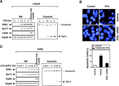
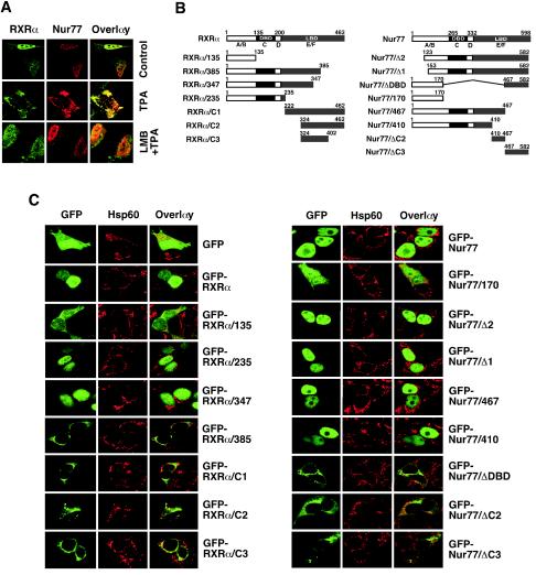
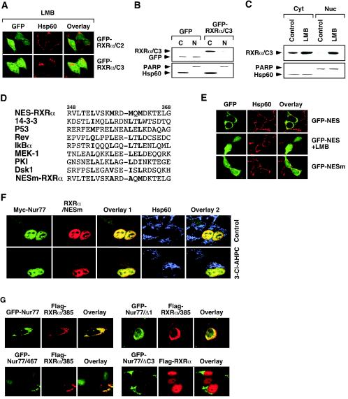
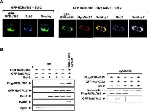
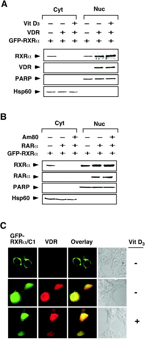
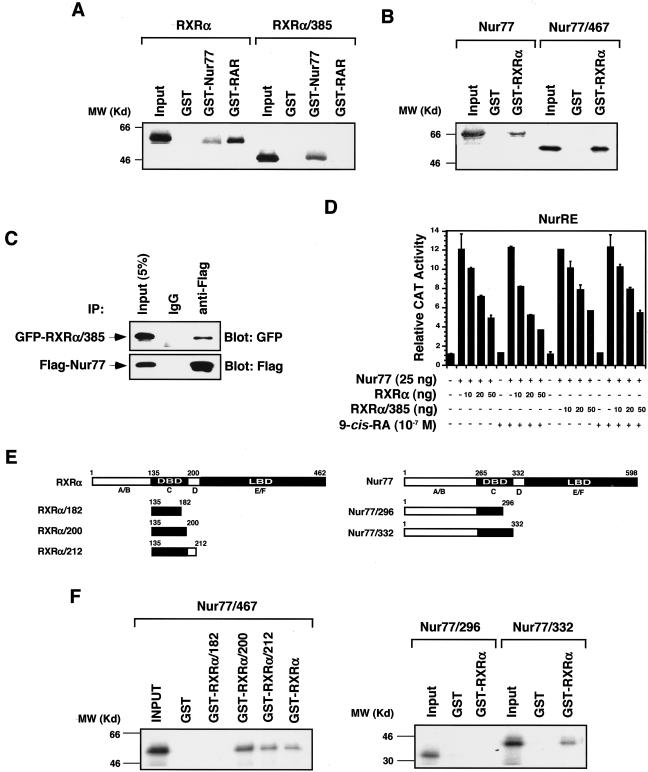
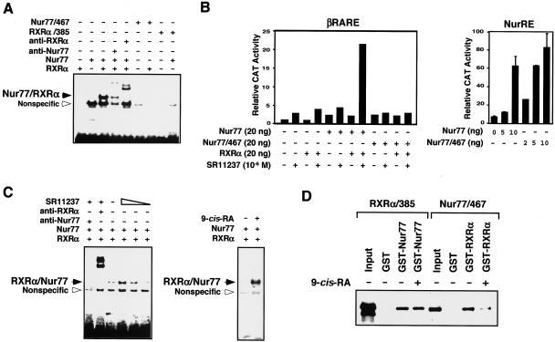
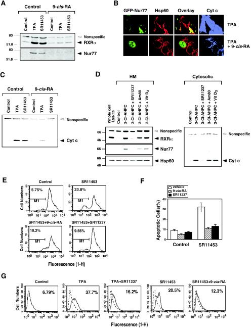
Retinoid X receptor (RXR) plays a central role in the regulation of intracellular receptor signaling pathways by acting as a ubiquitous heterodimerization partner of many nuclear receptors, including the orphan receptor Nur77 (also known as TR3 [corrected] or NGFI-B), which translocates from the nucleus to mitochondria, where it interacts with Bcl-2 to induce apoptosis. Here, we report that RXRalpha is required for nuclear export and mitochondrial targeting of Nur77 through their unique heterodimerization that is mediated by dimerization interfaces located in their DNA-binding domain. The effects of RXRalpha are attributed to a putative nuclear export sequence (NES) present in its carboxyl-terminal region. RXRalpha ligands suppress NES activity by inducing RXRalpha homodimerization or altering RXRalpha/Nur77 heterodimerization. The RXRalpha NES is also silenced by RXRalpha heterodimerization with retinoic acid receptor or vitamin D receptor. Consistently, we were able to show that the mitochondrial targeting of the RXRalpha/Nur77 heterodimer and its induction of apoptosis are potently inhibited by RXR ligands. Together, our results reveal a novel nongenotropic function of RXRalpha and its involvement in the regulation of the Nur77-dependent apoptotic pathway [corrected]


Figures

FIG. 1.
FIG. 1.
Nur77 and RXR comigrate from the nucleus to the cytoplasm. (A/B) RXRα (A) or Nur77 (B) targets mitochondria in response to apoptotic stimulus. LNCaP prostate cancer cells were treated with TPA (100 ng/ml) for 1 h and then immunostained with either anti-RXRα (Santa Cruz Biotechnology) (A) or anti-Nur77 (Active Motif) (B) antibody, followed by Cy3-conjugated secondary antibody (Sigma) to detect RXRα or Nur77 or with anti-Hsp60 (Santa Cruz Biotechnology), followed by FITC-conjugated secondary antibody (Sigma) to detect mitochondria. RXRα, Nur77, and mitochondria (Hsp60) were visualized by using confocal microscopy, and the images of RXRα or Nur77 with those of mitochondria were overlaid (overlay). About 80% of cells displayed mitochondrial targeting of RXRα and Nur77 when cells were treated with TPA. One of three similar experiments is shown. (C) Nur77 and RXRα comigrate from the nucleus to the cytoplasm. LNCaP cells were treated with or without TPA for 1 h and then immunostained with anti-RXRα antibody, followed by FITC-conjugated secondary antibody, or with anti-Nur77 antibody (Abgent, San Diego, Calif.), followed by Cy3-conjugated secondary antibody. RXRα and Nur77 were visualized by using confocal microscopy and the images were overlaid (overlay). Approximately 80% of TPA-treated cells showed RXRα colocalization with Nur77. One of three similar experiments is shown. (D) 3-Cl-AHPC induces mitochondrial localization of transfected RXRα and Nur77. Expression vectors for myc-Nur77 and RXRα weretransfected into LNCaP cells. Cells were then treated with 3-Cl-AHPC for 3 h and then immunostained with anti-myc antibody (9E10; Santa Cruz Biotechnology), followed by FITC-conjugated secondary antibody, anti-RXRα antibody, followed by Cy3-conjugated secondary antibody or anti-Hsp60 antibody, followed by Cy5-conjugated secondary antibody (Jackson Immunoresearch). Myc-Nur77, RXRα, and Hsp60 were visualized and the images were overlaid. Overlay 1 is the merger of myc-Nur77 and RXRα images, and overlay 2 is the merger of myc-Nur77, RXRα, and Hsp60 images. About 30% of transfected cells exhibited the colocalization presented. One of three similar experiments is shown. (E) Mitochondrial localization of RXRα is Nur77 dependent. LNCaP cells or LNCaP cells stably expressing Nur77 antisense RNA (Nur77/antisense) (40) were treated with or without TPA for 1 h and then immunostained with anti-RXRα antibody, followed by Cy3-conjugated secondary antibody. Approximately 70% of cells displayed the effect presented. One of two similar experiments is shown. (F) Effect of RXR siRNA on RXRα levels and Nur77 localization. LNCaP cells were transfected with or without RXRα siRNA or control siRNA for 72 h. Cell extracts were prepared and analyzed for RXRα expression by Western blotting. Cells were also analyzed for subcellular localization of RXRα and Nur77 by confocal microscopy. One of two similar experiments is shown. (G) Nur77/Δ1 prevents RXRα mitochondrial targeting. LNCaP cells were transfected with the GFP-Nur77/Δ1 expression vector and then treated with TPA for 1 h and immunostained with anti-RXRα antibody. RXRα and GFP-Nur77/Δ1 distributions were analyzed by confocal microscopy. About 80% of nontransfected cells showed cytoplasmic localization of RXRα after treatment with TPA, whereas >70% of cells transfected with GFP-Nur77/Δ1 showed RXRα nuclear localization with the same treatment. One of three similar experiments is shown. (H) 3-Cl-AHPC induces mitochondrial localization of RXRα and Nur77 in H460 lung cancer cells. H460 cells were treated with 3-Cl-AHPC (10−6 M) for 3 h and then immunostained with anti-RXRα antibody, followed by FITC-conjugated secondary antibody, anti-Nur77 followed by Cy3-conjugated secondary antibody, or anti-Hsp60 antibody, followed by Cy5-conjugated secondary antibody (Jackson Immunoresearch). RXRα, Nur77, and Hsp60 were visualized by confocal microscopy, and the images were overlaid. Overlay 1 represents the merger of RXRα and Nur77 images. Overlay 2 indicates merged RXRα, Nur77, and Hsp60 images. Approximately 80% of 3-Cl-AHPC-treated cells showed the patterns presented. One of three similar experiments is shown.
FIG. 2.
FIG. 2.
Time course analysis of mitochondrial targeting of RXRα and Nur77 and the release of cytochrome c from mitochondria. (A) Time course analysis of LNCaP cells in response to TPA. Cells were treated with TPA (100 ng/ml) for the indicated times. Mitochondrion-enriched HM and cytosolic fractions were prepared and analyzed for the presence of RXRα, Nur77, and cytochrome c as indicated. As a control, the whole-cell extract was also analyzed. Expression of mitochondrion-specific Hsp60 protein and nucleus-specific PARP protein was determined to control the purity of HM fractions. One of two similar experiments is shown. (B) Inhibition of RXRα expression suppresses the apoptotic effect of TPA. LNCaP cells were transfected with RXRα siRNA, followed by treatment with TPA (100 ng/ml) for 3 h. Cells were stained with DAPI and analyzed for nuclear morphological changes. Apoptotic cells were scored by examining 300 cells for apoptotic morphology from three different experiments. (C) Time course analysis of H460 cells in response to 3-Cl-AHPC. Cells were treated with 3-Cl-AHPC (10−6 M) and analyzed as described for panel A. One of two similar experiments is shown.
FIG. 3.
FIG. 3.
Identification of domains in RXRα and Nur77 required for their nuclear export. (A) Cytoplasmic localization of RXRα/Nur77 is mediated by CRM1-dependent nuclear export. LNCaP cells were treated with TPA (100 ng/ml) in the absence (control) or presence of LMB (2.5 ng/ml; Sigma) and analyzed by confocal microscopy as described for Fig. 1C. About 80% cells showed the cytoplasmic localization of RXRα and Nur77 after treatment with TPA, whereas >50% of cells showed nuclear localization of RXRα and Nur77 when pretreated with LMB. One of two similar experiments is shown. (B) Schematic representations of RXRα and Nur77 mutants. The DBD, LBD, and A to F domains are indicated. (C) Analysis of subcellular localization of Nur77 and RXRα mutants. The indicated plasmids were transfected into HEK293T cells and analyzed by confocal microscopy as described in Fig. 1. More than 90% of transfected cells showed diffused distribution of GFP and GFP-RXRα/135. Nuclear localization of GFP-RXRα, RXRα/235, and RXRα/347 was found in 80, 85, and 60% of transfected cells, respectively. More than 90% of transfected cells showed exclusive cytoplasmic localization of RXRα/385, RXRα/C2, and RXRα/C3, whereas cytoplasmic localization of RXRα/C1 was found in 70% of transfected cells. Nuclear localization of GFP-Nur77 and its mutants, GFP-Nur77/Δ2, GFP-Nur77/Δ1, GFP-Nur77/467, and GFP-Nur77/410, was found in >90% of transfected cells, whereas cytoplasmic localization of GFP-Nur77/ΔDBD, cytoplasmic localization of GFP-Nur77/ΔDBD, GFP-Nur77/ΔC2, and GFP-Nur77/ΔC3 was observed in 80, 60 and 70% of transfected cells, respectively. One of four similar experiments is shown.
FIG. 4.
FIG. 4.
Identification of a nuclear export sequence in RXRα. (A) Effect of LMB on subcellular localization of RXRα mutants. The indicated expression vector for GFP fusion proteins was transfected into HEK293T cells and analyzed by confocal microscopy. Approximately 70% of transfected cells displayed diffused distribution of RXRα/C2 and RXRα/C3 after treatment with LMB. One of four similar experiments is shown. (B) RXRα/C3 exclusively resides in the cytoplasm. GFP-RXRα/C3 was transfected into HEK293T cells, and its localization was analyzed by cellular fractionation, followed by Western blotting with anti-GFP antibody (Santa Cruz Biotechnology). One of three similar experiments is shown. (C) The effect of LMB on subcellular localization of RXRα/C3. RXRα/C3 was transfected into HEK293T cells, which were then treated with or without LMB (2.5 ng/ml) for 6 h. Localization of RXRα/C3 was analyzed as described in panel B. One of three similar experiments is shown. (D) Schematic representation of the RXRα NES. The identified RXRα NES is compared to known NESs identified in the indicated genes. The boldface letters indicate conserved amino acid residues. (E) The RXRα NES is capable of directing GFP to the cytoplasm. The putative RXRαNES (RVLTELVSKMRDMQMDKTELG) or its mutant (RVLTELVSKARDAQMDKTELG) (NESm) was fused to GFP, and the expression vectors were transfected into HEK293T cells. Cells were treated with or without LMB for 6 h and then stained for Hsp60 and analyzed by confocal microscopy. Approximately 90% of GFP-NES-transfected cells exhibited cytoplasmic localization, whereas <20% of GFP-NESm-transfected cells displayed the same cytoplasmic localization. One of three similar experiments is shown. (F) Mutation of the RXRα NES impairs the 3-Cl-AHPC-induced mitochondrial localization of RXRα/Nur77 heterodimer. Myc-Nur77 was cotransfected with RXRα/NESm into LNCaP cells, which were then treated with 3-Cl-AHPC (10−6 M) for 3 h. Cells were stained for Nur77 by anti-myc antibody and for RXRα/NESm by anti-RXRα antibody and analyzed by the confocal microscopy. About 90% of transfected cells showed nuclear localization of Myc-Nur77 and RXRα/NESm, even after treatment with 3-Cl-AHPC. (G) RXRα NES is required for cytoplasmic localization of Nur77. The indicated expression vectors were cotransfected into HEK293T cells. Their subcellular localization was analyzed by confocal microscopy as described in Fig. 1A. Less than 10% of transfected cells showed cytoplasmic localization of GFP-Nur77, Nur77/467, and Nur77/Δ1, which was increased to 30, 40, and 40% upon cotransfection with Flag-RXRα/385, respectively. About 80% of transfected cells showed nuclear localization of Flag-RXRα, with or without Nur77/ΔC3 cotransfection. One of three similar experiments is shown.
FIG. 5.
FIG. 5.
Role of Bcl-2 in mitochondrial targeting of RXRα and Nur77 mutants. (A) Mitochondrial targeting of RXRα/385 in HEK293T cells requires Nur77 and Bcl-2. Expression vectors of GFP-RXRα/385 and Bcl-2 were transfected, together with or without myc-Nur77 vector into HEK293T cells. Cells were then immunostained with anti-myc antibody (9E10; Santa Cruz Biotechnology), followed by Cy3-conjugated secondary antibody, or with anti-Bcl-2 antibody, followed by Cy5-conjugated secondary antibody. GFP-RXRα/385, myc-Nur77, and Bcl-2 were visualized by confocal microscopy. Overlay 1 represents the merger of the GFP-RXRα/385 and myc-Nur77 images, and overlay 2 is a merge of the GFP-RXRα/385, myc-Nur77, and Bcl-2 images. Approximately 70% of cells transfected with GFP-RXRα/385 and myc-Nur77 exhibited colocalization with Bcl-2, whereas <5% of cells transfected with GFP-RXRα/385 alone showed colocalization with Bcl-2. (B) Accumulation of RXRα/385 at mitochondria in HEK293T cells requires both Nur77 and Bcl-2. Flag-RXRα/385, GFP-Nur77/Δ2, and Bcl-2 were transfected into HEK293T cells as indicated. HM and cytosolic fractions were prepared and levels of Flag-RXRα/385, GFP-Nur77/Δ2, and Bcl-2 were determined by immunoblotting with anti-Flag (Sigma), anti-GFP, and anti-Bcl-2 antibody, respectively. For control, levels of Hsp60 and PARP were also determined. One of two similar experiments is shown.
FIG. 6.
FIG. 6.
Mitochondrial targeting and apoptosis effects of cytoplasmic RXRα and Nur77 mutants. (A) Cytoplasmic Nur77 mutants but not RXRα mutants induce apoptosis. The indicated GFP-RXRα or GFP-Nur77 mutant was transfected into LNCaP cells. After 48 h, cells were stained by DAPI and analyzed for nuclear morphological change by microscopy. Arrows indicate cells with extensive nuclear condensation or fragmentation. Percentages of apoptotic cells were determined by examining 200 GFP-positive cells for nuclear fragmentation and/or chromatin condensation. Bars represent averages ± the means from three experiments. (B) Cytoplasmic Nur77 mutant but not RXRα mutant induces cytochrome c (Cyt) release. GFP-Nur77/ΔDBD or GFP-RXRα/C1 expression vector was transfected into LNCaP cells. Cells were then stained for mitochondria (Hsp60) and cytochrome c and analyzed by confocal microscopy. Cytochrome c release was observed in 80% of cells showing Nur77/ΔDBD mitochondrial targeting, whereas it was not found in cells transfected with RXRα/C1. One of two similar experiments is shown.
FIG. 7.
FIG. 7.
Regulation of RXRα nuclear export by its ligands and homodimerization. (A and B) Analysis of subcellular localization of RXRα/C1 in the absence or presence of retinoids. (A) GFP-RXRα/C1 was transfected into HEK293T cells, which were then treated with the indicated retinoid, stained with Hsp60, and analyzed by confocal microscopy. The inhibitory effect of RXR ligands on the cytoplasmic localization of RXRα/C1 was observed in 80% of transfected cells, whereas >90% of transfected cells failed to respond to RAR ligands. (B) Nuclear and cytoplasmic extracts were also prepared and analyzed for expression of GFP-RXRα/C1 by Western blotting with anti-GFP antibody. One of three similar experiments is shown. (C) RXRα/C1 dimerization status determines its subcellular localization. GFP-RXRα/C1 was transfected into HEK293T cells, which were not treated or treated with 9-cis-RA (10−7 M). Nuclear and cytoplasmic extracts were prepared and analyzed by using nondenaturing PAGE and anti-GFP antibody. The same extracts were analyzed by denaturing PAGE for the expression of PARP and Hsp60 to ensure fraction purity. One of two similar experiments is shown. (D) Confocal microscopy analysis of RXR homodimerization-defective mutants. GFP-RXRα/385, GFP-RXRα/LLL, or GFP-RXRα/C1/C432R was transfected into HEK293T cells. Cells were treated with or without 9-cis-RA (10−7 M) and analyzed by confocal microscopy. Approximately 80% of transfected cells showed nuclear localization of GFP-RXRα, which was slightly increased to 85% after treatment with 9-cis-RA. Cytoplasmic localization of GFP-RXRα/385 (90%), GFP-RXRα/LLL (65%), and GFP-RXRα/C1/C432R (85%) was not affected by 9-cis-RA treatment. One of three similar experiments is shown.
FIG. 8.
FIG. 8.
Regulation of RXRα nuclear export by heterodimerization. (A and B) Regulation of RXR nuclear export by VDR (A) and RARα (B). Expression vector for VDR or RARα was transfected into HEK293T cells, together with GFP-RXRα expression vector. Cells were then treated with the indicated ligand, Vit D3 (10−7 M) or Am80 (10−6 M). Nuclear and cytoplasmic extracts were prepared and analyzed for expression of transfected GFP-RXRα by anti-GFP antibody. Expression of transfected VDR or RARα was also determined. One of two similar experiments is shown. (C) Confocal analysis of the effect of VDR expression on RXR localization. The GFP-RXRα/C1 expression vector was transfected into HEK293T cells together with or without the VDR expression vector. Cells were then treated with VD3 (10−7 M),stained with anti-VDR antibody (Santa Cruz Biotechnology), followed by Cy3-conjugated secondary antibody (Sigma) and analyzed by confocal microscopy. About 70% of transfected cells showed cytoplasmic localization of RXRα/C1, whereas >80% of cotransfected cells showed nuclear localization of RXRα/C1 and VDR, which was not enhanced by VD3 treatment. One of three similar experiments is shown.
FIG. 9.
FIG. 9.
C termini of RXRα and Nur77 are not required for RXRα/Nur77 interaction in solution. (A and B) GST pull-down assays for determination of RXRα/Nur77 heterodimerization. GST-Nur77, GST-RXRα, or GST control protein immobilized on glutathione-Sepharose (20 μl) was incubated with in vitro synthesized 35S-labeled Nur77, RXRα or their mutants (5 μl) as indicated. Bound proteins were analyzed by SDS-PAGE and autoradiography. One of three similar experiments is shown. (C) Coimmunoprecipitation assay for Nur77 and RXRα/385 interaction. Expression vectors for Flag epitope-tagged-Nur77 (Flag-Nur77) and GFP-RXRα/385 were cotransfected into HEK293T cells. The expressed Flag-Nur77 and GFP-RXRα/385 were then immunoprecipitated by using either anti-Flag antibody or control IgG, and immunoprecipitates were examined by Western blotting with anti-GFP antibody. The same membranes were also blotted with anti-Flag antibody to determine precipitation specificity and efficiency. Input represents 5% of total cell extract used in the precipitation assays. One of two similar experiments is shown. (D) Reportergene assay. (NurRE)2-tk-chloramphenicol acetyltransferase (CAT) (100 ng), β-galactosidase (β-Gal; 100 ng), and Nur77 (25 ng) expression vectors were transiently transfected into HEK293T cells together with or without RXRα or RXRα/385. Cells were treated with or without 9-cis-RA (10−7 M) as indicated. CAT activity was determined and normalized relative to β-Gal activity. Bars represent averages ± deviations from three different experiments. (E) Schematic representations of RXRα and Nur77 mutants. The DBD, LBD, and A to F domains are indicated. (F) GST pull-down assays for determination of RXRα/Nur77 heterodimerization. The indicated GST-RXRα, its mutants, or GST control protein was immobilized on glutathione-Sepharose (20 μl) and incubated with in vitro-synthesized 35S-labeled Nur77 or its mutants (5 μl) as indicated. Bound proteins were analyzed by SDS-PAGE and autoradiography. One of three similar experiments is shown.
FIG. 10.
FIG. 10.
9-cis-RA modulates RXRα/Nur77 heterodimerization and promotes DNA binding. (A) The C-terminal domains of Nur77 and RXRα are required for the formation of RXRα/Nur77 heterodimer on DNA. The indicated Nur77, RXRα, or their mutants were synthesized in vitro and analyzed for binding to the βRARE by gel shift assays. One of three similar experiments is shown. (B) Differential requirement of the Nur77 C terminus for transactivation of the Nur77 response element NurRE and the βRARE. (NurRE)2-tk-CAT (100 ng) or βRARE-tk-CAT (100 ng), β-Gal expression vector (100 ng), and the expression vector for Nur77 or a Nur77 mutant (20 ng) were transiently transfected into HEK293T cells with or without the RXRα expression vector. The CAT activity was determined and normalized relative to the β-Gal activity. One of three similar experiments is shown. (C) Effect of RXR ligands on binding of RXRα-Nur77 heterodimers to the βRARE. Equivalent amounts of in vitro synthesized Nur77 and RXRα were incubated alone or together with or without RXR ligand 9-cis-RA (10−7 M) or SR11237 (10−6 M) and analyzed by gel retardation assays with the βRARE as a probe. One of two similar experiments is shown. (D) RXR ligand modulates RXRα/Nur77 heterodimerization in solution. GST pull-down assays for determination of RXRα/Nur77 heterodimerization. The indicated GST fusions immobilized on glutathione-Sepharose (20 μl) were incubated with in vitro synthesized 35S-labeled receptor protein (5 μl) as indicated. Bound proteins were analyzed by SDS-PAGE and/or autoradiography. One of two similar experiments is shown.
FIG. 11.
FIG. 11.
Effect of RXR ligands on RXR mitochondrial targeting and apoptosis. (A) Effect of 9-cis-RA on mitochondrial localization of RXRα and Nur77. HM fractions were prepared from LNCaP cells treated with or without TPA or SR11453 (10−6 M) for 3 h with or without a 9-cis-RA (10−7 M) pretreatment for 12 h and then analyzed for expression of Nur77 or RXRα by immunoblotting. One of two similar experiments is shown. (B) 9-cis-RA inhibits Nur77-dependent release of cytochrome c from mitochondria. GFP-Nur77 expression vector was transfected into LNCaP cells, treated with or without 9-cis-RA for 12 h before TPA treatment (1 h). Cells were stained for mitochondria (Hsp60) and cytochrome c, and analyzed by confocal microscopy. 100% of cells displayed diffused cytochrome c staining when treated with TPA, whereas 60% of cells showed punctate cytochrome c staining when cells were cotreated with TPA and 9-cis-RA. (C) Inhibition of cytochrome c release by 9-cis-RA. LNCaP cells were treated with or without 9-cis-RA (10−7 M) for 12 h before treatment with TPA (100 ng/ml) or SR11453 (10−6 M) for 1 h. Cytosolic fractions were analyzed for cytochrome c by immunoblotting. A nonspecific band at ∼70 kDa served as a control for equal loading of proteins. One of three similar experiments is shown. (D) RAR and VDR ligands fail to inhibit mitochondrial localization of RXRα and Nur77 and cytochrome c release. LNCaP cells were treated with 3-Cl-AHPC (10−6 M) for 3 h after pretreatment with SR11237 (10−6 M), RAR ligand Am80 (10−6 M), or VDR ligand vitamin D3 (10−7 M). HM and cytosolic fractions were prepared and analyzed for mitochondrial localization of RXRα and Nur77, as well as the cytochrome c release described above. (E) RXR ligands prevent SR11453 induced mitochondrial membrane potential change (Δψm). LNCaP cells were pretreated with 9-cis-RA or SR11237 for 12 h before treatment with SR11453 (10−6 M) for 18 h. Cells were then incubated with Rh123 for 30 min and analyzed by FACScalibur cytometry. One of three similar experiments is shown. (F) Inhibition of apoptosis by RXR ligands. LNCaP cells were pretreated with 9-cis-RA or SR11237 for 12 h before treatment with SR11453 for 48 h. Apoptosis was determined by nuclear staining with DAPI. The percentages of cells fluorescing within the range of Rh123 were considered as depolarized (i.e., Δψm disrupted). Bars represent averages ± the standard deviations from two experiments. (G) LNCaP cells were pretreated 9-cis-RA or SR11237 for 12 h before treatment with SR11453 or TPA for 24 h. Apoptosis was determined by the TdT assay. One of two similar experiments is shown. Cyt c, cytochrome c.

References

    1. Aarnisalo, P., C. H. Kim, J. W. Lee, and T. Perlmann. 2002. Defining requirements for heterodimerization between the retinoid X receptor and the orphan nuclear receptor Nurr1. J. Biol. Chem. 277:35118-35123. - PubMed
    1. Baumann, C. T., P. Maruvada, G. L. Hager, and P. M. Yen. 2001. Nuclear cytoplasmic shuttling by thyroid hormone receptors. multiple protein interactions are required for nuclear retention. J. Biol. Chem. 276:11237-11245. - PubMed
    1. Bissonnette, R. P., T. Brunner, S. B. Lazarchik, N. J. Yoo, M. F. Boehm, D. R. Green, and R. A. Heyman. 1995. 9-cis retinoic acid inhibition of activation-induced apoptosis is mediated via regulation of fas ligand and requires retinoic acid receptor and retinoid X receptor activation. Mol. Cell. Biol. 15:5576-5585. - PMC - PubMed
    1. Bourguet, W., M. Ruff, P. Chambon, H. Gronemeyer, and D. Moras. 1995. Crystal structure of the ligand-binding domain of the human nuclear receptor RXR-α. Nature 375:377-382. - PubMed
    1. Bourguet, W., V. Vivat, J. M. Wurtz, P. Chambon, H. Gronemeyer, and D. Moras. 2000. Crystal structure of a heterodimeric complex of RAR and RXR ligand-binding domains. Mol. Cell 5:289-298. - PubMed

Publication types

MeSH terms

LinkOut - more resources