. 2004 Nov;24(22):9752-62.
doi: 10.1128/MCB.24.22.9752-9762.2004.

The TBC (Tre-2/Bub2/Cdc16) domain protein TRE17 regulates plasma membrane-endosomal trafficking through activation of Arf6

Affiliations

The TBC (Tre-2/Bub2/Cdc16) domain protein TRE17 regulates plasma membrane-endosomal trafficking through activation of Arf6

Lenka Martinu et al. Mol Cell Biol. 2004 Nov.

Abstract

TBC (Tre-2/Bub2/Cdc16) domains are predicted to encode GTPase-activating proteins (GAPs) for Rab family G proteins. While approximately 50 TBC proteins are predicted to exist in humans, little is known about their substrate specificity. Here we show that TRE17 (also called Tre-2 and USP6), a founding member of the TBC family, targets the Arf family GTPase Arf6, which regulates plasma membrane-endosome trafficking. Surprisingly, TRE17 does not function as a GAP for Arf6 but rather promotes its activation in vivo. TRE17 associates directly with Arf6 in its GDP- but not GTP-bound state. Mapping experiments pinpoint the site of interaction to the TBC domain of TRE17. Forced expression of TRE17 promotes the localization of Arf6 to the plasma membrane, leading to Arf6 activation, presumably due to facilitated access to membrane-associated guanine nucleotide exchange factors (GEFs). Furthermore, TRE17 cooperates with Arf6 GEFs to induce GTP loading of Arf6 in vivo. Finally, short interfering RNA-mediated loss of TRE17 leads to attenuated Arf6 activation. These studies identify TRE17 as a novel regulator of the Arf6-regulated plasma membrane recycling system and reveal an unexpected function for TBC domains.


Figures

FIG. 1.
FIG. 1.
TRE17 peptides traffic between plasma membrane and tubulo-vesicular compartment. (A) Domain structure of the three TRE17 isoforms TRE17(long), TRE17(onco), and TRE17(short). Also shown is mutant T17(447), encoding amino acids 1 to 447 of TRE17(long). The length of each (in amino acids) is indicated. TBC, Rab GAP homology domain; C and H, cysteine and histidine subdomains of the ubiquitin-specific protease domain, respectively. Dark bars at the C termini of TRE17(onco) and TRE17(short) represent unique sequences. (B) HA-tagged TRE17 isoforms were transfected into HeLa cells, visualized with anti-HA, and analyzed by confocal microscopy. Cells expressing TRE17(long) were treated with cytochalasin D (cyto D, 200 nM) for 30 min (right). Scale bar, 10 μm.
FIG. 2.
FIG. 2.
TRE17 localizes to the Arf6 tubular endosome. HeLa cells were cotransfected with HA-Arf6 (red) and GFP-TRE17(onco) (green) (A) or HA-TRE17(onco) (red) and GFP-PLCδ(PH) (green) (B). Cells were subjected to indirect immunofluorescence with anti-HA antibody and then analyzed by confocal microscopy. Scale bar, 5 μm.
FIG. 3.
FIG. 3.
TRE17(long) and Arf6 cooperate to induce a constitutively active Arf6 phenotype. HeLa cells were transfected with wild-type Arf6 (A) or Q67L (C) and then subjected to indirect immunofluorescence microscopy with anti-Arf6 antibody. For C, F-actin was visualized with fluorescein isothiocyanate-phalloidin (bottom). (B, D, and E), HA-TRE17(long) and wild-type Arf6 were cotransfected. In B, the two proteins were detected with anti-HA and anti-Arf6 antibodies, respectively. (D) Live cells were labeled with anti-major histocompatibility complex type I for 2 h prior to fixation. (E) F-actin was visualized with fluorescein isothiocyanate-phalloidin. Scale bar, 10 μm in A, C, and E; 5 μm in B and D. (F) Cells were transfected with wild-type Arf6 alone or with TRE17(long) or TRE17(onco). Numbers represent the percentage of cells coexpressing Arf6 and the indicated TRE17 peptide that contain vacuoles. The data represent the results of at least three independent experiments in which 100 cells were counted per experiment.
FIG. 4.
FIG. 4.
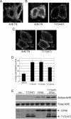
Alterations in tubular endosome morphology induced by TRE17 arise through activation of endogenous Arf6. HeLa cells were transfected with (A) HA-TRE17(long) and wild-type Arf6 or (B) HA-T17(447), encoding the N-terminal 447 amino acids of TRE17(long) (see Fig. 1 for domain structure) and then analyzed by confocal microscopy. (C) HeLa cells were transfected with HA-T17(447), and immunoelectron microscopy was performed with anti-HA antibody and secondary antibody conjugated to 1.4-nm gold particles. (D) The percentage of HA-T17(447)-expressing cells containing enlarged vesicles (as typified in B) was quantified in the absence and presence of coexpressed Arf6 T27N. The results are representative of three independent experiments in which at least 100 cells were counted per experiment. Scale bar, 10 μm.
FIG. 5.
FIG. 5.
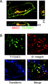
TRE17 vesicles specifically accumulate cargo of the Arf6 pathway. (A) Live HeLa cells expressing HA-T17(447) were incubated with anti-major histocompatibility complex type I for 2 h. Samples were fixed and subjected to indirect immunofluorescence microscopy, visualizing internalized anti-major histocompatibility complex type I (red) and T17(447) (green). (B) HeLa cells expressing T17(447) (green) were incubated with anti-β1-integrin (red) and Alexa Fluor 488-transferrin (blue) for 2 h prior to fixation. Scale bar, 10 μm.
FIG. 6.
FIG. 6.
TRE17 specifically activates Arf6 but not Arf1 in vivo. (A) Extracts were prepared from HeLa cells transfected with HA-Arf6 Q67L or T27N and then subjected to pulldowns (pdn) with GST-GGA3. Arf6 was detected by anti-HA immunoblotting. (B) HA-Arf6 was cotransfected with vector, HA-T17(447), or Flag-EFA6, and GGA3 pulldowns were performed as in A. EFA6 was detected with anti-Flag. Active Arf6, GGA3 pulldowns; total Arf6, whole-cell lysates. (C) HA-Arf1 was cotransfected with vector, HA-T17(447), or Myc-ARNO. GGA3 pulldown assays were performed. ARNO was detected with anti-Myc antibody. Active Arf1, GGA3 pulldowns; total Arf1, whole-cell lysates.
FIG. 7.
FIG. 7.
TBC domain of TRE17 binds directly to Arf6-GDP. (A) HA-tagged Arf6 Q67L (QL) or T27N (TN) was cotransfected with the indicated GST-tagged TRE17 allele into HeLa cells. TRE17 peptides were precipitated with glutathione-Sepharose beads, and associated Arf6 was detected by anti-HA blotting. WCL, whole-cell lysate. Pdn, pulldown. (B) Domain structure of the TRE17 mutants used in A. (C) HeLa cells were cotransfected with GST-T17(447) and wild-type HA-Arf6. Cells were incubated with or without 100 mM 2-deoxyglucose (2DG) for 2 h and then harvested as in A. (D) Purified wild-type Arf6 was incubated with MBP, MBP-TBC, or MBP-T17(447) conjugated to amylose beads. The beads were washed, and the associated Arf6 was detected by anti-Arf6 immunoblotting (top panel); use of comparable levels of MBP fusions was confirmed by anti-MBP blotting (middle panel). In the bottom panel, Arf6 was loaded with GTPγS or GDP in vitro prior to performing pulldowns with MBP or MBP-TBC.
FIG. 8.
FIG. 8.
TRE17 induces plasma membrane recruitment of Arf6 and cooperates with GEFs to promote activation. Arf6 T27N was expressed by itself (A) or with HA-T17(447) (B and C) in HeLa cells. (B) Representative image of plasma membrane-localized Arf6 T27N upon coexpression with T17(447). (C) Complete redistribution of Arf6 T27N to the plasma membrane was observed in a fraction of cells coexpressing T17(447). Scale bar, 10 μm. (D) Arf6 T27N was expressed by itself or with the indicated TRE17 peptide. The percentage of cells exhibiting plasma membrane (P.M.)-localized Arf6 T27N as typified in B was quantified. Results represent three independent experiments in which 100 cells were counted in each. (E) HA-Arf6 was cotransfected with the indicated plasmids into HeLa cells. GGA3 pulldown assays were performed as above.
FIG. 9.
FIG. 9.
TRE17 activates Arf6 independently of intrinsic GAP activity. (A) Sequence alignment of TBC proteins; only a portion of the TBC domain is shown. Numbers indicate the amino acid position of the first residue shown. The arginine residue that functions in catalysis is highlighted with an arrow; the corresponding residue in TRE17 is threonine (T150). Another conserved arginine residue required for activity is marked with an asterisk (corresponding to R106 in TRE17). (B) HeLa cells were transfected with a T17(447) mutant in which arginines 106 and 149 were mutated to lysines (RKRK). Live cells were incubated with anti-major histocompatibility complex type I antibody and then processed for indirect immunofluorescence. Scale bar, 10 μm. (C) Wild-type GST-T17(447) or the RKRK mutant was cotransfected with HA-Arf6 Q67L or T27N. T17(447) was precipitated with glutathione-Sepharose, and associated Arf6 was detected by anti-HA immunoblotting. (D) HA-Arf6 was cotransfected with wild-type T17(447) or the RKRK mutant. GGA3 pulldown assays were performed as above. Immunoblotting of whole-cell extracts confirmed uniform expression of the Arf6 and T17(447) mutants (data not shown).
FIG. 10.
FIG. 10.
Arf6 activity is attenuated in HeLa cells depleted of TRE17. (A) HeLa cells were transfected with a construct targeting TRE17 (T17−) or a negative control plasmid (Cont) and then selected in neomycin. RNA was isolated from pooled populations, and reverse transcription-PCR was performed with primers against TRE17 (top panel) or glyceraldehyde-3-phosphate dehydrogenase (bottom panel). (B) T17− and control cells were transfected with HA-Arf6 and then subjected to GGA3 pulldowns.

Similar articles

Cited by

References

    1. Albert, S., and D. Gallwitz. 1999. Two new members of a family of Ypt/Rab GTPase activating proteins. Promiscuity of substrate recognition. J. Biol. Chem. 274:33186-33189. - PubMed
    1. Albert, S., and D. Gallwitz. 2000. Msb4p, a protein involved in Cdc42p-dependent organization of the actin cytoskeleton, is a Ypt/Rab-specific GAP. Biol. Chem. 381:453-456. - PubMed
    1. Albert, S., E. Will, and D. Gallwitz. 1999. Identification of the catalytic domains and their functionally critical arginine residues of two yeast GTPase-activating proteins specific for Ypt/Rab transport GTPases. EMBO J. 18:5216-5225. - PMC - PubMed
    1. Ausubel, F. M., R. Brent, R. E. Kingston, D. D. Moore, J. G. Seidman, J. A. Smith, and K. Struhl. 1997. Curr. Prot. Mol. Biol. II 16.16.11-16.16.15.
    1. Bernards, A. 2003. GAPs galore! A survey of putative Ras superfamily GTPase activating proteins in man and Drosophila. Biochim. Biophys. Acta 1603:47-82. - PubMed

Publication types

MeSH terms