. 2004 Nov;24(22):9823-34.
doi: 10.1128/MCB.24.22.9823-9834.2004.

Architectural role of mitochondrial transcription factor A in maintenance of human mitochondrial DNA

Affiliations

Architectural role of mitochondrial transcription factor A in maintenance of human mitochondrial DNA

Tomotake Kanki et al. Mol Cell Biol. 2004 Nov.

Abstract

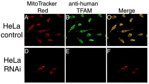
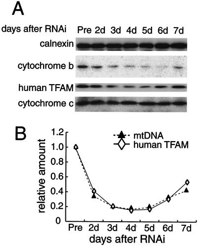
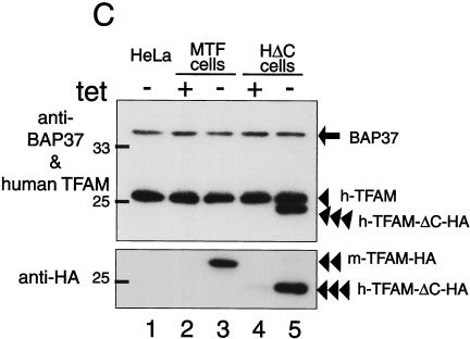
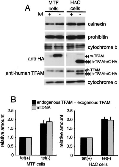
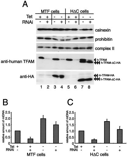
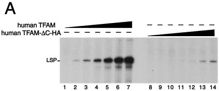
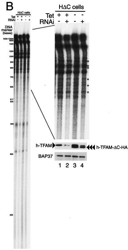
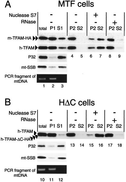
Mitochondrial transcription factor A (TFAM), a transcription factor for mitochondrial DNA (mtDNA) that also possesses the property of nonspecific DNA binding, is essential for maintenance of mtDNA. To clarify the role of TFAM, we repressed the expression of endogenous TFAM in HeLa cells by RNA interference. The amount of TFAM decreased maximally to about 15% of the normal level at day 3 after RNA interference and then recovered gradually. The amount of mtDNA changed closely in parallel with the daily change in TFAM while in organello transcription of mtDNA at day 3 was maintained at about 50% of the normal level. TFAM lacking its C-terminal 25 amino acids (TFAM-DeltaC) marginally activated transcription in vitro. When TFAM-DeltaC was expressed at levels comparable to those of endogenous TFAM in HeLa cells, mtDNA increased twofold, suggesting that TFAM-DeltaC is as competent in maintaining mtDNA as endogenous TFAM under these conditions. The in organello transcription of TFAM-DeltaC-expressing cells was no more than that in the control. Thus, the mtDNA amount is finely correlated with the amount of TFAM but not with the transcription level. We discuss an architectural role for TFAM in the maintenance of mtDNA in addition to its role in transcription activation.


Figures

FIG. 1.
FIG. 1.
Immunofluorescent images of HeLa cells treated by RNAi. Human TFAM in HeLa cells was identified with anti-human TFAM antibodies (B and E). Mitochondria were stained with MitoTracker Red (A and D). Panels C and F are merged images of panels A plus B and D plus E, respectively.
FIG. 2.
FIG. 2.
Parallel decreases and increases in human TFAM and mtDNA after RNAi. (A) Calnexin (as a standard of cell amount), cytochrome b, human TFAM, and cytochrome c in HeLa cells were analyzed by Western blotting 2 to 7 days after RNAi. The relative amount of mtDNA was measured by quantitative PCR. (B) Representative daily changes in the relative amounts of human TFAM (⋄) and mtDNA (▴).
FIG.3.
FIG.3.
Tetracycline-regulated expression of TFAM. (A) The scheme of recombinant TFAM molecules. Human and mouse TFAM have two HMG boxes (white square) and a C-tail region (gray bar). An HA.11 epitope tag is indicated by a gray square. The position of the RNAi target is indicated by a dotted line. (B) MTF cells and HΔC cells were cultured with doxycycline [tet (+)] or without doxycycline [tet (−)]. Mitochondria were stained with MitoTracker Red (panels a, d, and g), and recombinant TFAM was stained with anti-HA antibodies (panels b, e, and h). The merged images are shown in panels c, f, and i. (C) Western blotting analysis of TFAM molecules. Total cell lysates were used for Western blotting. BAP37 (arrow) is shown as an internal standard for the amount of protein applied in the samples. Antibodies for BAP-37 and human TFAM were added together for detecting the two proteins (upper panel). Then the membrane was reprobed with anti-HA antibody (lower panel). Endogenous human TFAM (arrowhead), exogenous mouse TFAM-HA (double arrowheads), and exogenous human TFAM-ΔC-HA (triple arrowheads) are indicated.
FIG.3.
FIG.3.
Tetracycline-regulated expression of TFAM. (A) The scheme of recombinant TFAM molecules. Human and mouse TFAM have two HMG boxes (white square) and a C-tail region (gray bar). An HA.11 epitope tag is indicated by a gray square. The position of the RNAi target is indicated by a dotted line. (B) MTF cells and HΔC cells were cultured with doxycycline [tet (+)] or without doxycycline [tet (−)]. Mitochondria were stained with MitoTracker Red (panels a, d, and g), and recombinant TFAM was stained with anti-HA antibodies (panels b, e, and h). The merged images are shown in panels c, f, and i. (C) Western blotting analysis of TFAM molecules. Total cell lysates were used for Western blotting. BAP37 (arrow) is shown as an internal standard for the amount of protein applied in the samples. Antibodies for BAP-37 and human TFAM were added together for detecting the two proteins (upper panel). Then the membrane was reprobed with anti-HA antibody (lower panel). Endogenous human TFAM (arrowhead), exogenous mouse TFAM-HA (double arrowheads), and exogenous human TFAM-ΔC-HA (triple arrowheads) are indicated.
FIG. 4.
FIG. 4.
Amount of mitochondrial proteins and mtDNA in MTF and HΔC cells. (A) Total cell lysates of cells cultured with doxycycline [tet (+)] or without doxycycline [tet (−)] were used for Western blotting. Prohibitin and cytochrome c are mitochondrial proteins encoded by the nuclear genome. Cytochrome b is a mitochondrial protein encoded by mtDNA. Endogenous human TFAM (arrowhead), mouse TFAM-HA (double arrowheads), and human TFAM-ΔC-HA (triple arrowheads) are indicated. Calnexin, a microsomal protein, is shown as an internal standard. (B) Quantification of relative amounts of TFAM (black bar) and mtDNA (gray bar). Signal intensities on the Western blots were measured for estimating the number of TFAM molecules, endogenous TFAM, and exogenous TFAM. The amount of mtDNA was measured by quantitative PCR. Error bars indicate ±1 standard deviation from the mean of three independent experiments.
FIG. 5.
FIG. 5.
(A) MTF and HΔC cells were cultured with doxycycline [tet (+)] or without doxycycline [tet (−)]. Three days after RNAi treatment (RNAi +) or without RNAi treatment (RNAi −), the cells were collected and analyzed. The expression of proteins in MTF and HΔC cells was analyzed by Western blotting. Endogenous human TFAM (arrowhead), exogenous mouse TFAM-HA (double arrowheads), and exogenous human TFAM-ΔC-HA (triple arrowheads) are indicated. (B and C) The amount of mtDNA was measured in cells with and without doxycycline and RNAi treatments, as indicated. Error bars indicate ± 1 standard deviation from the mean of three independent experiments.
FIG. 6.
FIG. 6.
In vitro and in organello transcription assays. (A) TFAM lacking its C-terminal tail only supports low levels of transcription from LSP. A template containing the light-strand promoter (LSP, 85 fmol) of human mtDNA was used for in vitro runoff transcription assays. The reactions were performed with the following pure recombinant proteins: human mitochondrial RNA polymerase (250 fmol), human TFB2 M (500 fmol), and wild-type human TFAM with an N-terminal His tag or human ΔC-tail TFAM with an N-terminal His tag and C-terminal HA tag (human TFAM-ΔC-HA) in increasing amounts (0.02, 0.05, 0.2, 0.5, 1.6, 5, and 15 pmol). (B) A representative image of five independent experiments is shown. The HΔC cells were cultured with and without doxycycline and treated or not by RNAi, and mitochondria were isolated from these cells 3 days after RNAi treatment. The newly synthesized transcripts were labeled with [α-32P]UTP for 30 min in mitochondria and analyzed by urea-polyacrylamide gel electrophoresis. The approximate size of nucleotides is shown based on the [γ-32P]ATP-labeled DNA size markers (left panel). The part showing bands larger than 600 bases was enlarged (right upper panel). The asterisks indicate measured bands used for estimation. The isolated mitochondria (5 μg of protein) were analyzed by Western blotting to check the protein amount (anti-BAP37 antibody, right lower panel) and the efficiency of RNAi (anti-human TFAM antibody, right middle panel).
FIG. 6.
FIG. 6.
In vitro and in organello transcription assays. (A) TFAM lacking its C-terminal tail only supports low levels of transcription from LSP. A template containing the light-strand promoter (LSP, 85 fmol) of human mtDNA was used for in vitro runoff transcription assays. The reactions were performed with the following pure recombinant proteins: human mitochondrial RNA polymerase (250 fmol), human TFB2 M (500 fmol), and wild-type human TFAM with an N-terminal His tag or human ΔC-tail TFAM with an N-terminal His tag and C-terminal HA tag (human TFAM-ΔC-HA) in increasing amounts (0.02, 0.05, 0.2, 0.5, 1.6, 5, and 15 pmol). (B) A representative image of five independent experiments is shown. The HΔC cells were cultured with and without doxycycline and treated or not by RNAi, and mitochondria were isolated from these cells 3 days after RNAi treatment. The newly synthesized transcripts were labeled with [α-32P]UTP for 30 min in mitochondria and analyzed by urea-polyacrylamide gel electrophoresis. The approximate size of nucleotides is shown based on the [γ-32P]ATP-labeled DNA size markers (left panel). The part showing bands larger than 600 bases was enlarged (right upper panel). The asterisks indicate measured bands used for estimation. The isolated mitochondria (5 μg of protein) were analyzed by Western blotting to check the protein amount (anti-BAP37 antibody, right lower panel) and the efficiency of RNAi (anti-human TFAM antibody, right middle panel).
FIG. 7.
FIG. 7.
Mitochondria were prepared from MTF (A) and HΔC (B) cells cultured without doxycycline. The mitochondria were solubilized with 0.5% NP-40 (total) and separated into pellet (P1) and supernatant (S1). The P1 fractions were treated with nuclease S7 or DNase-free RNase A and then separated again into pellet (P2) and supernatant (S2). Each sample was analyzed by Western blotting with anti-human TFAM, anti-P32, anti-mtSSB, and anti-HA antibodies. mtDNA in total, P1, and S1 were detected by PCR. Human TFAM (arrowhead), mouse TFAM-HA (double arrowheads), and human TFAM-ΔC-HA (triple arrowheads) are indicated.

References

    1. Alam, T. I., T. Kanki, T. Muta, K. Ukaji, Y. Abe, H. Nakayama, K. Takio, N. Hamasaki, and D. Kang. 2003. Human mitochondrial DNA is packaged with TFAM. Nucleic Acids Res. 31:1640-1645. - PMC - PubMed
    1. Albring, M., J. Griffith, and G. Attardi. 1977. Association of a protein structure of probable membrane derivation with HeLa cell mitochondrial DNA near its origin of replication. Proc. Natl. Acad. Sci. USA 74:1348-1352. - PMC - PubMed
    1. Barat, M., D. Rickwood, C. Dufresne, and J. C. Mounolou. 1985. Characterization of DNA-protein complexes from the mitochondria of Xenopus laevis oocytes. Exp. Cell Res. 157:207-217. - PubMed
    1. Bowmaker, M., M. Y. Yang, T. Yasukawa, A. Reyes, H. T. Jacobs, J. A. Huberman, and I. J. Holt. 2003. Mammalian mitochondrial DNA replicates bidirectionally from an initiation zone. J. Biol. Chem. 278:50961-50969. - PubMed
    1. Caron, F., C. Jacq, and J. Rouviere-Yaniv. 1979. Characterization of a histone-like protein extracted from yeast mitochondria. Proc. Natl. Acad. Sci. USA 76:4265-4269. - PMC - PubMed

Publication types

MeSH terms

LinkOut - more resources