. 2004 Nov;24(22):10000-15.
doi: 10.1128/MCB.24.22.10000-10015.2004.

Extracellular signal-regulated kinase 1c (ERK1c), a novel 42-kilodalton ERK, demonstrates unique modes of regulation, localization, and function

Affiliations

Extracellular signal-regulated kinase 1c (ERK1c), a novel 42-kilodalton ERK, demonstrates unique modes of regulation, localization, and function

Daniel M Aebersold et al. Mol Cell Biol. 2004 Nov.

Erratum in

Abstract

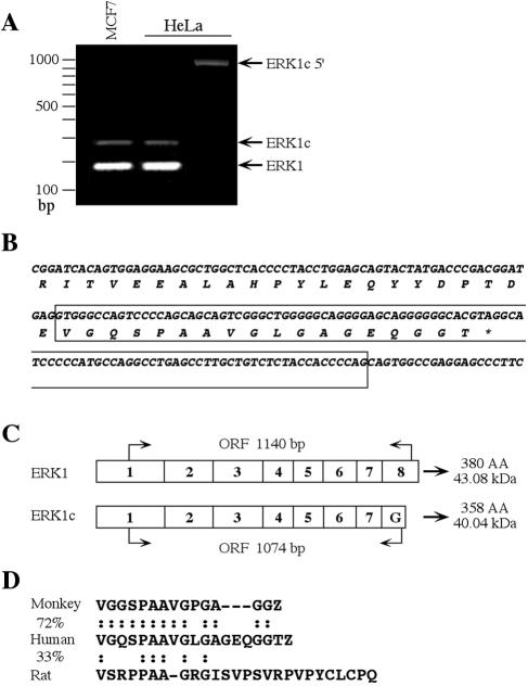
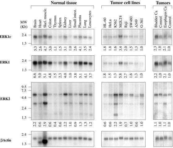
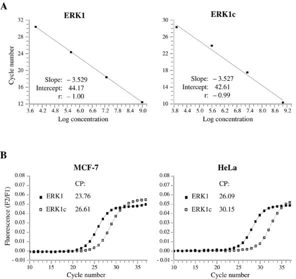
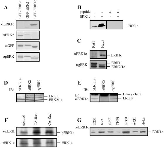
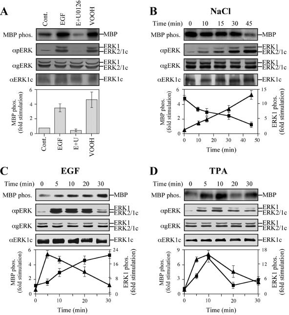
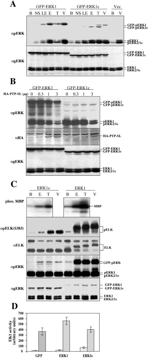
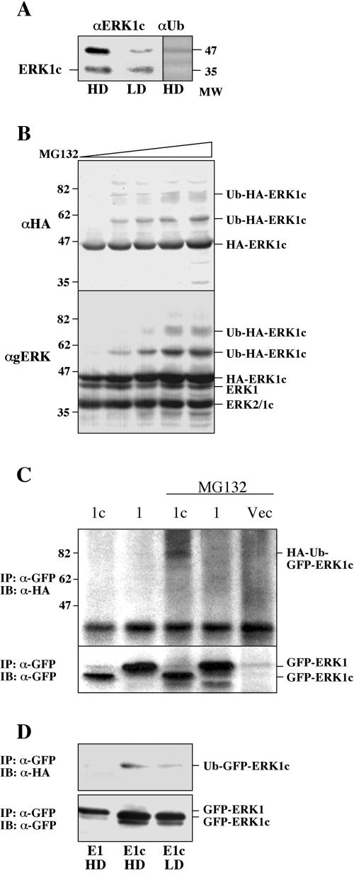
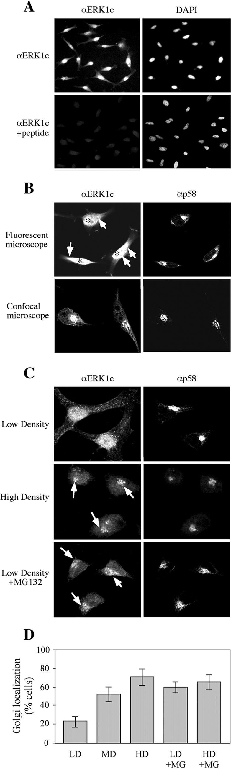
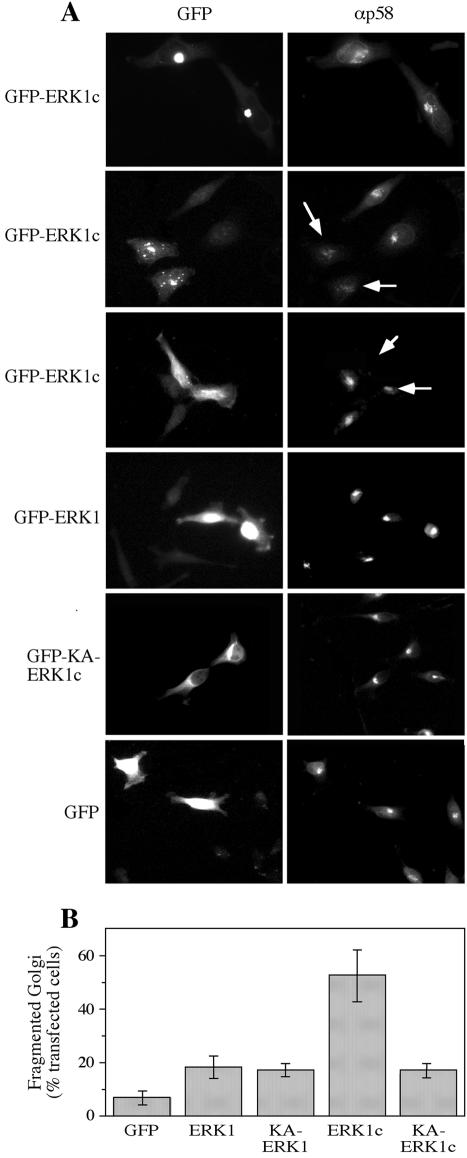
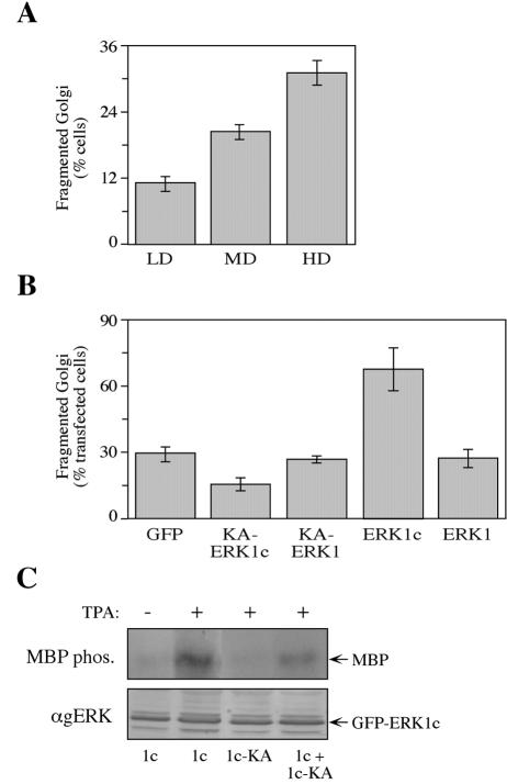
Extracellular signal-regulated kinases (ERKs) are signaling molecules that regulate many cellular processes. We have previously identified an alternatively spliced 46-kDa form of ERK1 that is expressed in rats and mice and named ERK1b. Here we report that the same splicing event in humans and monkeys causes, due to sequence differences in the inserted introns, the production of an ERK isoform that migrates together with the 42-kDa ERK2. Because of the differences of this isoform from ERK1b, we named it ERK1c. We found that its expression levels are about 10% of ERK1. ERK1c seems to be expressed in a wide variety of tissues and cells. Its activation by MEKs and inactivation by phosphatases are slower than those of ERK1, which is probably the reason for its differential regulation in response to extracellular stimuli. Unlike ERK1, ERK1c undergoes monoubiquitination, which is increased with elevated cell density concomitantly with accumulation of ERK1c in the Golgi apparatus. Elevated cell density also causes enhanced Golgi fragmentation, which is facilitated by overexpression of native ERK1c and is prevented by dominant-negative ERK1c, indicating that ERK1c mediates cell density-induced Golgi fragmentation. The differential regulation of ERK1c extends the signaling specificity of MEKs after stimulation by various extracellular stimuli.


Figures

FIG. 1.
FIG. 1.
Cloning of the human ERK1c. (A) RT-PCR cloning of ERK1c. The MCF7 lane shows RT-PCR with oligonucleotide primers ERK1-Exon7-S and ERK1-Exon8-AS and total RNA from MCF-7 cells as a template. The left HeLa lane shows the same RT-PCR mixture as in the MCF7 lane, except the RNA was from HeLa cells. The right HeLa lane shows RT-PCR with oligonucleotide primers ERK1-Exon1-S and ERK1c-insert-AS and total RNA from HeLa cells. The positions of the relevant fragments and DNA markers (in base pairs) are indicated at the sides of the gel. (B) cDNA and amino acid sequences of the C-terminal region of ERK1c. The unique ERK1c insert is boxed. (C) Exon organization of ERK1c and ERK1. The length of the open reading frames (ORFs), the number of amino acids (AA), and the predicted molecular mass are indicated. (D) Sequence alignment of human ERK1c, monkey ERK1c, and rat ERK1b proteins. Amino acids that are identical in the different proteins (:) and the percentage identity between the proteins are indicated. Gaps introduced to maximize alignment are indicated by the dashes.
FIG. 2.
FIG. 2.
Distribution of ERK1c mRNA in cells and tissues. Northern blot analysis was performed on human tissues (MTN blots; Clontech), human cancer cell lines (MTN blots; Clontech), and human tumor blots (ResGen). The specific tissue, cell line, or tumor is indicated (Skel. muscle, skeletal muscle; Small intest., small intestine; Bladder Ca, bladder cancer). The probes used for detection were generated as described in Materials and Methods. The following probes were used: ERK1c unique sequence (ERK1c), human ERK1 N terminus (ERK1), human ERK2 N terminus (ERK2), and actin. Blots were hybridized in ULTRAhyb hybridization buffer (catalog no. 8670; Ambion) following the manufacturer's protocol. The positions of the RNA markers are indicated to the left of the gels. Relative expression was calculated by using a densitometer (model 690; Bio-Rad) and is indicated under the gels.
FIG. 3.
FIG. 3.
Relative expression of ERK1c and ERK1 in MCF7 and HeLa cells. (A) Real-time PCR was performed in a LightCycler (Roche) with the FastStart DNA Master Hybridization Probes kit (Roche), following the manufacturer's instructions for the composition of the reaction mixture. The fluorescent hybridization probes for sequence-specific detection (synthesized by Tib Molbiol) were used. Real-time PCR was performed by a touchdown procedure (stepwise decrease of the annealing temperature for the amplification primers). To produce the cDNA, total RNA was reverse transcribed using the first-strand cDNA synthesis kit for RT-PCR (Roche) with random hexamers as primers, according to the manufacturer's recommendations. (B) To generate standard curves, template dilution series (cDNA mixture in four subsequent 1:50 dilution steps) were added to the reaction mixture. Results were processed using the LightCycler software package (version 3.5.28). The crossing points (CP) are indicated.
FIG. 4.
FIG. 4.
Characterization of the endogenous ERK1c protein. (A) Testing the specificity of the anti-ERK1c Ab. HEK-293 cells were transfected with the indicated constructs, and extracts (containing cytosolic and nuclear proteins; 50 μg) were immunoblotted with four different Abs: anti-ERK1c Ab (αERK1c), anti-ERK2 Ab (αERK2), anti-GFP Ab (αGFP), or anti-ERK1/2 Ab (αgERK). (B) Competing the anti-ERK1c Ab with the antigenic peptide. HeLa cell extract (containing cytosolic and nuclear proteins; 50 μg) from nonstimulated HeLa cells transfected with ERK1c (+) or not transfected (−) were subjected to immunoblotting with anti-ERK1c Ab (ERK1c) in the absence (−) or presence (+) of the antigenic peptide (25 μg/ml). (C) ERK1c is not expressed in rat cells. HeLa and Rat1 cell extracts (50 μg) were loaded on SDS-polyacrylamide gels and immunoblotted with the indicated Abs. (D) ERK1c migrates together with ERK2 on SDS-polyacrylamide gels. HeLa cell extracts were loaded on one lane (100 μg) of the SDS-polyacrylamide gel and transferred to a nitrocellulose membrane. After the transfer, the membrane was cut into three pieces, and each piece was immunoblotted (IB) with a different Ab as indicated. (E) Immunoprecipitation of ERK1c from HeLa cells. An anti-ERK1c Ab was used to immunoprecipitate (IP) ERK1c from HeLa cell extract (500 μg), followed by immunoblotting (IB) with the indicated Abs. (F) ERK1c is phosphorylated on its Thr and Tyr residues upon activation. HeLa cells were transfected with constitutively activated Ras (CA-Ras), constitutively activated Rac (CA-Rac), or a vector control. ERK1c was immunoprecipitated with the anti-ERK1c Ab and immunoblotted with anti-pERK and anti-ERK1c Abs. (G) ERK1c expression in several human tumor cell lines. Cell lysate (50 μg) from a nonstimulated human glioblastoma brain tumor cell line, U-251 MG, U87 PTEN-deficient glioma cells, human choriocarcinoma cell line jeg-3, a natural human monocytic cell line (THP1), Jurkat T lymphocytes, A431 epidermal carcinoma cells, or HeLa cells were subjected to immunoblotting with anti-ERK1c Ab. The amounts of ERK1 and ERK2 detected in this experiment by anti-ERK Ab were similar in all cells. The experiments were reproduced three times.
FIG. 5.
FIG. 5.
(A) Kinetics of ERK1c activation. Stimulation and inhibition of MBP phosphorylation (MBP phos.) by ERK1c are shown. HeLa cells were serum starved for 16 h and then stimulated with either EGF (50 ng/ml, 10 min), EGF plus U0126 (E+U) (15-min pretreatment with 5 μM U0126, followed by 10 min of EGF [50 ng/ml]), or VOOH (100 μM Na3VO4 and 200 μM H2O2, 15 min) or left untreated as a control (Cont.). After harvesting, extracts were either immunoprecipitated with anti-ERK1c Ab (αERK1c) and subjected to an in vitro MBP phosphorylation (MBP phos.), or they were immunoblotted with anti-ERK1c, anti-pERK, or anti-gERK Ab. The bar graph shows the means ± standard errors (error bars) from three different experiments. (B) Time course of ERK1c activation by osmotic shock. HeLa cells were serum starved for 16 h and then stimulated with 0.7 M NaCl for the indicated times. ERK1c activity was examined and immunoblotting was performed as described above for panel A. Squares represent ERK1c activity, while the triangles represent the phosphorylation (phos.) of ERK1 and ERK2. The results in the graph show the means ± standard errors from three different experiments. (C and D) Time course of ERK1c activation by EGF and TPA. HeLa cells were treated as described above for panel B, except that EGF (50 ng/ml) or TPA (250 nM) was used for stimulation.
FIG. 6.
FIG. 6.
Biochemical properties of ERK1c. (A) Phosphorylation of exogenous ERK1c in response to extracellular stimuli. COS7 cells were transfected with GFP-ERK1 or GFP-ERK1c. Two days later, the cells were either not starved (NS) or the cells were starved and stimulated with either EGF for 14 h (10 ng/ml, 12 h) (LE), EGF (50 ng/ml, 10 min) (E), TPA (250 nM, 15 min) (T), VOOH (Na3VO4 [100 μM] and H2O2 [200 μM], 20 min) (V), or left untreated (B) as control. Cell extracts were analyzed by immunoblotting with the indicated Abs. Thedata are from a representative experiment (the experiment was performed three times). (B) Differential dephosphorylation of ERK1 and ERK1c by PTP-SL. COS7 cells were cotransfected with plasmids (1 μg each) containing either HA-ERK1 or HA-ERK1c, together with the indicated amounts of plasmid containing wild-type PTP-SL. After serum starvation, the cells were stimulated with EGF (50 ng/ml, 10 min) and harvested. Cytosolic extracts were subjected to an immunoblot analysis with the indicated Abs (anti-HA Ab [αHA] used to demonstrate increasing PTP-SL). The data are from a representative experiment (the experiment was three times). (C) Phosphorylation of MBP and Elk1 by ERK1c. COS7 cells were transfected with GFP-ERK1 or GFP-ERK1c. After serum starvation, the cells were stimulated with EGF (50 ng/ml, 10 min) (E), TPA (250 nM, 15 min) (T), or VOOH (18 min) (V) or left untreated (B). The GFP-ERKs were immunoprecipitated with anti-GFP Ab and subjected to an in vitro kinase assay with MBP or Elk1 as the substrate. The phosphorylation (phos.) of MBP was detected by autoradiography on an X-ray film (Agfa). The phosphorylation of Elk1 was detected by anti-pElk1(S383) Ab and also by upshift of Elk1 detected by anti-Elk1 Ab. The phosphorylation and amount of the GFP-ERKs were determined by an immunoblot analysis with the indicated Abs. The positions of phospho-MBP (pMBP), GFP-ERKs, GFP-phospho-ERKs (GFP-pERKs), and Abs are indicated at the sides of the gels. These results were from a representative experiment (the experiment was performed four times). (D) Effect of ERK1c on Elk1 transcriptional activity. HEK-293 cells were transfected with pFR-Luc (reporter), pFA2-Elk1 (fusion transactivator), pRenilla (reporter of transfection yield), together with GFP-ERK1 (ERK1), GFP-ERK1c (ERK1c), or GFP empty vector. Serum-starved cells were stimulated with EGF (50 ng/ml) for 14 h. Luciferase and Renilla luminescence were monitored as described in Materials and Methods. The results represent the means and standard errors (error bars) of three experiments.
FIG. 7.
FIG. 7.
Monoubiquitination of ERK1c. (A) Appearance of a 50-kDa ERK1c protein in dense cells. HeLa cells were grown until they were confluent and either harvested (medium density) or left for another 36 h (high density [HD]) and then harvested. Cell extracts (100 μg) were subjected to immunoblotting with the indicated Abs. These results were from a representative experiment (the experiment was performed five times). In parallel, ERK1c from the high-density cellextracts was purified on MonoQ, heparin, and Superdex columns. An aliquot of the purified ERK1c was immunoblotted with antiubiquitin Ab (αUb). MW, molecular weight in thousands. (B) Appearance of putative mono- and diubiquitinated ERK1c upon MG132 treatment. HeLa cells were transfected with HA-ERK1c. Forty-eight hours later, the cells were treated with MG132 (0, 0.1, 0.3, 1.0, and 3 μg/ml) for 8 h, and the expression of HA-ERK1c and endogenous ERKs was tested by immunoblotting with the indicated Abs. These results were from a representative experiment (the experiment was performed two times). (C) Monoubiquitination of GFP-ERK1c but not ERK1c in response to treatment with MG132. COS7 cells were cotransfected with HA-ubiquitin and GFP constructs of ERK1 (lanes 1) or ERK1c (1c) or vector (Vec) alone. After 2 days, the cells were treated with MG132 (1 μg/ml, 12 h) or left untreated, and the GFP proteins were immunoprecipitated (IP) by anti-GFP Ab (α-GFP). The amounts of GFP-containing proteins and HA-ubiquitin were analyzed by immunoblotting (IB) with anti-GFP or anti-HA Ab. The positions of GFP-ERK1, GFP-ERK1c, and HA-ubiquitin bound to GFP-ERK1c (HA-ERK1c) are indicated. These results were reproduced three times. (D) Immunoprecipitation of HA-ubiquitinated GFP-ERK1c from cells at different densities. HeLa cells were cotransected with GFP-ERK1c or GFP-ERK1 together with HA-ubiquitin (HA-Ub) and grown at low and high densities as described above. GFP-ERK1 containing cells were grown at high density. The cells were then harvested, and cell lysates were subjected to immunoprecipitation (IP) with monoclonal anti-GFP Ab. HA-ubiquitin and GFP-ERKs were detected by Western blotting (IB) with the appropriate Ab.
FIG.8.
FIG.8.
Immunostaining of ERK1c. (A) Subcellular localization of ERK1c. HeLa cells were grown on 18-mm microslides in 12-well plates for 24 h. The cells were stained with anti-ERK1c Ab (αERK1c) and DAPI as described in Materials and Methods. The specificity of staining was confirmed by competition with the antigenic peptide (25 μg/ml). (B) ERK1c is localized in the nucleus and Golgi apparatus. HeLa cells were stained as described above with anti-ERK1c and anti-p58 Abs. Visualization with a regular fluorescence microscope revealed nuclear staining (asterisks) and perinuclear staining (arrows) that corresponded to the Golgi staining. With a confocal microscope, we detected sections in which only the Golgi staining by anti-ERK1c and anti-p58 Abs was apparent. (C) Staining of ERK1c in cells at different densities or MG132-treated cell cultures. HeLa cells (20,000 cells/well in the Low Density panels and 120,000 cells/well in the High Density panel) were seeded on 18-mm microslides in 12-well plates. Twenty-four hours after plating, the cells were either treated with MG132 (1 μg/ml, 6 h) or left untreated and then were fixed and stained with anti-ERK1c and anti-p58 Abs. The staining was visualized with a confocal microscope as described above for panel B. The positions of the Golgi apparatus in the stained cells are indicated by the white arrows. (D) Accumulation of ERK1c in the Golgi apparatus is dependent on cell density. HeLa cells were grown at three different densities: 20,000 cells/well (low density [LD]), 60,000 cells/well (medium density [MD]), and 120,000 cells/well (high density [HD]). HeLa cells were seeded on 18-mm microslides (in 12-well plates), which were treated as described in the legend to Fig. 8C. The percentage of cells with apparent Golgi localization of ERK1c was determined by counting 200 cells in each slide. The values are the means ± standard errors (error bars) for three experiments.
FIG. 9.
FIG. 9.
Overexpression of GFP-ERK1c results in Golgi fragmentation. (A) Localization and effect on Golgi apparatus of the GFP-ERK constructs. HeLa cells were transfected with GFP-ERK1c, GFP-ERK1, GFP-KA-ERK1c, or GFP and grown on microslides in 12-well plates. Fourteen hours after transfection, the cells were fixed and stained with anti-p58 Ab (αp58) and examined with a fluorescence microscope. Fragmentation or lack of Golgi apparatus is indicated by the white arrows. (B) Quantitation of Golgi fragmentation. The HeLa cells (200 cells in each slide) in panel A were counted, and the percentage of cells with fragmented Golgi apparatus out of cells expressing GFP-ERK1c is presented. The values are the means ± standard errors (error bars) for three experiments.
FIG. 10.
FIG. 10.
Inhibition of cell density-dependent Golgi fragmentation by the dominant-negative KA-ERK1c. (A) Golgi fragmentation increased with elevated cell density. HeLa cells were seeded on 18-mm microslides at low density (LD) (20,000 cell/well of a 12-well plate), medium density (MD) (60,000 cells/well), and high density (HD) (120,000 cells/well). Twenty-four hours after plating, the cells were fixed and stained with anti-p58 Ab. The percentage of cells with fragmented Golgi apparatus was determined by counting 200 cells in each slide. The values are the means ± standard errors (error bars) for three experiments. (B) Overexpression of KA-ERK1c inhibits cell density-dependent Golgi fragmentation. HeLa cells were seeded on 18-mm microslides at high density (120,000 cells/well) and transfected with GFP, GFP-KA-ERK1c, GFP-KA-ERK1, GFP-ERK1c, or GFP-ERK1. Twenty-four hours after transfection, the cells were fixed and stained with anti-p58 Ab. The percentage of cells with fragmented Golgi apparatus out of cells expressing the exogenous construct is presented. The values are the means ± standard errors (error bars) for three experiments. (C) KA-ERK1c has a dominant-negative effect. HEK-293 cells were transfected with GFP-ERK1c (1c), GFP-KA-ERK1c (1c-KA), or HA-ERK1c together with GFP-KA-ERK1c (1c + 1c-KA). The cells were serum starved and stimulated with TPA (+) (250 nM, 15 min) or left untreated (−). After harvesting, the GFP proteins were immunoprecipitated with anti-GFP Ab, subjected to an in vitro MBP phosphorylation (MBP phos.) and concomitantly immunoblotted with anti-gERK Abs (αgERK) to confirm equal immunoprecipitation of the GFPs.

References

    1. Acharya, U., A. Mallabiabarrena, J. K. Acharya, and V. Malhotra. 1998. Signaling via mitogen-activated protein kinase kinase (MEK1) is required for Golgi fragmentation during mitosis. Cell 92:183-192. - PubMed
    1. Bott, C. M., S. G. Thorneycroft, and C. J. Marshall. 1994. The sevenmaker gain-of-function mutation in p42 MAP kinase leads to enhanced signalling and reduced sensitivity to dual specificity phosphatase action. FEBS Lett. 352:201-205. - PubMed
    1. Boucher, M. J., D. Jean, A. Vezina, and N. Rivard. 2004. Dual role of MEK/ERK signaling in senescence and transformation of intestinal epithelial cells. Am. J. Physiol. Gastrointest. Liver Physiol. 286:G736-G746. (First published 30 December 2003; http://ajpgi.physiology.org/cgi/content/full/286/5/G736.) - PubMed
    1. Boulton, T. G., and M. H. Cobb. 1991. Identification of multiple extracellular signal-regulated kinases (ERKs) with antipeptide antibodies. Cell Regul. 2:357-371. - PMC - PubMed
    1. Boulton, T. G., S. H. Nye, D. J. Robbins, N. Y. Ip, E. Radziejewska, S. D. Morgenbesser, R. A. DePinho, N. Panayotatos, M. H. Cobb, and G. D. Yancopoulos. 1991. ERKs: a family of protein-serine/threonine kinases that are activated and tyrosine phosphorylated in response to insulin and NGF. Cell 65:663-675. - PubMed

Publication types

MeSH terms

LinkOut - more resources