. 2004 Nov 24;23(23):4560-70.
doi: 10.1038/sj.emboj.7600464. Epub 2004 Oct 28.

Functional characterization of wild-type and mutant human sialin

Affiliations

Functional characterization of wild-type and mutant human sialin

Pierre Morin et al. EMBO J. .

Abstract

The modification of cell surface lipids or proteins with sialic acid is essential for many biological processes and several diseases are caused by defective sialic acid metabolism. Sialic acids cleaved off from degraded sialoglycoconjugates are exported from lysosomes by a membrane transporter, named sialin, which is defective in two allelic inherited diseases: infantile sialic acid storage disease (ISSD) and Salla disease. To develop a functional assay of human sialin, we redirected the protein to the plasma membrane by mutating a dileucine-based internalization motif. Cells expressing the plasmalemmal construct accumulated neuraminic acid at acidic pH by a process equivalent to lysosomal efflux. The assay was used to determine how pathogenic mutations affect transport. Interestingly, while two missense mutations and one small, in-frame deletion associated with ISSD abolished transport, the mutation causing Salla disease (R39C) slowed down, but did not stop, the transport cycle, thus explaining why the latter disorder is less severe. Since neurological symptoms predominate in Salla disease, our results suggest that sialin is rate-limiting to specific sialic acid-dependent processes of the nervous system.


Figures
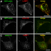
Figure 1
Figure 1
Intracellular localization of GFP- and V5-tagged sialin. Confocal sections of HeLa cells transiently expressing WT GFP-sialin (A, B) and/or sialin-V5 (B, C) show that the recombinant proteins colocalize extensively with each other and with the endogenous late endosomal and lysosomal marker LAMP1. Scale bar, 20 μm.
Figure 2
Figure 2
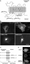
Role of the DRTPLL motif in intracellular targeting of sialin. (A) Topological model of sialin showing potential dileucine-based sorting signals, pathogenic mutations and potential N-glycosylation sites. (B) The intracellular distribution of WT GFP-sialin and dileucine mutants in transiently transfected HeLa cells was analysed by epifluorescence microscopy. (C) Schematic representation of the CD4 chimeras. From top to bottom: CD4ΔCt, CD4-Nsia and CD4-Nsia L22G/L23G. (D) Steady-state localization of the CD4 constructs. Cells were fixed, permeabilized and incubated with SIM.2 monoclonal antibodies. CD4-Nsia is found on intracellular compartments, whereas the other constructs are mainly at the plasma membrane. Scale bars, 20 μm.
Figure 3
Figure 3
Mutation of the DRTPLL motif results in cell surface expression. HEK293 cells transiently expressing GFP (lanes 1–3), WT GFP-sialin (lanes 4–6) or GFP-sialin L22G/L23G (lanes 7–9) were treated with an impermeant biotinylation reagent. After cell lysis, biotinylated proteins were purified on streptavidin–agarose beads. In lanes 10–12, biotinylation was omitted to verify the selectivity of the affinity chromatography. Equal amounts of cell lysate (tot) and streptavidin-unbound material (ub) were analysed by SDS–PAGE and immunoblotting using anti-GFP antibodies. Streptavidin-bound proteins (bo) were derived from an 18-fold higher amount of material. The position and molecular mass (kDa) of protein standards is shown on the left.
Figure 4
Figure 4
Sialin mediates a pH gradient-driven sialic acid transport. (A) HEK 293 cells expressing sialin at the cell surface (GFP-sialin L22G/L23G construct) take up [3H]Neu5Ac from acidic (pH 5.6, filled bars), but not from neutral (pH 7.4, open bars) extracellular medium. Perforation of the plasma membrane with 20 μM digitonin released the radioactivity accumulated into GFP-sialin L22G/L23G-transfected cells (striped bar). (B) Saturation kinetics of sialin-mediated transport. Mock- (□) and GFP-sialin L22G/L23G-transfected (▪) cells were incubated for 15 min with increasing concentrations of Neu5Ac at pH 5.6. Nonlinear regression yielded KM and Vmax values of 2.16±0.16 mM and 245±9.7 pmol/min per 106 cells in this experiment. (C) [3H]Neu5Ac uptake by mock- (open symbols) and GFP-sialin L22G/L23G-transfected (filled symbols) cells was performed in MES (squares) or MOPS (triangles) buffer adjusted to several pH values. Transport was stimulated at acidic pH. (D) At pH 5.6, dissipation of the transmembrane pH gradient by 5 μM nigericin (striped bars) inhibited sialin L22G/L23G-mediated transport by more than 90%. For all experiments, error bars indicate s.e.m.
Figure 5
Figure 5
Effect of pathogenic mutations on sialin activity. (A) The R39C, P334R, H183R, ΔSSLRN and K136E mutations did not impair surface expression of GFP-sialin L22G/L23G in HeLa cells. Scale bar, 20 μm. (B) HEK 293 cells expressing GFP-sialin L22G/L23G without (control) or with pathogenic mutations were analysed for [3H]Neu5Ac uptake at pH 7.4 (open bars) or 5.0 (filled bars). Only the R39C and K136E mutants showed partial activity. (C) Surface biotinylation of transfected HEK 293 cells and quantitative immunoblotting analysis revealed similar amounts of GFP-sialin L22G/L23G at the cell surface, whatever the pathogenic mutation. The number of independent measurements is indicated in brackets.
Figure 6
Figure 6
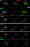
Effect of pathogenic mutations on sialin intracellular localization. HeLa cells transiently expressing WT or mutant (R39C, ΔSSLRN, K136E, H183R and P334R) GFP-sialin with an intact DRTPLL motif were fixed, stained with anti-LAMP1 antibodies and analysed by epifluorescence microscopy. GFP-sialin, LAMP1 and merged images (sialin, green; LAMP1, red) are shown from left to right. The WT and P334R proteins fully colocalize with LAMP1. Other mutants partially colocalize with LAMP1 (arrowheads), but they are also found in a perinuclear compartment and in LAMP1-negative puncta (arrows). Images are 3D reconstructions of the cells after deconvolution. Insets are magnifications of the rectangles shown on merged images. Scale bar, 20 μm.

References

    1. Aula N, Jalanko A, Aula P, Peltonen L (2002) Unraveling the molecular pathogenesis of free sialic acid storage disorders: altered targeting of mutant sialin. Mol Genet Metab 77: 99–107 - PubMed
    1. Aula N, Kopra O, Jalanko A, Peltonen L (2004) Sialin expression in the CNS implicates extralysosomal function in neurons. Neurobiol Dis 15: 251–261 - PubMed
    1. Aula N, Salomaki P, Timonen R, Verheijen F, Mancini G, Mansson JE, Aula P, Peltonen PL (2000) The spectrum of SLC17A5-gene mutations resulting in free sialic acid-storage diseases indicates some genotype–phenotype correlation. Am J Hum Genet 67: 832–840 - PMC - PubMed
    1. Aula P, Gahl WA (2001) Disorders of free sialic acid storage. In The Metabolic and Molecular Bases of Inherited Disease, Scriver CR, Beaudet AL, Sly WS, Valle D (eds), pp 5109–5120. New York: McGraw-Hill
    1. Autio-Harmainen H, Oldfors A, Sourander P, Renlund M, Dammert K, Simila S (1988) Neuropathology of Salla disease. Acta Neuropathol (Berl) 75: 481–490 - PubMed

Publication types