. 2004 Nov 24;23(23):4527-37.
doi: 10.1038/sj.emboj.7600458. Epub 2004 Oct 28.

Three myosin V structures delineate essential features of chemo-mechanical transduction

Affiliations

Three myosin V structures delineate essential features of chemo-mechanical transduction

Pierre-Damien Coureux et al. EMBO J. .

Abstract

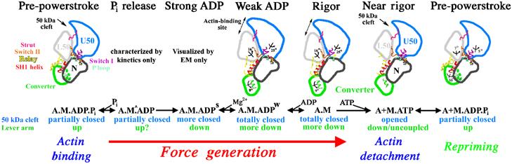
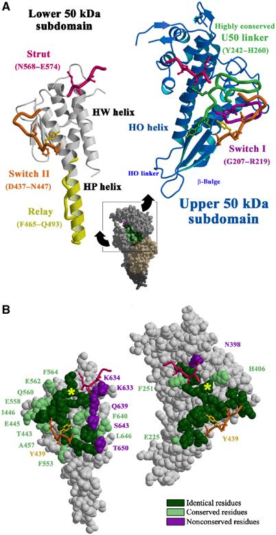
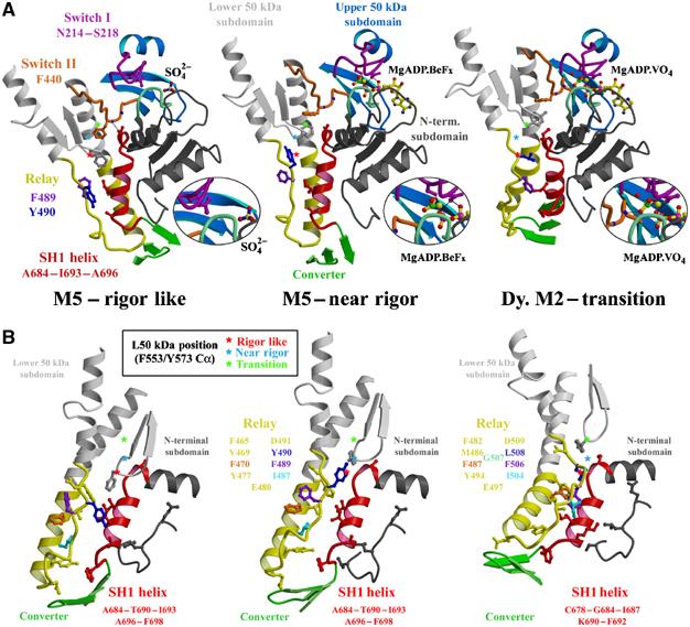
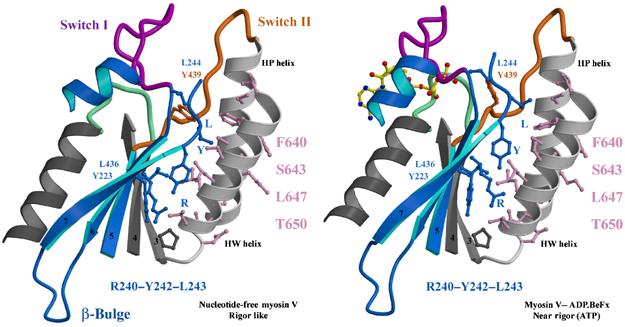
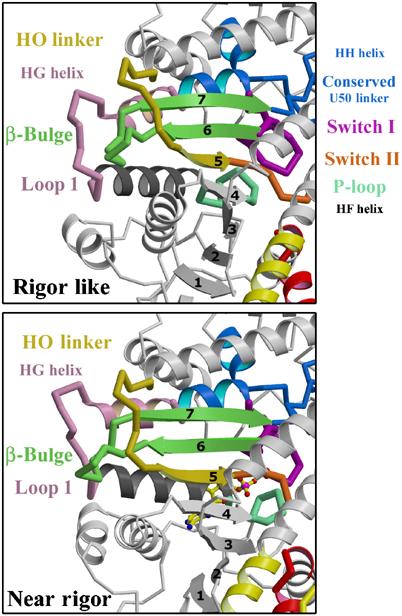
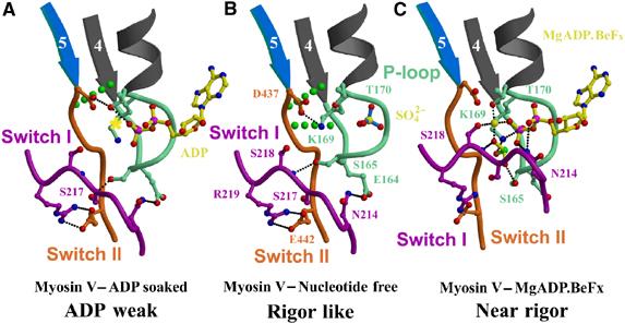
The molecular motor, myosin, undergoes conformational changes in order to convert chemical energy into force production. Based on kinetic and structural considerations, we assert that three crystal forms of the myosin V motor delineate the conformational changes that myosin motors undergo upon detachment from actin. First, a motor domain structure demonstrates that nucleotide-free myosin V adopts a specific state (rigor-like) that is not influenced by crystal packing. A second structure reveals an actomyosin state that favors rapid release of ADP, and differs from the rigor-like state by a P-loop rearrangement. Comparison of these structures with a third structure, a 2.0 angstroms resolution structure of the motor bound to an ATP analog, illuminates the structural features that provide communication between the actin interface and nucleotide-binding site. Paramount among these is a region we name the transducer, which is composed of the seven-stranded beta-sheet and associated loops and linkers. Reminiscent of the beta-sheet distortion of the F1-ATPase, sequential distortion of this transducer region likely controls sequential release of products from the nucleotide pocket during force generation.


Figures

Figure 1
Figure 1
Chemo-mechanical transduction in the actin–myosin ATPase cycle. Structural transitions in the actomyosin ATPase cycle are depicted in terms of characterized subdomain positions. For each state, the biochemical composition, the status of the large 50 kDa cleft and lever arm position are noted. The depiction begins with the pre-powerstroke bound to actin. An uncharacterized structural isomerization (seen kinetically (Rosenfeld and Sweeney, 2004), and denoted as M^) opens an exit route for phosphate. This is followed by an essentially irreversible isomerization (Rosenfeld and Sweeney, 2004) to create a state seen in cryo-EM (Whittaker et al, 1995) and characterized kinetically (strong ADP binding) (Rosenfeld et al, 2000). Most of force generation and lever arm movement on actin are partitioned between these two as yet unresolved structural transitions. Based on the new structure presented in this paper (weak ADP binding; Figure 7), the next isomerization (Rosenfeld et al, 2000) destroys Mg2+ binding, has a lever arm swing associated with it and greatly weakens ADP affinity. ADP then dissociates, forming the rigor state. Binding of ATP to the rigor state opens the 50 kDa cleft, causing myosin to dissociate from actin in the post-rigor state. The post-rigor state isomerizes into the pre-powerstroke conformation, repriming the lever arm and allowing hydrolysis of ATP. The hydrolysis products are trapped until the myosin rebinds to actin, which induces the product release transitions. Note that the attributes of the myosin structural states characterized to date at high resolution are described in Supplementary Table 7.
Figure 2
Figure 2
Overall view of the myosin V structures. The myosin V MDE NF structure (Coureux et al, 2003) (red) and the myosin V MDE ADP-BeFx structure (green) are compared to various myosin V and Dictyostelium myosin II structures by superimposing atoms of the lower 50 kDa subdomains so that the degree of cleft closure between the U50 and L50 subdomains can be visualized. To compare the structural states of these motor domains, we have selected 393 Cα atoms corresponding to 117, 167 and 109 atoms of the N-terminal, U50 and L50 subdomains. Using these atoms, the r.m.s. differences between myosin V MDE NF (in red) and myosin V MD NF (in gray) are only 0.507, 0.407, 0.520 and 0.593 Å for molecules A, B, C and D, respectively, while the r.m.s. differences between myosin V MDE NF and myosin II MD NF (Reubold et al, 2003) (in blue) is 1.298 Å. Using these same atoms, an r.m.s. difference of only 0.840 Å is obtained between the two post-rigor states myosin V MDE ADP-BeFx and myosin II MD ADP-BeFx (in purple; PDB code: 1MMD), while an r.m.s. difference of 3.047 Å is obtained for the two myosin V MDE structures in the rigor-like (in red) and post-rigor state (in green). Note that the selected Cα residues are (70–78, 85–94, 97–102, 107–116, 119–128, 135–161, 167–183, 655–668, 673–686), (199–224, 237–265, 304–340, 354–364, 366–380, 388–422, 575–582, 587–592) and (440–467, 493–501, 504–513, 519–531, 535–540, 546–569, 635–653) for the N-terminal, U50 and L50 subdomains, respectively.
Figure 3
Figure 3
Cleft closure. (A) The lower and upper 50 kDa subdomains of myosin V MDE have been pulled apart exposing the surface interactions allowing cleft closure. Note (in green on the right) the U50 highly conserved linker that interacts with the HW and HP helices (white on the left) of the L50 subdomain. Switch II (orange) and the strut (pink) are two connectors between the subdomains that help mediate the interactions between these two surfaces and they are shown on both sides. In particular, a hydrophobic residue of switch II (Y439, yellow ball and stick) is a serine or alanine in all myosin II isoforms. This difference may account in part for the difference in the kinetics of cleft closure for the two molecules. (B) A surface CPK representation of the residues involved in this interface is presented in the same orientation as in A). Depending on the conservation in the sequence of these residues in the myosin superfamily, different colors are used (absolutely conserved (green), conservative changes (pale green) and nonconserved (purple)). A yellow star indicates how to reposition the two surfaces to reconstruct the interface.
Figure 4
Figure 4
Switch II controls converter position by inducing conformational changes at the interface between the relay and the SH1 helix. (A) The region around switch II for myosin V MDE rigor-like and post-rigor states and Dictyostelium myosin II pre-powerstroke state are shown, positioned by overlaying the N-terminal subdomains. Changes in the switch II conformation that depend on the nucleotide state of the active site (see the magnification of this region in the inset) control the movements of the L50 subdomain (gray). In order to demonstrate this movement, stars denote the Cα position of one residue of the L50 subdomain (F553 in myosin V; Y573 in Dictyostelium myosin II) in each of the three states. The star is red, cyan and green for the rigor-like, post-rigor and pre-powerstroke states, respectively. Note that from the rigor-like position, the star moves ‘up' in the post-rigor state, changing the interface between the relay and the SH1 helix, but without drastically changing the overall conformation of the relay. This results in minimal change in position of the converter (green) and thus the lever arm. The additional upward movement seen in the pre-powerstroke state structure results in steric hindrance between the relay and the SH1 helix. To accommodate the clash, the relay helix bends and positions the converter and lever arm in the pre-powerstroke conformation. In the rigor-like structure, switch II directly interacts with the beginning of the SH1 helix, contributing, along with the greater relay–SH1 helix interactions, to stabilization of this region. This would likely create a more rigid coupling of the converter to the motor domain, which would be necessary to bear force in an actomyosin rigor state. (B) The interface between the SH1 helix and the relay is shown for myosin V rigor-like and post-rigor states and Dictyostelium myosin II pre-powerstroke state, positioned by overlaying the SH1 helix. Colored residues of the relay show how they change the orientation of their side chain to accommodate the sliding at the interface between the two connectors. Note, in particular, the differences between how the beginning of the SH1 helix interacts with hydrophobic side chains of the relay and the L50 subdomain while in the rigor-like state and how the beginning of the SH1 helix interacts with the L50 subdomain near residue F553. In the post-rigor state, the upward movement of this subdomain weakens these interactions leading to a state from which the SH1 helix may unwind more easily. In the pre-powerstroke state, a bending in the relay allows additional contacts between the SH1 helix and the relay to be created via several conserved hydrophobic side chains of the relay.
Figure 5
Figure 5
Sliding on hydrophobic patches. The surfaces of the HP and HW helices of the L50 subdomain make variable contacts with the seven-stranded β-sheet, as shown for myosin V MDE rigor-like and post-rigor states (positioned by overlaying the L50 subdomains). Note in particular the sliding of two residues of the β-sheet (Y223 and L436) on the surface of the HW helix near T650, as well as three residues (R240, Y242 and L243; labelled RYL) found near the beginning of the highly conserved U50 linker involved in closing the cleft.
Figure 6
Figure 6
The transducer. The transducer is the central region of the motor domain near the nucleotide-binding site that includes the last three strands of the seven-stranded β-sheet that undergo distortion between the rigor-like and post-rigor states and the structural elements that accommodate this distortion. Among these elements are the previously studied loop, commonly referred to as loop 1 (residues 184–191), and the β-bulge (pale green) found at the end of the last two β-strands. Another of these elements is a linker that follows the HO helix, which provides a pathway of communication to the actin interface. We thus refer to this linker as the HO linker (residues 424–430); it leads to the fifth β-strand that is followed by switch II. Switch I and the P-loop are connected via three parallel α-helices that interact with the β-sheet. Loop 1 connects the ends of two of these helices (HF and HG). When the β-sheet undergoes distortion, these helices rotate and translate relative to each other.
Figure 7
Figure 7
Active site. Interactions between the nucleotide-binding elements are shown for the M5 MDE in the rigor-like state (A), the ADP weak state (B) and the post-rigor state with MgADP.BeFx bound (C). The yellow star in the ADP weak state marks the position of the Mg2+ in the post-rigor state. In the rigor-like state, switch II interacts with the fourth β-strand and the P-loop of the N-terminal subdomain (broken green lines represent two hydrogen bonds found between the main-chain hydrogen of the amide of Ile438 and Gly163 and the carbonyls of Gly161 and Ile438, respectively). These bonds are progressively lost when P-loop and switch II rearrange upon nucleotide binding.

References

    1. Bernstein SI, Milligan RA (1997) Fine tuning a molecular motor: the location of alternative domains in the Drosophila myosin head. J Mol Biol 271: 1–6 - PubMed
    1. Collaborative Computational Project No. 4 (1994) The CCP4 suite: programs for protein crystallography. Acta Crystallogr D 50: 760–763 - PubMed
    1. Coureux P-D, Wells AL, Menetrey J, Yengo CM, Morris CA, Sweeney HL, Houdusse A (2003) A structural state of the myosin V motor without bound nucleotide. Nature 425: 419–423 - PubMed
    1. De La Cruz EM, Wells AL, Safer D, Ostap EM, Sweeney HL (1999) The kinetic mechanism of myosin V. Proc Natl Acad Sci USA 96: 13726–13731 - PMC - PubMed
    1. Fisher AJ, Smith CA, Thoden JB, Smith R, Sutoh K, Holden HM, Rayment I (1995) X-ray structures of the myosin motor domain of Dictyostelium discoideum complexed with MgADP.BeFx and MgADP.AlF4−. Biochemistry 34: 8960–8972 - PubMed

Publication types

LinkOut - more resources