Comparative Study
. 2004 Nov 10;23(22):4451-61.
doi: 10.1038/sj.emboj.7600455. Epub 2004 Oct 28.

Autophosphorylation of ataxia-telangiectasia mutated is regulated by protein phosphatase 2A

Affiliations
Comparative Study

Autophosphorylation of ataxia-telangiectasia mutated is regulated by protein phosphatase 2A

Aaron A Goodarzi et al. EMBO J. .

Abstract

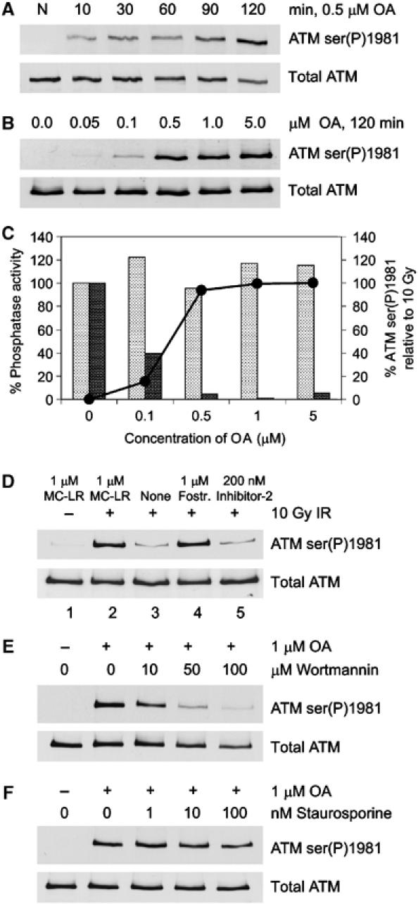
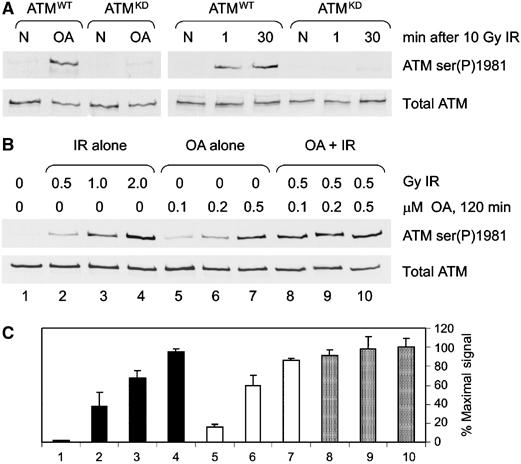
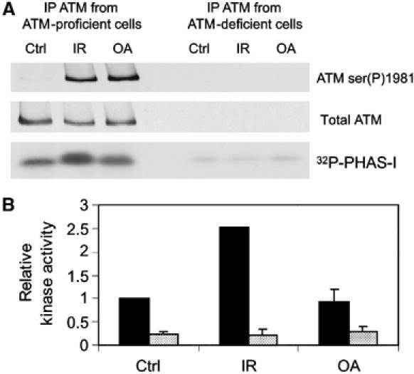
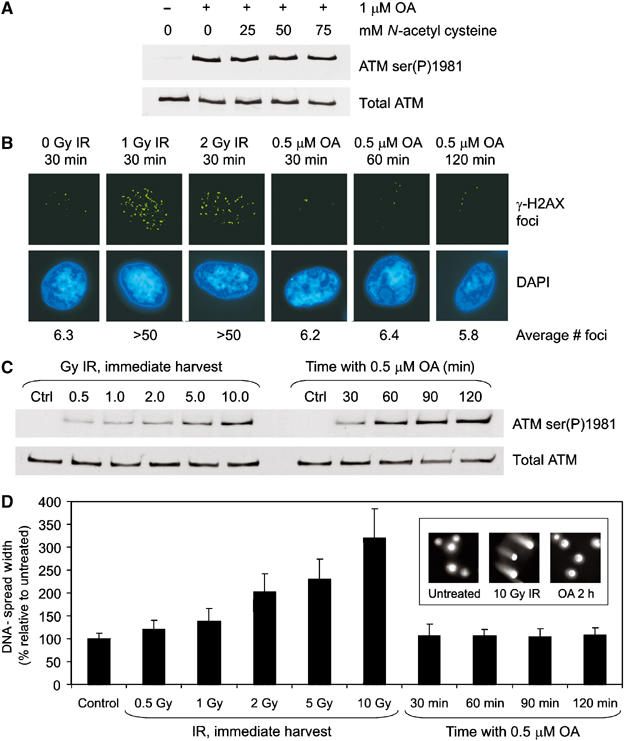
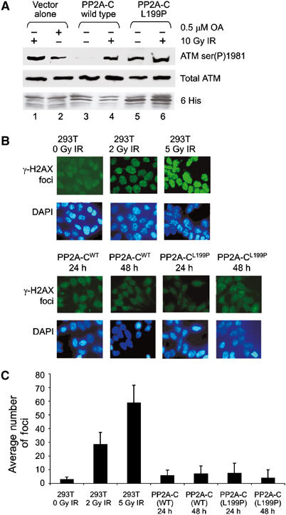
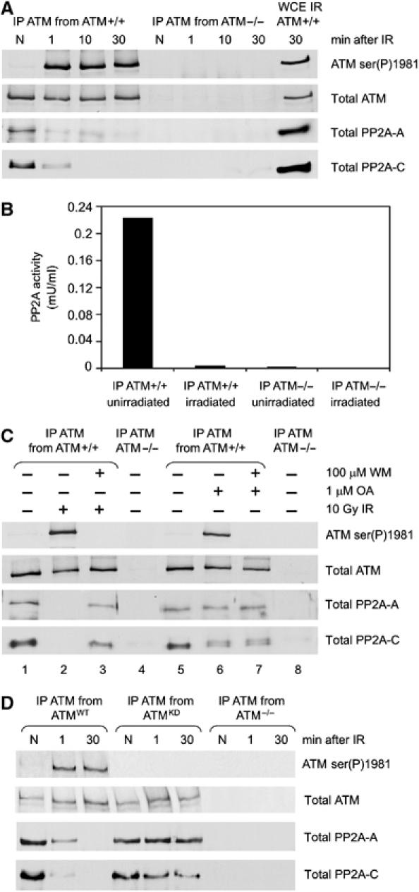
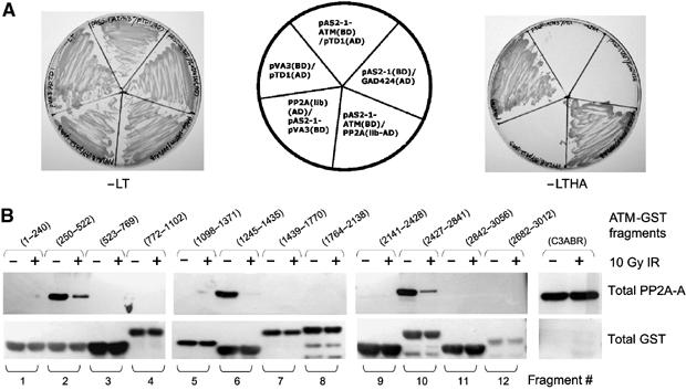
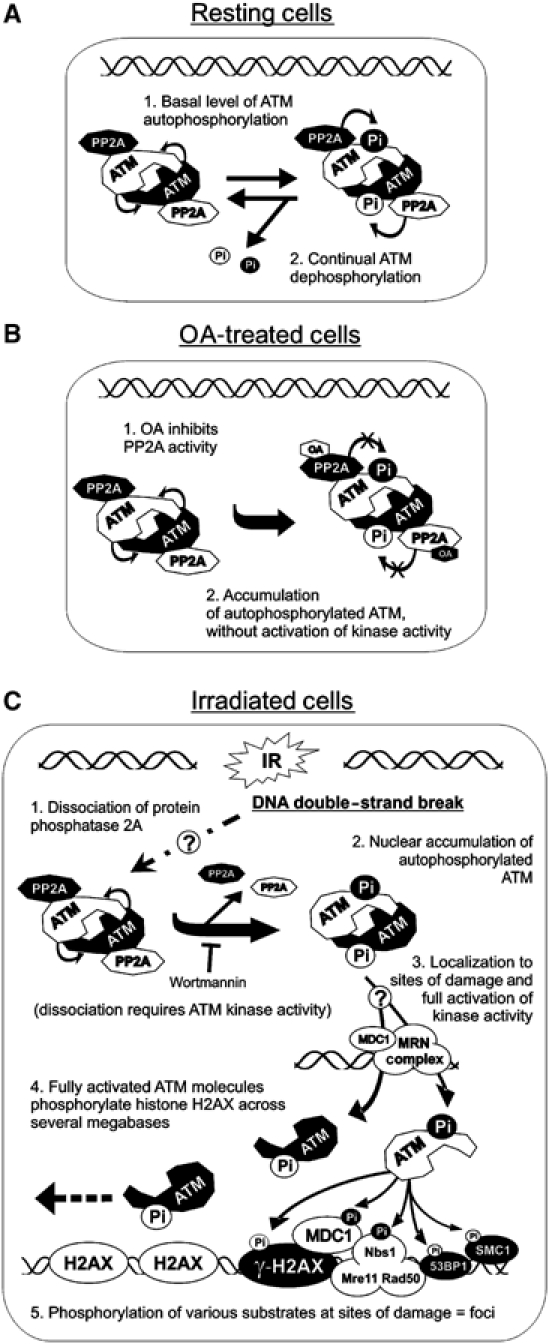
Ionizing radiation induces autophosphorylation of the ataxia-telangiectasia mutated (ATM) protein kinase on serine 1981; however, the precise mechanisms that regulate ATM activation are not fully understood. Here, we show that the protein phosphatase inhibitor okadaic acid (OA) induces autophosphorylation of ATM on serine 1981 in unirradiated cells at concentrations that inhibit protein phosphatase 2A-like activity in vitro. OA did not induce gamma-H2AX foci, suggesting that it induces ATM autophosphorylation by inactivation of a protein phosphatase rather than by inducing DNA double-strand breaks. In support of this, we show that ATM interacts with the scaffolding (A) subunit of protein phosphatase 2A (PP2A), that the scaffolding and catalytic (C) subunits of PP2A interact with ATM in undamaged cells and that immunoprecipitates of ATM from undamaged cells contain PP2A-like protein phosphatase activity. Moreover, we show that IR induces phosphorylation-dependent dissociation of PP2A from ATM and loss of the associated protein phosphatase activity. We propose that PP2A plays an important role in the regulation of ATM autophosphorylation and activity in vivo.


Figures

Figure 1
Figure 1
Inhibition of PP2A-like protein phosphatase activity induces phosphorylation of ATM at serine 1981. (A) C35ABR cells were untreated (N) or incubated with 0.5 μM OA for the indicated times. Whole cell extracts were resolved by sodium dodecyl sulfate–polyacrylamide gel electrophoresis (SDS–PAGE) and immunoblotted for phosphorylation of ATM on serine 1981 (ATM ser(P)1981) and total ATM as shown. (B) C35ABR cells were incubated for 2 h with increasing concentrations of OA as indicated and processed as described in panel A. (C) The extracts shown in panel B were assayed for the percent of PP1 (light bars) or PP2A-like (dark bars) protein phosphatase activity remaining. The solid line with black circles indicates the quantitated ATM ser(P)1981 signals induced by the indicated concentrations of OA relative to those produced by 10 Gy IR. (D) C35ABR cells were either unirradiated or irradiated with 10 Gy IR and harvested immediately. Cells were lysed in NETN buffer with or without protein phosphatase inhibitors as indicated. Whole cell extracts were incubated at 30°C for 30 min, resolved by SDS–PAGE and immunoblotted for ATM ser(P)1981 and total ATM. (E, F) C35ABR cells were treated with increasing amounts of wortmannin (E) or staurosporine (F) for 30 min before incubation with 1 μM OA for an additional 2 h. Whole cell extracts were resolved by SDS–PAGE and immunoblotted for ATM ser(P)1981 and total ATM as described above.
Figure 2
Figure 2
OA induces ATM autophosphorylation and synergizes with IR. (A) AT1ABR cells transfected with wild-type ATM (ATMWT) or kinase-dead ATM (ATMKD) were either untreated (N), treated with 0.5 μM OA for 2 h (OA) or irradiated with 10 Gy IR and harvested immediately (1) or 30 min later. Extracts were prepared and immunoblotted as described in Figure 1. (B) C35ABR cells were treated with IR and/or OA as indicated and processed as described in panel A. (C) The immunoblot signals from representative experiments were quantitated and expressed as a percentage of the maximal signal.
Figure 3
Figure 3
OA does not cause an increase in ATM protein kinase activity. (A) C35ABR (ATM-proficient) and L3 (ATM-deficient) cells were either untreated (Ctrl), irradiated with 10 Gy IR or treated with 1 μM OA for 2 h. ATM was immunoprecipitated, and one-half of the immunoprecipitation (IP) was used to immunoblot for ATM serine 1981 phosphorylation and total ATM, while the other half was assayed for ATM protein kinase activity with PHAS-I (phosphorylated, heat and acid stable, stimulated by insulin) as a substrate. Protein kinase reactions were resolved by SDS–PAGE and analyzed by autoradiography. (B) PHAS-I bands were excised and the incorporation of 32P was analyzed by Cerenkov counting. Black bars correspond to kinase activity of ATM immunoprecipitated from C35ABR cells, while shaded bars correspond to kinase activity of ATM immunoprecipitated from L3 cells. This experiment was repeated in AT1ABR cells expressing either ATMWT or ATMKD. As predicted, IR but not OA induced a significant increase in ATM kinase activity, and no ATM kinase activity was observed when ATM was immunoprecipitated when cells expressing ATMKD were irradiated (Supplementary Figure 2).
Figure 4
Figure 4
OA does not induce detectable DNA damage. (A) C35ABR cells were treated with increasing amounts of NAC for 30 min before incubation with 1 μM OA for 2 h. Extracts were processed as described in Figure 1A. (B) Immunofluorescence microscopy of γ-H2AX (green, upper panel) in normal human fibroblast (Hs68) cells that were either untreated (0 Gy IR), treated with 1 or 2 Gy IR and harvested 30 min later, or treated with 0.5 μM OA for 30–120 min as indicated. Nuclei are indicated by 4′,6-diamidino-2-phenylindole (DAPI) staining (blue, lower panel). Foci were counted in at least 25 cells and average values are indicated. (C) Hs68 cells were treated with DMSO (Ctrl), IR or OA as indicated and immunoblotted for ATM. (D) Cells prepared for panel C were analyzed by the neutral comet assay. DNA-spread width, defined as the lateral width of cell nucleus plus the lateral width of any comet halo, was measured for at least 50 cells per condition. The average DNA-spread width was expressed as a percentage of the DMSO control. The inset shows representative nuclei from untreated and cells treated with 10 Gy IR or incubated for 2 h with 0.5 μM OA.
Figure 5
Figure 5
Expression of a dominant-negative mutant of PP2A-C induces autophosphorylation of ATM at serine 1981. (A) 293T cells were transiently transfected with either His-tagged wild-type (wt) PP2A-C or dominant-negative PP2A-C (L199P). Cells were either irradiated with 10 Gy IR or incubated with 0.5 μM OA for 2 h as indicated. Extracts were immunoblotted for ATM ser(P)1981, total ATM and His expression. (B) Untransfected 293T cells were either untreated or irradiated as indicated. 293T cells were transfected with either wild-type PP2A-C or PP2A-C (L199P) and examined 24 or 48 h later. All cells were analyzed for induction of γ-H2AX foci formation (green) as described but without deconvolution. The lower panel shows DAPI-stained nuclei. (C) The number of γ-H2AX foci in each condition shown in panel B was quantitated and averaged.
Figure 6
Figure 6
The scaffolding and catalytic subunits of PP2A interact with ATM in unirradiated cells, and dissociate after irradiation. (A) C35ABR (ATM-proficient) and L3 (ATM-deficient) cells were either unirradiated (N) or irradiated with 10 Gy and harvested at the times indicated. Whole cell extracts were prepared and ATM was immunoprecipitated and immunoblotted for ATM serine 1981, total ATM, the PP2A-A scaffolding A subunit and PP2A-C catalytic subunit, as shown. A 10 μg portion of whole cell extract from irradiated C35ABR cells was immunoblotted as a positive control. (B) ATM was immunoprecipitated from C35ABR (ATM+/+) or L3 cells (ATM−/−) that were untreated or treated with 10 Gy IR and harvested immediately, and immunoprecipitates were assayed for PP2A activity. No PP1 protein phosphatase activity was detected. (C) C35ABR cells were treated with 100 μM wortmannin or an equivalent volume of DMSO for 30 min, as indicated, followed by either no treatment, 10 Gy IR (and harvested 10 min later) or 1 μM OA for 2 h, as indicated. L3 cells were treated with DMSO only. Extracts, immunoprecipitation and immunoblotting were carried out as in panel A. (D) AT1ABR cells transfected with wild-type ATM (ATMWT), kinase-dead ATM (ATMKD) or no ATM (ATM−/−) were unirradiated (N) or irradiated with 10 Gy and harvested at the times indicated. Whole cell extracts were prepared and processed as in panel A.
Figure 7
Figure 7
ATM interacts with PP2A-A. (A) A yeast two-hybrid screen. Yeast cells were cotransformed with the pAS2-1-ATM (residues 2138–3056) bait vector and pGAD424 empty vector or PP2A-A clone isolated from library and positive control (pVA3 and pTD1), and interactions were tested by growth selection on synthetic dropout media lacking the amino acids Leu and Trp (−LT, left) to select for plasmids, or on plates lacking Leu, Trp, His and Ade (−LTHA, right) for growth selection. (B) Localization of the region of ATM that binds to PP2A-A. Extracts from either unirradiated or irradiated cells were incubated with glutathione agarose beads containing GST-ATM fusion proteins (5 μg). After binding, the beads were analyzed by SDS–PAGE followed by Western blotting with anti-PP2A-A antibody (top panel) or anti-GST antibody (bottom panel).
Figure 8
Figure 8
A model for the regulation of ATM by PP2A. (A) In the absence of DNA damage, (1) dimers of ATM undergo a basal level of autophosphorylation (at serine 1981) that is (2) removed by bound PP2A. (B) The addition of OA to cells (1) inhibits PP2A protein phosphatase activity, allowing (2) autophosphorylated ATM to accumulate in the absence of DSBs and without an increase in protein kinase activity. (C) IR causes (1) dissociation of PP2A from ATM in a mechanism that requires the protein kinase activity of ATM, leading to (2) accumulation of autophosphorylated ATM. Independently, IR causes the recruitment of the MRN complex (possibly with MDC1 and other proteins) to sites of DNA damage. Autophosphorylated ATM is then (3) recruited to the DSB, possibly resulting in full activation of ATM protein kinase activity, followed by (4) phosphorylation of histone H2AX across several megabases of DNA and (5) localized phosphorylation of ATM substrates at IR-induced foci.

Similar articles

Cited by

References

    1. Ali A, Zhang J, Bao S, Liu I, Otterness D, Dean NM, Abraham RT, Wang XF (2004) Requirement of protein phosphatase 5 in DNA-damage-induced ATM activation. Genes Dev 18: 249–254 - PMC - PubMed
    1. Bakkenist CJ, Kastan MB (2003) DNA damage activates ATM through intermolecular autophosphorylation and dimer dissociation. Nature 421: 499–506 - PubMed
    1. Bakkenist CJ, Kastan MB (2004) Phosphatases join kinases in DNA-damage response pathways. Trends Cell Biol 14: 339–341 - PubMed
    1. Banin S, Moyal L, Shieh S, Taya Y, Anderson CW, Chessa L, Smorodinsky NI, Prives C, Reiss Y, Shiloh Y, Ziv Y (1998) Enhanced phosphorylation of p53 by ATM in response to DNA damage. Science 281: 1674–1677 - PubMed
    1. Borthwick EB, Zeke T, Prescott AR, Cohen PT (2001) Nuclear localization of protein phosphatase 5 is dependent on the carboxy-terminal region. FEBS Lett 491: 279–284 - PubMed

Publication types

MeSH terms