

LAP2alpha and BAF collaborate to organize the Moloney murine leukemia virus preintegration complex
- PMID: 15510219
- PMCID: PMC533042
- DOI: 10.1038/sj.emboj.7600452
LAP2alpha and BAF collaborate to organize the Moloney murine leukemia virus preintegration complex
Abstract
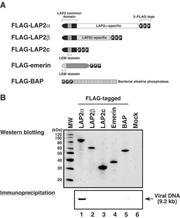
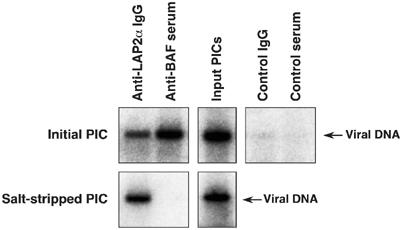
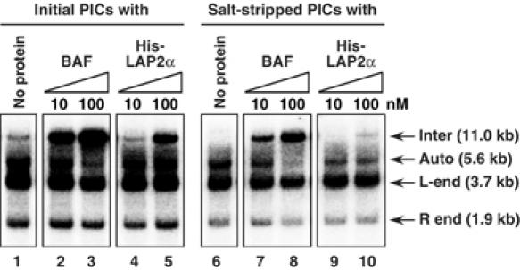
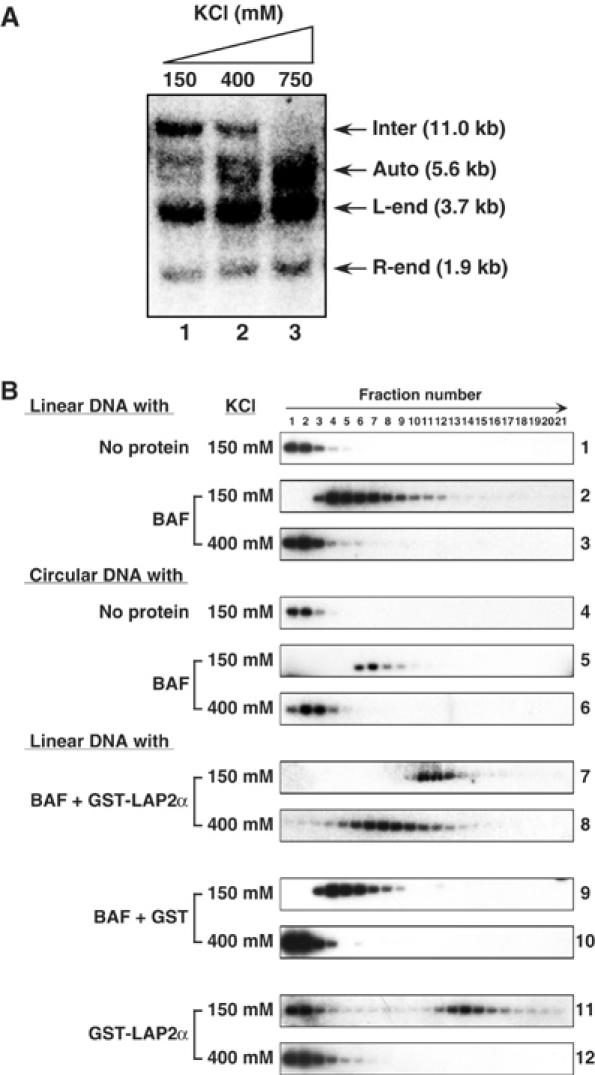
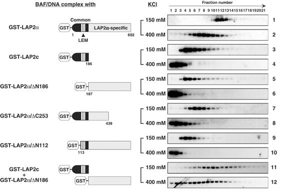
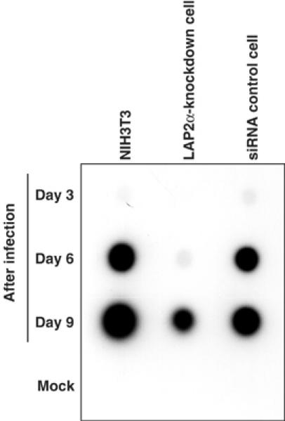
Integration of viral DNA into the host genome is an essential step in retroviral replication. The viral DNA made by reverse transcription is a component of the preintegration complex (PIC) that also contains the viral integrase protein, the enzyme that integrates the viral DNA. Several other viral and cellular proteins are present in the PIC, but their functional roles are less well established. Barrier-to-autointegration factor (BAF) is a cellular protein component of the PIC that blocks autointegration of the viral DNA and stimulates intermolecular integration. In uninfected cells, BAF interacts with members of the LEM family of inner nuclear membrane and nucleoplasmic proteins. Here, we demonstrate that one of the LEM proteins, lamina-associated polypeptide 2alpha (LAP2alpha), is a component of the PIC. LAP2alpha stabilizes the association of BAF with the PIC to stimulate intermolecular integration and suppress autointegration. To further understand the role of LAP2alpha, we established LAP2alpha-knockdown cell lines. Depletion of LAP2alpha significantly inhibited viral replication. Our results demonstrate a critical contribution of LAP2alpha to the nucleoprotein organization of the PIC and to viral replication.
Figures


Similar articles
-
Regulatory mechanisms by which barrier-to-autointegration factor blocks autointegration and stimulates intermolecular integration of Moloney murine leukemia virus preintegration complexes.J Virol. 2002 Dec;76(23):12376-80. doi: 10.1128/jvi.76.23.12376-12380.2002. J Virol. 2002. PMID: 12414981 Free PMC article.
-
BAF: roles in chromatin, nuclear structure and retrovirus integration.Trends Cell Biol. 2004 May;14(5):261-6. doi: 10.1016/j.tcb.2004.03.004. Trends Cell Biol. 2004. PMID: 15130582 Review.
-
The inner-nuclear-envelope protein emerin regulates HIV-1 infectivity.Nature. 2006 Jun 1;441(7093):641-5. doi: 10.1038/nature04682. Epub 2006 May 7. Nature. 2006. PMID: 16680152
-
Functional disruption of the moloney murine leukemia virus preintegration complex by vaccinia-related kinases.J Biol Chem. 2010 Jul 30;285(31):24032-43. doi: 10.1074/jbc.M110.116640. Epub 2010 May 28. J Biol Chem. 2010. PMID: 20511217 Free PMC article.
-
Barrier-to-autointegration factor--a BAFfling little protein.Trends Cell Biol. 2007 Apr;17(4):202-8. doi: 10.1016/j.tcb.2007.02.004. Epub 2007 Feb 21. Trends Cell Biol. 2007. PMID: 17320395 Review.
Cited by
-
The LEM domain proteins emerin and LAP2alpha are dispensable for human immunodeficiency virus type 1 and murine leukemia virus infections.J Virol. 2008 Jun;82(12):5860-8. doi: 10.1128/JVI.00076-08. Epub 2008 Apr 9. J Virol. 2008. PMID: 18400857 Free PMC article.
-
Transient and stable knockdown of the integrase cofactor LEDGF/p75 reveals its role in the replication cycle of human immunodeficiency virus.J Virol. 2006 Feb;80(4):1886-96. doi: 10.1128/JVI.80.4.1886-1896.2006. J Virol. 2006. PMID: 16439544 Free PMC article.
-
Mouse mammary tumor virus integration site selection in human and mouse genomes.J Virol. 2008 Feb;82(3):1360-7. doi: 10.1128/JVI.02098-07. Epub 2007 Nov 21. J Virol. 2008. PMID: 18032509 Free PMC article.
-
Immunity of replicating Mu to self-integration: a novel mechanism employing MuB protein.Mob DNA. 2010 Feb 1;1(1):8. doi: 10.1186/1759-8753-1-8. Mob DNA. 2010. PMID: 20226074 Free PMC article.
-
The role of chromatin in retroviral preintegration complex function.J Biol Chem. 2025 Aug;301(8):110440. doi: 10.1016/j.jbc.2025.110440. Epub 2025 Jul 3. J Biol Chem. 2025. PMID: 40615040 Free PMC article. Review.
References
-
- Asante-Appiah E, Skalka AM (1999) HIV-1 integrase: structural organization, conformational changes, and catalysis. Adv Virus Res 52: 351–369 - PubMed
-
- Berger R, Theodor L, Shoham J, Gokkel E, Brok-Simoni F, Avraham KB, Copeland NG, Jenkins NA, Rechavi G, Simon AJ (1996) The characterization and localization of the mouse thymopoietin/lamina-associated polypeptide 2 gene and its alternatively spliced products. Genome Res 6: 361–370 - PubMed
-
- Bowerman B, Brown PO, Bishop JM, Varmus HE (1989) A nucleoprotein complex mediates the integration of retroviral DNA. Genes Dev 3: 469–478 - PubMed
-
- Brown PO (1997) Integration. In Retroviruses, Coffin JM, Hughes SH, Varmus HE (eds), pp 161–203. Cold Spring Harbor, NY: Cold Spring Harbor Laboratory Press - PubMed
-
- Brown PO, Bowerman B, Varmus HE, Bishop JM (1987) Correct integration of retroviral DNA in vitro. Cell 49: 347–356 - PubMed
Publication types
MeSH terms
Substances
LinkOut - more resources
Full Text Sources
Other Literature Sources
