. 2004 Nov;2(11):e367.
doi: 10.1371/journal.pbio.0020367. Epub 2004 Oct 26.

Tuberculous granuloma formation is enhanced by a mycobacterium virulence determinant

Affiliations

Tuberculous granuloma formation is enhanced by a mycobacterium virulence determinant

Hannah E Volkman et al. PLoS Biol. 2004 Nov.

Abstract

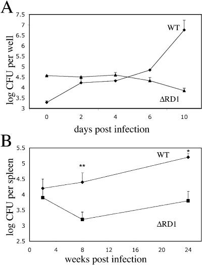
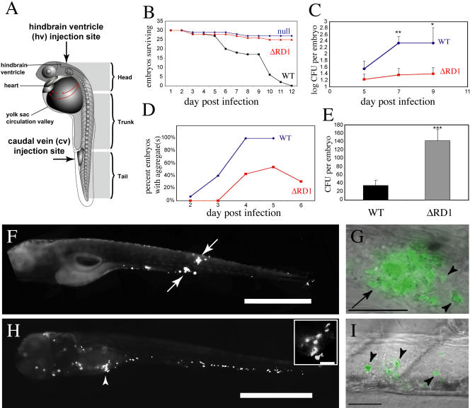
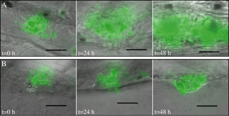
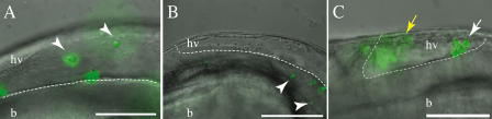
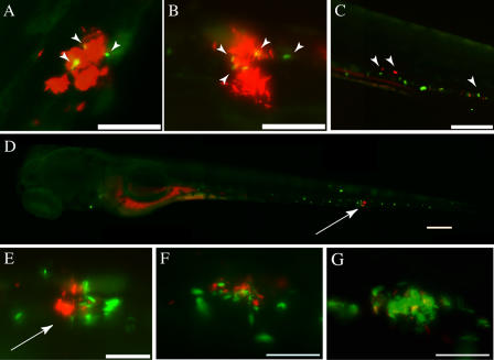
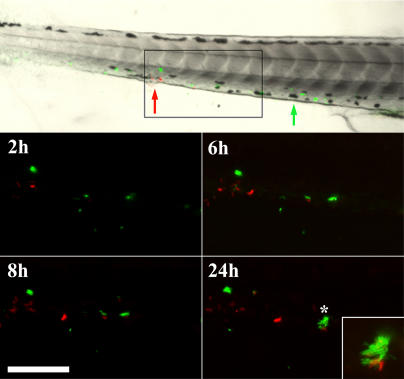
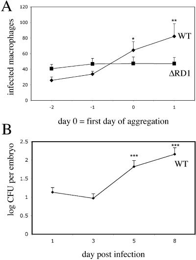
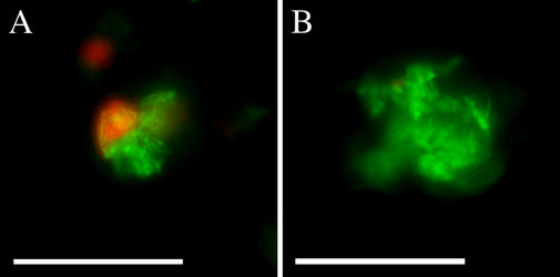
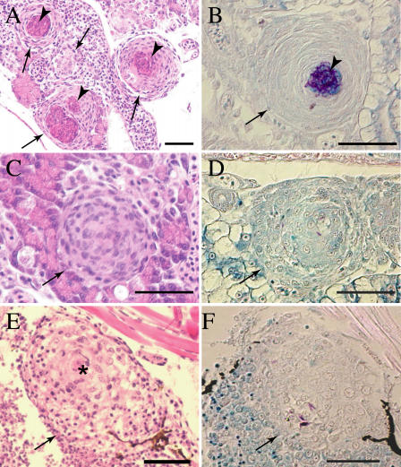
Granulomas are organized host immune structures composed of tightly interposed macrophages and other cells that form in response to a variety of persistent stimuli, both infectious and noninfectious. The tuberculous granuloma is essential for host containment of mycobacterial infection, although it does not always eradicate it. Therefore, it is considered a host-beneficial, if incompletely efficacious, immune response. The Mycobacterium RD1 locus encodes a specialized secretion system that promotes mycobacterial virulence by an unknown mechanism. Using transparent zebrafish embryos to monitor the infection process in real time, we found that RD1-deficient bacteria fail to elicit efficient granuloma formation despite their ability to grow inside of infected macrophages. We showed that macrophages infected with virulent mycobacteria produce an RD1-dependent signal that directs macrophages to aggregate into granulomas. This Mycobacterium-induced macrophage aggregation in turn is tightly linked to intercellular bacterial dissemination and increased bacterial numbers. Thus, mycobacteria co-opt host granulomas for their virulence.


Conflict of interest statement

The authors have declared that no conflicts of interest exist.

Figures

Figure 1
Figure 1. The RD1 Regions in M. tuberculosis and M. marinum Are Homologous and Syntenic
The white arrows represent the RD1 region deleted from M. tuberculosis. The black arrow represents a predicted open reading frame not present in M. tuberculosis. Rv3874 and Rv3875 are also known as cfp-10 and esat-6, respectively. Numbers represent the percent amino acid identities between the corresponding proteins of the two organisms.
Figure 2
Figure 2. M. marinum ΔRD1 Is Attenuated In Vitro and In Vivo
(A) Growth of M. marinum WT and ΔRD1 in J774 cells. Each time point represents the average of triplicate values. Error bars are ± standard error of the mean (SEM). (B) WT and ΔRD1 bacterial numbers in frog spleens. Each time point represents the average colony counts from 3–5 frogs. Error bars are ± SEM (* p ≤ 0.05, ** p = 0.016, unpaired Student's t-test). Infecting doses were 5.8 × 10⁵ CFU for WT and 1.2 × 10⁶ CFU for ΔRD1.
Figure 3
Figure 3. ΔRD1 Is Attenuated in Zebrafish Larvae
(A) Diagram of the zebrafish embryo/larva. Arrows indicate the two injection sites used in this study. (B) Survival of embryos infected with ΔRD1 (410 CFU) or WT bacteria (250 CFU) and null-injected embryos. (C) Whole embryo bacterial counts of WT- and ΔRD1-infected embryos. Infecting doses: 32 CFU for WT, 36 CFU for ΔRD1. Error bars are ± SEM (** p = 0.0075 comparing 7-d postinfection WT to 7-d postinfection ΔRD1; * p = 0.05 comparing 9-d postinfection WT to 9-d postinfection ΔRD1, unpaired Student's t-test). (D) Time of aggregate formation, showing delayed aggregation in the ΔRD1-infected embryos (n = 13) as compared to WT-infected embryos (n = 15). Infecting doses: 131 CFU for WT, 301 CFU for ΔRD1. (E) Whole embryo bacterial counts of WT- and ΔRD1-infected embryos on day of aggregate formation. Infecting doses: 36 CFU for WT, 78 CFU for ΔRD1. Error bars are ± SEM (*** p = 0.0008, unpaired Student's t-test; ΔRD1 n = 28, WT n = 29). (F) Fluorescent image of WT-infected embryo at 6 d postinfection with two aggregates (arrows). Scale bar, 200 μm. (G) WT-infected embryos with higher magnification overlay of fluorescent and DIC images showing an aggregate (arrow) with individual infected macrophages that are migrating toward aggregate (arrowheads). Scale bar, 50 μm. (H) Fluorescent image of ΔRD1-infected embryo at 6 d postinfection that has not formed any aggregates. Note the numerous infected macrophages throughout the head, body, and tail. Arrowhead and close-up insert (scale bar, 50 μm) show infected macrophages close to each other, but not aggregating. Scale bar, 200 μm. (I) ΔRD1-infected embryo under higher-magnification overlay of DIC and fluorescent images showing three individual infected macrophages (arrowheads). Scale bar, 50 μm.
Figure 4
Figure 4. Progression of Aggregates a WT Aggregate (A), and a ΔRD1 Aggregate (B)
(A) WT aggregates shown on the first day of aggregate formation (t = 0 h); 24 h after aggregate formation (t = 24 h); and 48 h after aggregate formation (t = 48 h). (B) ΔRD1 aggregates shown at the same time points as in (A). A 60× water lens was used for all photomicrographs except the image in (A) t = 48 h, which was taken with a 40× lens. Scale bar represents 50 μm.
Figure 5
Figure 5. Normal Macrophage Chemotaxis to Initial Sites of ΔRD1 Infection
Overlay of DIC and fluorescent images showing the hindbrain ventricle (hv) of infected embryos. The hindbrain ventricle/brain (hv/b) boundary indicated by a white dashed line. (A) ΔRD1-infected embryo 4 h postinfection with individual infected macrophages marked by arrowheads. (B) ΔRD1-infected embryo 5 h postinfection in which individual infected macrophages (arrowheads) have migrated from the hindbrain ventricle and into the brain. (C) WT-infected embryo 24 h postinfection with macrophages beginning to aggregate (white arrow) in the hindbrain ventricle. A second out-of-focus aggregate is to the left (yellow arrow). Scale bar, 100 μm.
Figure 6
Figure 6. Superinfection with WT Bacteria Rescues ΔRD1 Aggregation Defect
(A and B) Embryos with aggregates at 3d postinfection with 85 CFU red-fluorescent WT bacteria are shown 4 h after superinfection with green-fluorescent strains of either ΔRD1 (134 CFU) (A) or WT (169 CFU) (B) bacteria. Superinfecting strains were injected at sites distant from the aggregates, and pictures were taken outside of injection regions. Arrowheads indicate macrophages infected with superinfecting strain. Scale bar, 100 μm. (C) Embryo infected with 171 CFU green-fluorescent ΔRD1 for 4 d shown 4 h post-superinfection with 364 CFU of red-fluorescent ΔRD1. Arrowheads point to macrophages infected with each of the bacterial strains. Scale bar, 200 μm. (D) Embryo infected with 171 CFU green-fluorescent ΔRD1 for 4 d shown 4 h after superinfection with 363 CFU of red-fluorescent WT bacteria. Arrow points to macrophage aggregate. Scale bar, 200 μm. (E) Higher magnification image of aggregate (arrow) in (D) showing green fluorescent ΔRD1 and red fluorescent WT bacteria. Arrowhead points to WT-infected macrophage outside the aggregate. Scale bar, 50 μm. (F and G) Embryo infected with green fluorescent ΔRD1, superinfected with red fluorescent WT (as in D and E) shown at 24 h post-superinfection (F), and the same aggregate at 48 h post-secondary infection (G). Scale bars, 50 μm. All panels are fluorescent images.
Figure 7
Figure 7. Superinfection with WT Bacteria Rescues ΔRD1 Aggregation Defect over Time
Embryos were injected with fluorescent ΔRD1 (green) at 1 d postfertilization. 3 d post-primary infection, embryos were injected with fluorescent WT (red) and followed for 24 h post-secondary infection. Approximate injection sites are shown with green and red arrows for ΔRD1 and WT bacteria, respectively. Box in top panel indicates the magnified field in fluorescent images. Inset panel at 24-h time point is a magnified image of the starred aggregate. Scale bar, 125 μm.
Figure 8
Figure 8. Macrophage Aggregation Correlates with Bacterial Dissemination during WT Infection
(A) Enumeration of infected macrophages in embryos by fluorescent and DIC microscopy after infection with green-fluorescent bacteria. Infecting doses: 151 CFU for WT, 301 CFU for ΔRD1. Time points are in reference to day of aggregate formation, which is set at 0. 15 WT infected embryos and 13 ΔRD1 embryos were monitored. The graph represents all 15 WT embryos, but only the 7/13 ΔRD1 infected embryos that formed aggregates over the course of the experiment. Error bars are ± SEM. (*p = 0.0136 comparing WT day 0 and WT day –2; ** p = 0.0053 comparing WT day 1 and WT day –2, unpaired Student's t-test). (B) Whole embryo bacterial counts following WT infection (*** p ≤ 0.0003, 5 d postinfection and 8 d postinfection, respectively, compared to 3 d postinfection, unpaired Student's t-test).
Figure 9
Figure 9. WT Aggregates Are More Likely to Have TUNEL-Positive Cells Than ΔRD1 Aggregates
Representative fluorescent images of aggregates following TUNEL staining of 6-d postfertilization embryos infected with 71 green-fluorescent WT (A), or 474 green-fluorescent ΔRD1 (B) bacteria. TUNEL staining is imaged with red fluorescence, and colocalization with green-fluorescent bacteria appears yellow. Scale bar, 100 μm.
Figure 10
Figure 10. ΔRD1 Infection Is Associated with Persistent Defects in Granuloma Organization
Tissue histology of 32-d postfertilization fish infected with either 21 WT (A–D) or 9 ΔRD1 (E–F) bacteria (doses were not significantly different p = 0.15) at 1 d postfertilization. Arrows indicate granulomas and loose aggregates, arrowheads indicate caseum. Hematoxylin and eosin staining are shown in (A), (C), and (E), and modified acid-fast staining is shown in (B), (D), and (F). (A) Organized caseating WT granulomas (arrow) with central caseum (arrowhead). (B) WT granuloma showing mycobacteria predominantly in caseum with a few within epithelioid cells. (C) Noncaseating but highly organized WT M. marinum-induced granulomas showing the expected few bacteria within cells in (D). (E) Large, loose, and poorly organized macrophage aggregate of ΔRD1-infected fish with evidence of epithelioid transformation only in the center (denoted by *). (F) A few mycobacteria in the ΔRD1 aggregates. Scale bar, 100 μm. Images in (A–D) were taken with a 40× lens, whereas those in (E) and (F) were taken with a 20× lens.

Similar articles

Cited by

References

    1. Adams DO. The granulomatous inflammatory response. A review. Am J Pathol. 1976;84:164–191. - PMC - PubMed
    1. Behr MA, Small PM. A historical and molecular phylogeny of BCG strains. Vaccine. 1999;17:915–922. - PubMed
    1. Bouley DM, Ghori N, Mercer KL, Falkow S, Ramakrishnan L. Dynamic nature of host-pathogen interactions in Mycobacterium marinum granulomas. Infect Immun. 2001;69:7820–7831. - PMC - PubMed
    1. Chan K, Knaak T, Satkamp L, Humbert O, Falkow S, et al. Complex pattern of Mycobacterium marinum gene expression during long-term granulomatous infection. Proc Natl Acad Sci U S A. 2002;99:3920–3925. - PMC - PubMed
    1. Cosma CL, Sherman DR, Ramakrishnan L. The secret lives of the pathogenic mycobacteria. Annu Rev Microbiol. 2003;57:641–676. - PubMed

Publication types

MeSH terms