

Reduced NAA in motor and non-motor brain regions in amyotrophic lateral sclerosis: a cross-sectional and longitudinal study
- PMID: 15512902
- PMCID: PMC2744639
- DOI: 10.1080/14660820410017109
Reduced NAA in motor and non-motor brain regions in amyotrophic lateral sclerosis: a cross-sectional and longitudinal study
Abstract
Objectives: After replication of previous findings we aimed to: 1) determine if previously reported (1)H MRSI differences between ALS patients and control subjects are limited to the motor cortex; and 2) determine the longitudinal metabolic changes corresponding to varying levels of diagnostic certainty.
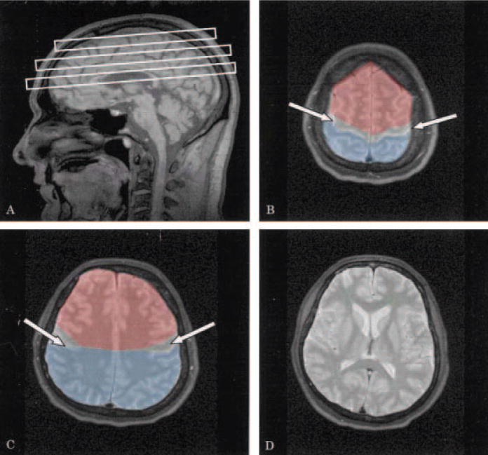
Methods: Twenty-one patients with possible/suspected ALS, 24 patients with probable/definite ALS and 17 control subjects underwent multislice (1)H MRSI co-registered with tissue-segmented MRI to obtain concentrations of the brain metabolites N-acetylaspartate (NAA), creatine, and choline in the left and right motor cortex and in gray matter and white matter of non-motor regions in the brain.
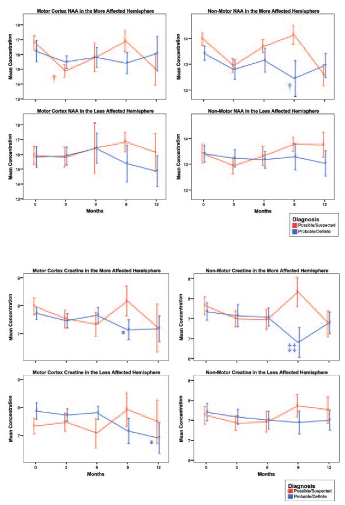
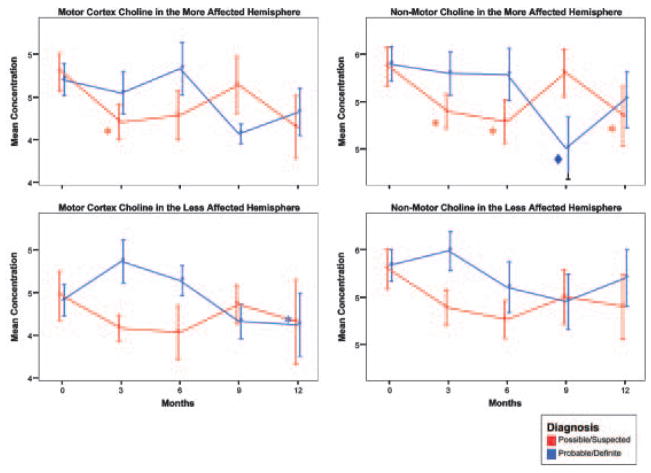
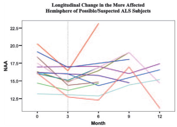
Results: In the more affected hemisphere, reductions in the ratios, NAA/Cho and NAA/Cre+Cho were observed both within (12.6% and 9.5% respectively) and outside (9.2% and 7.3% respectively) the motor cortex in probable/definite ALS. However, these reductions were significantly greater within the motor cortex (P<0.05 for NAA/Cho and P<0.005 for NAA/Cre+Cho). Longitudinal changes in NAA were observed at three months within the motor cortex of both possible/suspected ALS patients (P<0.005) and at nine months outside the motor cortex of probable/definite patients (P<0.005). However, there was no clear pattern of progressive change over time.
Conclusions: NAA ratios are reduced in the motor cortex and outside the motor cortex in ALS, suggesting widespread neuronal injury. Longitudinal changes of NAA are not reliable, suggesting that NAA may not be a useful surrogate marker for treatment trials.
Figures
Similar articles
-
Early detection and longitudinal changes in amyotrophic lateral sclerosis by (1)H MRSI.Neurology. 2002 Mar 12;58(5):773-9. doi: 10.1212/wnl.58.5.773. Neurology. 2002. PMID: 11889242 Free PMC article.
-
Decreased N-acetylaspartate in motor cortex and corticospinal tract in ALS.Neurology. 1998 Jun;50(6):1800-5. doi: 10.1212/wnl.50.6.1800. Neurology. 1998. PMID: 9633731
-
Proton magnetic resonance spectroscopy of the motor cortex in 70 patients with amyotrophic lateral sclerosis.Arch Neurol. 2001 May;58(5):729-35. doi: 10.1001/archneur.58.5.729. Arch Neurol. 2001. PMID: 11346367
-
ALS surrogate markers. MRS.Amyotroph Lateral Scler Other Motor Neuron Disord. 2004 Sep;5 Suppl 1:111-4. doi: 10.1080/17434470410019861. Amyotroph Lateral Scler Other Motor Neuron Disord. 2004. PMID: 15512889 Review.
-
[Proton MR spectroscopy studies of the brain in ALS patients].Neurol Neurochir Pol. 2001;35(1 Suppl):61-70. Neurol Neurochir Pol. 2001. PMID: 11732281 Review. Polish.
Cited by
-
Cerebral degeneration in amyotrophic lateral sclerosis: A prospective multicenter magnetic resonance spectroscopy study.Neurol Clin Pract. 2019 Oct;9(5):400-407. doi: 10.1212/CPJ.0000000000000674. Neurol Clin Pract. 2019. PMID: 31750025 Free PMC article.
-
Gray matter perfusion correlates with disease severity in ALS.Neurology. 2010 Mar 9;74(10):821-7. doi: 10.1212/WNL.0b013e3181d3e2dd. Epub 2010 Feb 10. Neurology. 2010. PMID: 20147656 Free PMC article.
-
MR spectroscopy and imaging-derived measurements in the supplementary motor area for biomarkers of amyotrophic lateral sclerosis.Neurol Sci. 2021 Oct;42(10):4257-4263. doi: 10.1007/s10072-021-05107-3. Epub 2021 Feb 17. Neurol Sci. 2021. PMID: 33594539
-
Investigating metabolic dysregulation in serum of triple transgenic Alzheimer's disease male mice: implications for pathogenesis and potential biomarkers.Amino Acids. 2024 Feb 5;56(1):10. doi: 10.1007/s00726-023-03375-1. Amino Acids. 2024. PMID: 38315232 Free PMC article.
-
Whole-brain and regional brain atrophy in amyotrophic lateral sclerosis.AJNR Am J Neuroradiol. 2007 Feb;28(2):255-9. AJNR Am J Neuroradiol. 2007. PMID: 17296989 Free PMC article.
References
-
- Moffett JR, Namboodiri MA, Cangro CB, Neale JH. Immunohistochemical localization of N-acetylaspartate in rat brain. Neuroreport. 1991;2:131–134. - PubMed
-
- Kalra S, Cashman NR, Genge A, Arnold DL. Recovery of N-acetylaspartate in corticomotor neurons of patients with ALS after riluzole therapy. Neuroreport. 1998;9:1757–1761. - PubMed
-
- De Stefano N, Matthews PM, Arnold DL. Reversible decreases in N-acetylaspartate after acute brain injury. Magn Reson Med. 1995;34:721–727. - PubMed
-
- Rooney WD, Miller RG, Gelinas D, Schuff N, Maudsley AA, Weiner MW. Decreased N-acetylaspartate in motor cortex and corticospinal tract in ALS. Neurology. 1998;50:1800–1805. - PubMed
-
- Ellis CM, Simmons A, Andrews C, Dawson JM, Williams SC, Leigh PN. A proton magnetic resonance spectroscopic study in ALS: correlation with clinical findings. Neurology. 1998;51:1104–1109. - PubMed
Publication types
MeSH terms
Substances
Grants and funding
LinkOut - more resources
Full Text Sources
Other Literature Sources
Medical
Miscellaneous