Comparative Study
. 2005 Feb 1;562(Pt 3):697-706.
doi: 10.1113/jphysiol.2004.077289. Epub 2004 Oct 28.

Capacitative calcium entry supports calcium oscillations in human embryonic kidney cells

Affiliations
Comparative Study

Capacitative calcium entry supports calcium oscillations in human embryonic kidney cells

Gary St J Bird et al. J Physiol. .

Abstract

Treatment of human epithelial kidney (HEK293) cells with low concentrations of the muscarinic agonist methacholine results in the activation of complex and repetitive cycling of intracellular calcium ([Ca(2+)](i)), known as [Ca(2+)](i) oscillations. These oscillations occur with a frequency that depends on the concentration of methacholine, whereas the magnitude of the [Ca(2+)](i) spikes does not. The oscillations do not persist in the absence of extracellular Ca(2+), leading to the conclusion that entry of Ca(2+) across the plasma membrane plays a significant role in either their initiation or maintenance. However, treatment of cells with high concentrations of GdCl(3), a condition which limits the flux of calcium ions across the plasma membrane in both directions, allows sustained [Ca(2+)](i) oscillations to occur. This suggests that the mechanisms that both initiate and regenerate [Ca(2+)](i) oscillations are intrinsic to the intracellular milieu and do not require entry of extracellular Ca(2+). This would additionally suggest that, under normal conditions, the role of calcium entry is to sustain [Ca(2+)](i) oscillations. By utilizing relatively specific pharmacological manoeuvres we provide evidence that the Ca(2+) entry that supports Ca(2+) oscillations occurs through the store-operated or capacitative calcium entry pathway. However, by artificial introduction of a non-store-operated pathway into the cells (TRPC3 channels), we find that other Ca(2+) entry mechanisms can influence oscillation frequency in addition to the store-operated channels.


Figures
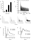
Figure 1
Figure 1. Characterization of the [Ca2+]i signalling response to MeCh in HEK293 cells loaded with the calcium sensitive dye fura-5F
HEK293 cells loaded with fura-5F were treated with low concentrations of MeCh in HBSS containing 1.8 mm extracellular calcium. A, concentration–response relationship for MeCh in terms of the percentage of total cells responding (mean ± s.e.m., 0.1 μm, n = 3, 93 cells total; 0.5 μm, n = 3, 134 cells total; 1.0 μm, n = 7, 256 cells total; 5.0 μm, n = 9, 298 cells total). B, typical calcium response of a field of HEK293 cells to 5 μm MeCh. The 5 min data binning periods are indicated. C, examples of diverse 5 μm MeCh-induced calcium responses in single cells. D, effect of 1 μm (^) and 5 μm (•) MeCh (added at arrow) on oscillation frequency. E, effect of 1 μm (^) and 5 μm (•) MeCh (added at arrow, t = 0) on calcium spike magnitude. The data in D and E were extracted from the same experimental data performed at each MeCh concentration on the same day, and are means ± s.e.m. of 4 (for 1.0 μm) or 3 (for 5.0 μm) independent experiments, with a total of 69 (1.0 μm) or 62 (5.0 μm) cells.
Figure 2
Figure 2. Effect of removing extracellular calcium on MeCh-induced [Ca2+]i oscillations
A, typical calcium response of a field of fura-5F-loaded HEK293 cells to 5 μm MeCh in the absence of extracellular calcium. The 5 min data binning periods are indicated. B, time course of 5 μm MeCh-induced calcium oscillation frequency in the presence (1.8 mm) or absence (nominally Ca2+-free with or without 200 μm BAPTA) of extracellular calcium. MeCh (5 μm) added at arrow. Means ± s.e.m. of: + Ca2+, 102 cells, 3 experiments; − Ca2+, 101 cells, 3 experiments; + BAPTA, 108 cells, 3 experiments.
Figure 3
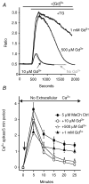
Figure 3. Effects of GdCl3 on the bi-directional flux of calcium ions across the plasma membrane in thapsigargin- and MeCh-treated cells
A, effect of different concentrations of GdCl3 on the thapsigargin-induced calcium transient in HEK293 cells loaded with fura-5F and in the absence of extracellular calcium. All calcium traces are the average of 84–96 total cells from 3 independent experiments. B, effects of different concentrations of GdCl3 on the frequency of 5 μm MeCh-induced calcium oscillations in the absence of extracellular calcium. MeCh added at arrow (mean ± s.e.m. of 98–114 cells from 3 independent experiments).
Figure 4
Figure 4. Gd3+ (1 mM) affects neither spike shape nor amplitude
A, examples of traces showing Ca2+ spiking under control conditions (black trace), and in the absence of extracellular Ca2+ in the presence of 1 mm Gd3+ (grey trace). B, summary of spike amplitudes under the same conditions as in A. Means ± s.e.m. of 99 (control) and 76 (1 mm Gd3+) observations from 3 independent experiments.
Figure 5
Figure 5. Characterization of the calcium entry process sustaining MeCh-induced calcium oscillations
For all experiments in this figure, 1.8 mm extracellular Ca2+ was present throughout. A, effect of 1 μm GdCl3 on the sustained 5 μm MeCh-induced calcium oscillation frequency. HEK293 cells loaded with fura-5F were stimulated (at arrow) with 5 μm MeCh in the presence of extracellular calcium, and in the absence (control, •) and presence (^) of 1 μm GdCl3 (mean ± s.e.m. of 141–152 cells from 4 independent experiments). B, effect of 30 μm 2-APB on the 5 μm MeCh-induced calcium response. HEK293 cells loaded with fura-5F were stimulated (at arrow) with 5 μm MeCh in the presence of extracellular calcium, and in the absence (control, •) and presence (^) of 30 μm 2-APB. The effect of 2-APB on the MeCh-induced calcium response was also tested in the presence of 1 mm GdCl3 and in the absence of extracellular calcium (▴: Gd-insulation condition). All data are mean ± s.e.m. of 94–104 cells from 3 independent experiments. C, effect of depolarizing HEK293 cells with 65 mm K+ on the 5 μm MeCh-induced calcium response. The effect of 65 mm K+ on the MeCh-induced calcium response was also tested in the presence of 1 mm GdCl3 and in the absence of extracellular calcium (▴: Gd-insulation condition). All data are mean ± s.e.m. of 98–109 cells from 3 independent experiments. D, effect of 1 μm GdCl3 on percentage responsive cells compared with control from experiments in A. E, effect of 30 μm 2-APB on percentage responsive cells for experiments described in B. F, effect of 65 mm K+ on percentage responsive cells for experiments described in C.
Figure 6
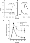
Figure 6. Effect of cell density on the response of HEK293 cells to arachidonic acid (AA) and MeCh
A, as described in the Methods section, HEK293 cells were plated at low and high density. Fura-5F-loaded HEK293 cells were then treated with 30 μm AA in the presence of 1.8 mm extracellular calcium. The data presented are from a single experiment performed on the same day, with each trace the mean response from 30 cells. The data are representative of 5 similar experiments, covering a total of approximately 150 cells. B, the [Ca2+]i signal in low-density cells is comprised of Ca2+ release as well as Ca2+ entry across the plasma membrane. The protocol was as for the low-density cells in A, except that the medium contained no added Ca2+ initially, and was restored to 1.8 mm where indicated. The trace is a mean response from 13 cells, representative of 3 experiments with a total of approximately 45 cells. C, effect of 5 μm MeCh (added at arrow) on HEK293 cells grown at low cell density cell population. The control response to 5 μm MeCh (^) in the presence of 1.8 mm extracellular calcium was compared with the response in nominally Ca2+-free calcium conditions (^), and that in the presence of 1 μm GdCl3 and 1.8 mm extracellular calcium (▴). Data are mean ± s.e.m. of 88–90 cells from 3 independent experiments.
Figure 7
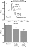
Figure 7. Effect of 5 μm [MeCh] on intracellular [Ca2+] pools
A, following a 25 min incubation of fura-5F-loaded HEK293 cells under control conditions (continous line), or with 5 μm MeCh in the presence (dotted line) or absence (grey continuous line) of extracellular calcium, cells were treated with 20 μm ionomycin (+ 3 mm BAPTA) (data shown are mean responses from 26 to 28 cells in a single experiment, representative of 4 independent experiments). B, summarized data for the amplitude of the ionomycin-induced [Ca2+]i transient under the same conditions as in A. Means ± s.e.m. of 100–106 observations; *P < 0.001 compared with control; **P < 0.001 compared with MeCh +[Ca2+]o.
Figure 8
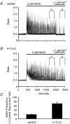
Figure 8. A non-capacitative calcium entry process can support MeCh-induced [Ca2+]i oscillations in HEK293 cells
Wild type HEK293 (A) or H-T3-G (B) cells were treated with 5 μm MeCh (as indicated) in the presence of 1.8 mm extracellular calcium. After 25 min, and at a point when calcium entry is critical for sustaining this response, the cells were treated with 1 μm GdCl3 to block CCE. C, the percentage of the control oscillation frequency after Gd3+ addition (frequency of b/frequency of a × 100%) for each cell type (mean ± s.e.m. of 92–93 cells from 3 independent experiments).

Comment in

  • What's in store for Ca2+ oscillations?
    Taylor CW, Tovey SC. Taylor CW, et al. J Physiol. 2005 Feb 1;562(Pt 3):645. doi: 10.1113/jphysiol.2004.078592. Epub 2004 Nov 11. J Physiol. 2005. PMID: 15539392 Free PMC article. No abstract available.

References

    1. Berridge MJ. Inositol trisphosphate and calcium oscillations. Adv Second Messenger Phosphoprotein Res. 1992;26:211–223. - PubMed
    1. Berridge MJ. Inositol trisphosphate and calcium signalling. Nature. 1993;361:315–325. - PubMed
    1. Berridge MJ. Capacitative calcium entry. Biochem J. 1995;312:1–11. - PMC - PubMed
    1. Berridge MJ. Elementary and global aspects of calcium signalling. J Physiol. 1997;499:291–306. - PMC - PubMed
    1. Bird GJS, Wedel BJ, Lièvremont J-P, Trebak M, Aziz O, Vazquez G, Putney JW., Jr Mechanisms of phospholipase C-regulated calcium entry. Current Mol Med. 2004;4:291–301. - PubMed

Publication types

LinkOut - more resources