. 2004 Nov 9;101(45):15870-5.
doi: 10.1073/pnas.0407074101. Epub 2004 Oct 28.

A locking mechanism preventing radical damage in the absence of substrate, as revealed by the x-ray structure of lysine 5,6-aminomutase

Affiliations

A locking mechanism preventing radical damage in the absence of substrate, as revealed by the x-ray structure of lysine 5,6-aminomutase

Frederick Berkovitch et al. Proc Natl Acad Sci U S A. .

Abstract

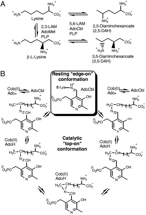
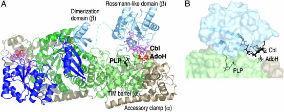
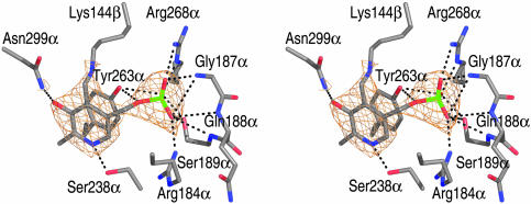
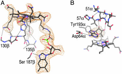
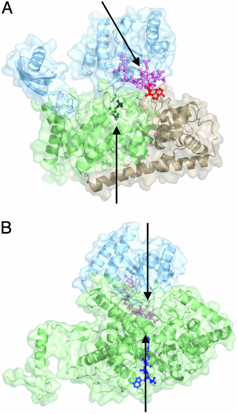
Lysine 5,6-aminomutase is an adenosylcobalamin and pyridoxal-5'-phosphate-dependent enzyme that catalyzes a 1,2 rearrangement of the terminal amino group of dl-lysine and of l-beta-lysine. We have solved the x-ray structure of a substrate-free form of lysine-5,6-aminomutase from Clostridium sticklandii. In this structure, a Rossmann domain covalently binds pyridoxal-5'-phosphate by means of lysine 144 and positions it into the putative active site of a neighboring triosephosphate isomerase barrel domain, while simultaneously positioning the other cofactor, adenosylcobalamin, approximately 25 A from the active site. In this mode of pyridoxal-5'-phosphate binding, the cofactor acts as an anchor, tethering the separate polypeptide chain of the Rossmann domain to the triosephosphate isomerase barrel domain. Upon substrate binding and transaldimination of the lysine-144 linkage, the Rossmann domain would be free to rotate and bring adenosylcobalamin, pyridoxal-5'-phosphate, and substrate into proximity. Thus, the structure embodies a locking mechanism to keep the adenosylcobalamin out of the active site and prevent radical generation in the absence of substrate.


Figures

Fig. 1.
Fig. 1.
Aminomutases in the bacterial lysine fermentation pathway. (A) 5,6-LAM and 2,3-LAM catalyze similar reactions and act on similar substrates. Both enzymes require PLP, but 5,6-LAM is AdoCbl-dependent, whereas 2,3-LAM is an AdoMet-dependent iron-sulfur enzyme. The natural substrates of 5,6-LAM include dl-lysine and β-l-lysine. 2,3-LAM acts on l-lysine and does not accept d-lysine as a substrate. (B) Proposed mechanism of 5,6-LAM, modified from ref. . The boxed step represents the state of the enzyme observed in this study. The unboxed steps are proposed to occur while 5,6-LAM is in the hypothetical top-on conformation (see the Introduction).
Fig. 2.
Fig. 2.
Overall structure of 5,6-LAM and location of cofactors. (A) Ribbon diagram of the α2β2 5,6-LAM tetramer with the accessory clamp of α in brown, the TIM barrel of α in green, and the Rossmann domain and the dimerization domain of β in blue. The dashed line represents the disordered loop connecting the two domains of β. The second αβ unit is represented in darker colors. AdoH is shown in red sticks, Cbl in pink sticks and sphere, and PLP in black sticks. With the exception of Figs. 1, 2, and 3B, all figures were prepared by using pymol (42). (B) Relative positions of PLP and AdoCbl. PLP is inserted into the C terminus of the TIM barrel by Lys-144β, which anchors the Rossmann domain in an off-center conformation on the top corner of the TIM barrel. His-133β (the lower axial ligand to Co in Cbl), Lys-144β (which forms the imine bond to PLP), PLP, Cbl, and AdoH are all depicted in black sticks. The secondary structural elements of the Rossmann domain that contain His-133β and Lys-144β are shown in a ribbon representation. Opaque domain surfaces are shown and are colored as in Fig. 1 A.
Fig. 3.
Fig. 3.
PLP bound in the putative active-site of 5,6-LAM. Stereoview of the putative active site of 5,6-LAM. Lys-144β forms an imine bond to PLP; all other protein–PLP interactions are made by residues of the TIM barrel. A simulated annealing composite 2 FoFc omit electron density map (orange mesh), contoured at 1.5 σ, is shown around the PLP. Unless otherwise noted, the coloring scheme for all stick or ball-and-stick diagrams is as follows: gray, C; red, O; blue, N; green, P.
Fig. 4.
Fig. 4.
The AdoCbl-binding site. (A) View of Cbl within a 1.8-Å radius simulated annealing composite 2 FoFc omit electron density map contoured at 1.0 σ. Hydrogen bonds between a propionamide side chain of the corrin ring and the backbone N and side chain of Thr-191β and the backbone N of Gln-192β are omitted for clarity. (B) View of AdoH within a 1.8-Å radius simulated annealing 2 FoFc omit electron density map contoured at 1.0 σ. Tyr-193α is part of the TIM barrel domain; otherwise, all protein-AdoH contacts are made by residues of the accessory clamp.
Fig. 5.
Fig. 5.
Edge-on vs. top-on enzyme conformations. (A) Structure of the substrate-free form of 5,6-LAM with the Rossmann domain in an edge-on conformation above the TIM barrel. Protein domains and cofactors are colored as in Fig. 2 A. Arrows represent the axes of the TIM barrel and Rossmann domains. (B) Structure of substrate-bound MCM (Protein Data Bank ID code 1REQ) with the Rossmann domain sitting directly on top of the TIM barrel (top-on). The substrate fragment, desulfo-coenzyme A (dark blue), threads through the TIM barrel domain, effecting the closure of the TIM barrel to the more compact structure shown. The Ado moiety of AdoCbl was not observed. We propose that the substrate-bound 5,6-LAM adopts a subunit arrangement like that of substrate-bound MCM, with the Rossmann domain and AdoCbl docked directly onto the center of the TIM barrel (see Results).

Similar articles

Cited by

References

    1. Toraya, T. (2003) Chem. Rev. 103, 2095–2127. - PubMed
    1. Banerjee, R. (2003) Chem. Rev. 103, 2083–2094. - PubMed
    1. Frey, P. A. & Abeles, R. H. (1966) J. Biol. Chem. 241, 2732–2733. - PubMed
    1. Hay, B. P. & Finke, R. G. (1988) Polyhedron 7, 1469–1481.
    1. Hay, B. P. & Finke, R. G. (1987) J. Am. Chem. Soc. 109, 8012–8018.

Publication types

MeSH terms

Associated data

LinkOut - more resources