. 2005 Mar 15;386(Pt 3):445-51.
doi: 10.1042/BJ20041627.

A mitochondrial pool of sphingomyelin is involved in TNFalpha-induced Bax translocation to mitochondria

Affiliations

A mitochondrial pool of sphingomyelin is involved in TNFalpha-induced Bax translocation to mitochondria

Helene Birbes et al. Biochem J. .

Abstract

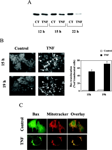
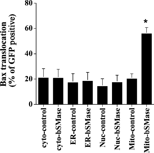
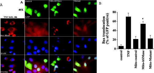
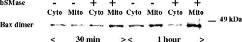
We recently showed that targeting bSMase (bacterial sphingomyelinase) specifically to mitochondria caused accumulation of ceramide in mitochondria, and induced cytochrome c release and cell death [Birbes, El Bawab, Hannun and Obeid (2001) FASEB J., 15, 2669-2679]. In the present study, we investigated the role of this mitochondrial pool of ceramide in response to a receptor-mediated event, namely TNFalpha (tumour necrosis factor alpha), and the involvement of this mitochondrial pool of ceramide in Bax translocation to mitochondria, an event that precedes cytochrome c release. Treatment of MCF7 cells with TNFalpha caused an increase in ceramide levels in the mitochondrial fraction which accompanied Bax translocation to mitochondria. Targeting bSMase to mitochondria specifically resulted in Bax translocation to mitochondria, suggesting that the mitochondrial ceramide pool is involved in Bax translocation. Moreover, in a reconstituted cell-free system, treatment of isolated mitochondria with bSMase enhanced Bax association with mitochondrial membranes. Collectively, these results suggest that the generation of ceramide in mitochondria in response to TNFalpha is sufficient to induce Bax translocation to mitochondria and subsequent cytochrome c release and cell death.


Figures

Figure 1
Figure 1. TNFα-induced mitochondrial translocation of Bax in MCF7 cells
(A) Western blot analysis of cytosolic extracts (5 μg) from control (CT) and TNFα-treated (TNF) cells after 12, 15 and 22 h of incubation. Cells (1.7×10⁶) were treated with TNFα (3 nM) for the indicated times, and cytosolic extracts for analysis of Bax levels were obtained as described in the Experimental section. (B) Immunocytochemical analysis of Bax in control and TNFα-treated cells. Cells (1.7×10⁶) were treated with TNFα (3 nM) for the indicated times, and analysis of Bax localization by indirect immunofluorescence was performed as described in the Experimental section. A clear translocation of Bax was observed after 15 h of TNFα treatment. The punctate pattern was more pronounced after 19 h of treatment. Ten to 15 random fields from each experimental condition were counted, and the percentage of cells that showed translocation of Bax over the total number of counted cells is represented in the histogram. Results are means±S.D. for three different experiments. (C) Localization of Bax after TNFα treatment. Cells (1.7×10⁶) were treated with TNFα (3 nM) for 15 h. At 20 min before fixation, the cells were incubated with 25 nM MitoTracker Red, and analysis of Bax localization and mitochondrial patterns were performed as described in the Experimental section.
Figure 2
Figure 2. Mitochondrial ceramide generation induces Bax translocation
MCF7 cells were co-transfected with 3.5 μg of the different pCMV/GFP controls vector or pCMV/bSMase-GFP vectors plus 0.35 μg of pEGFP-N1 vector. At 48 h after transfection, cells were fixed, immunostained with Bax mouse monoclonal antibody, and visualized with rhodamine-conjugated secondary antibody. Cells were then mounted, and observed by phase-contrast and fluorescent microscopy. In each field, total GFP-positive cells were counted, and the results are expressed as the percentage of GFP-positive cells having Bax translocation to mitochondria. Different fields (at least 300 cells) were counted in each experiment. Data are from one experiment performed in duplicate, representative of at least three separate experiments. Asterisks indicate a significant difference (P<0.05) compared with the control. For statistical analysis, Student's t test for paired sample means was used. Cyto, cytoplasm; ER, endoplasmic reticulum; Nuc, nuclei; Mito, mitochondria.
Figure 3
Figure 3. Overexpression of the mitochondria-targeted bSMase-D295G mutant did not cause Bax translocation
MCF7 cells were treated with 3 nM TNFα for 18 h or were co-transfected with 3.5 μg of pCMV/GFP/Mito empty vector plus 0.35 μg of pEGFP-N1 vector, or with 3.5 μg of pCMV/bSMase-GFP/Mito vector plus 0.35 μg of pEGFP-N1 vector, or 3.5 μg of pCMV/bSMase-D295G-GFP/Mito mutant vector plus 0.35 μg of pEGFP-N1. At 48 h after transfection, cells were fixed, immunostained with anti-Bax mouse monoclonal antibody, and visualized with rhodamine-conjugated secondary antibody. Analysis and quantification of Bax translocation was performed by indirect immunofluorescence as described in the Experimental section. Data are from one experiment performed in duplicate, representative of at least three separate experiments. Asterisks indicate a significant difference (P<0.05) compared with the control. For statistical analysis, Student's t test for paired sample means was used.
Figure 4
Figure 4. Treatment of mitochondria with bSMase induces Bax association with mitochondria
Mitochondrial fraction was prepared by fractionation as described in the Experimental section, and treated with or without 300 m-units of bSMase for 1 h at 37 °C. Mitochondrial fraction (5 μg of protein) was then incubated with 25 μg of cytosolic fraction for the indicated times at 37 °C. At the end of incubation, the mitochondrial and cytosolic fractions were separated by centrifugation (5000 g for 15 min) and Bax translocation was evaluated by Western blotting as described in the Experimental section, but in the absence of SDS. Cyto, cytoplasm; Mito, mitochondria.

References

    1. Hsu Y. T., Wolter K. G., Youle R. J. Cytosol-to-membrane redistribution of Bax and Bcl-XL during apoptosis. Proc. Natl. Acad. Sci. U.S.A. 1997;94:3668–3672. - PMC - PubMed
    1. Gross A., Jockel J., Wei M. C., Korsmeyer S. J. Enforced dimerization of BAX results in its translocation, mitochondrial dysfunction and apoptosis. EMBO J. 1998;17:3878–3885. - PMC - PubMed
    1. Goping I. S., Gross A., Lavoie J. N., Nguyen M., Jemmerson R., Roth K., Korsmeyer S. J., Shore G. C. Regulated targeting of BAX to mitochondria. J. Cell Biol. 1998;143:207–215. - PMC - PubMed
    1. Desagher S., Osen-Sand A., Nichols A., Eskes R., Montessuit S., Lauper S., Maundrell K., Antonsson B., Martinou J. C. Bid-induced conformational change of Bax is responsible for mitochondrial cytochrome c release during apoptosis. J. Cell Biol. 1999;144:891–901. - PMC - PubMed
    1. Li P., Nijhawan D., Budihardjo I., Srinivasula S. M., Ahmad M., Alnemri E. S., Wang X. Cytochrome c and dATP-dependent formation of Apaf-1/caspase-9 complex initiates an apoptotic protease cascade. Cell. 1997;91:479–489. - PubMed

Publication types

MeSH terms