. 2004 Nov;186(22):7783-95.
doi: 10.1128/JB.186.22.7783-7795.2004.

Functional characterization of a catabolic plasmid from polychlorinated- biphenyl-degrading Rhodococcus sp. strain RHA1

Affiliations

Functional characterization of a catabolic plasmid from polychlorinated- biphenyl-degrading Rhodococcus sp. strain RHA1

René Warren et al. J Bacteriol. 2004 Nov.

Abstract

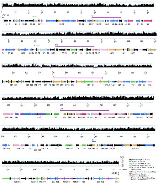
Rhodococcus sp. strain RHA1, a potent polychlorinated-biphenyl (PCB)-degrading strain, contains three linear plasmids ranging in size from 330 to 1,100 kb. As part of a genome sequencing project, we report here the complete sequence and characterization of the smallest and least-well-characterized of the RHA1 plasmids, pRHL3. The plasmid is an actinomycete invertron, containing large terminal inverted repeats with a tightly associated protein and a predicted open reading frame (ORF) that is similar to that of a mycobacterial rep gene. The pRHL3 plasmid has 300 putative genes, almost 21% of which are predicted to have a catabolic function. Most of these are organized into three clusters. One of the catabolic clusters was predicted to include limonene degradation genes. Consistent with this prediction, RHA1 grew on limonene, carveol, or carvone as the sole carbon source. The plasmid carries three cytochrome P450-encoding (CYP) genes, a finding consistent with the high number of CYP genes found in other actinomycetes. Two of the CYP genes appear to belong to novel families; the third belongs to CYP family 116 but appears to belong to a novel class based on the predicted domain structure of its reductase. Analyses indicate that pRHL3 also contains four putative "genomic islands" (likely to have been acquired by horizontal transfer), insertion sequence elements, 19 transposase genes, and a duplication that spans two ORFs. One of the genomic islands appears to encode resistance to heavy metals. The plasmid does not appear to contain any housekeeping genes. However, each of the three catabolic clusters contains related genes that appear to be involved in glucose metabolism.


Figures

FIG. 1.
FIG. 1.
Analysis of RHA1 telomere fragments. RHA1 cells were lysed in an agarose plug with (+) or without (−) proteinase K treatment. Agarose plugs containing RHA1 DNA were subjected to PFGE directly (lanes 1, 2, 5, and 6) or after PstI digestion (lanes 3, 4, 7, and 8). Electrophoresis was conducted for 6 h with a voltage of 6 V/cm and a pulse time that was increased from 2 to 10 s as the electrophoresis progressed. Lanes M to 4 and 9 were stained with ethidium bromide. Lanes 5 to 10 represent Southern blots with a probe derived from the right telomere of pRHL3. The experiment shown in lanes 9 and 10 was performed by using conditions of higher stringency. Lane M, 1-kb plus DNA ladder size marker (Invitrogen, Carlsbad, Calif.). The position of intact linear plasmid DNA containing pRHL3 is indicated on the left. The estimated sizes of the fragments detected by hybridization are indicated on the right.
FIG. 2.
FIG. 2.
Sequence analysis of actinomycete invertron telomeres. (A) Alignment of rhodococcal invertron telomere nucleotide sequences. The nucleotide sequences are derived from each of the three RHA1 invertrons (except for pRHL2-L), as well as pHG201 of R. opacus MR11, pHG204 of R. opacus MR22 (31), pBD2 of R. erythropolis (60), and pHG207 of R. sp. strain MR2253 (30). Strictly conserved nucleotides are indicated with hodococcus asterisks. The two sets of inverted repeats are indicated with arrows. The GCTXCGC central motif is boxed. (B) Radial view of best maximum-parsimony tree obtained by PHYLIP analyses of actinomycete telomeres. The first 800 nucleotides of each telomere were aligned. Sequences were taken from each of the plasmids in 2a, as well as the following invertrons: S. clavuligerus pSCL1 (65), S. coelicolor A3 SCP1, S. violaceoruber pSV2 (59), S. rochei 7434AN4 pSLA2-L (28), Planobispora rosea pPR1 and pPR2 (47), and M. celatum pCLP (46).
FIG. 3.
FIG. 3.
Analysis of RHA1 transformants containing origin of replication of pRHL3. Transformants were analyzed by PFGE (A) and Southern hybridization with a probe derived from the tsr gene (B). Lanes were loaded with the following: M, a chromosome size marker derived from Saccharomyces cerevisiae; 1, wild-type RHA1; and 2 to 4, independent transformants of RHA1 containing pCHB79. The positions of the RHA1 chromosome and each plasmid are indicated on the left. An arrow on the right indicates the deduced position of pCHB79, which corresponds to the origin of electrophoresis.
FIG. 4.
FIG. 4.
Replication origin region of pRHL3. (A) Subcloning of the origin of replication. Subclone plasmids are labeled, and their inserts are represented by horizontal thick bars. The results of transformation experiments are presented on the right: +, clones that yielded transformants of RHA1; −, clones that yielded no transformants. The six-digit numbers indicate the nucleotide positions of restriction sites on pRHL3. (B) Annotated ORFs in the replication origin region. ORFs are numbered according to Fig. 5 and are represented by horizontal arrows; rep1 is presented by a shaded arrow. A closed vertical arrowhead and an open box indicate the respective locations of the direct repeats and AT-rich region described in the text.
FIG. 5.
FIG. 5.
Physical map and G+C content of pRHL3. The G+C content is depicted in a histogram in which each vertical bar indicates the G+C composition calculated over a 100-bp interval by using a sliding window of 10 bp. The bottom bar depicts predicted ORFs grouped into eight functional categories (a color code is provided in the lower right corner of the figure). The orientation of each ORF is indicated by an arrowhead. The symbols between the upper and lower bars indicate the respective positions of genomic islands (GI1 to -4; fuchsia-colored bars), IS elements (light blue bar), TIRs (dark blue arrows), and possible duplications (light blue and green arrows).

References

    1. Altschul, S. F., W. Gish, W. Miller, E. W. Myers, and D. J. Lipman. 1990. Basic local alignment search tool. J. Mol. Biol. 215:403-410. - PubMed
    1. Apweiler, R., T. K. Attwood, A. Bairoch, A. Bateman, E. Birney, M. Biswas, P. Bucher, L. Cerutti, F. Corpet, M. D. R. Croning, R. Durbin, L. Falquet, W. Fleischmann, J. Gouzy, H. Hermjakob, N. Hulo, I. Jonassen, D. Kahn, A. Kanapin, Y. Karavidopoulou, R. Lopez, B. Marx, N. J. Mulder, T. M. Oinn, M. Pagni, F. Servant, C. J. A. Sigrist, and E. M. Zdobnov. 2001. The InterPro database, an integrated documentation resource for protein families, domains, and functional sites. Nucleic Acids Res. 29:37-40. - PMC - PubMed
    1. Bachrach, G., M. J. Colston, H. Bercovier, D. Bar-Nir, C. Anderson, and K. G. Papavinasasundaram. 2000. A new single-copy mycobacterial plasmid, pMF1, from Mycobacterium fortuitum which is compatible with the pAL5000 replicon. Microbiology 146:297-303. - PubMed
    1. Batzoglou, S. B. B. Jaffe, K. Stanley, J. Butler, S. Gnerre, E. Mauceli, B. Berger, J. P. Mesirov, and E. S. Lander. 2002. ARACHNE: a whole-genome shotgun assembler. Genome Res. 12:177-189. - PMC - PubMed
    1. Beggs, M. L., J. T. Crawford, and K. D. Eisenach. 1995. Isolation and sequencing of the replication region of Mycobacterium avium plasmid pLR7. J. Bacteriol. 177:4836-4840. - PMC - PubMed

Publication types

Substances