

Differential roles of CCL2 and CCR2 in host defense to coronavirus infection
- PMID: 15518805
- PMCID: PMC7111831
- DOI: 10.1016/j.virol.2004.09.006
Differential roles of CCL2 and CCR2 in host defense to coronavirus infection
Abstract
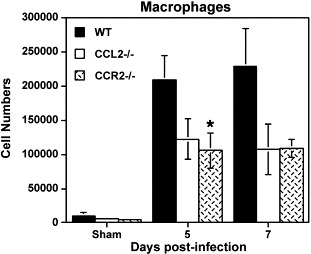
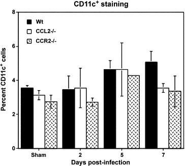
The CC chemokine ligand 2 (CCL2, monocyte chemoattractant protein-1) is important in coordinating the immune response following microbial infection by regulating T cell polarization as well as leukocyte migration and accumulation within infected tissues. The present study examines the consequences of mouse hepatitis virus (MHV) infection in mice lacking CCL2 (CCL2(-/-)) in order to determine if signaling by this chemokine is relevant in host defense. Intracerebral infection of CCL2(-/-) mice with MHV did not result in increased morbidity or mortality as compared to either wild type or CCR2(-/-) mice and CCL2(-/-) mice cleared replicating virus from the brain. In contrast, CCR2(-/-) mice displayed an impaired ability to clear virus from the brain that was accompanied by a reduction in the numbers of antigen-specific T cells as compared to both CCL2(-/-) and wild-type mice. The paucity in T cell accumulation within the central nervous system (CNS) of MHV-infected CCR2(-/-) mice was not the result of either a deficiency in antigen-presenting cell (APC) accumulation within draining cervical lymph nodes (CLN) or the generation of virus-specific T cells within this compartment. A similar reduction in macrophage infiltration into the CNS was observed in both CCL2(-/-) and CCR2(-/-) mice when compared to wild-type mice, indicating that both CCL2 and CC chemokine receptor 2 (CCR2) contribute to macrophage migration and accumulation within the CNS following MHV infection. Together, these data demonstrate that CCR2, but not CCL2, is important in host defense following viral infection of the CNS, and CCR2 ligand(s), other than CCL2, participates in generating a protective response.
Figures


Similar articles
-
Lack of CCR2 results in increased mortality and impaired leukocyte activation and trafficking following infection of the central nervous system with a neurotropic coronavirus.J Immunol. 2001 Oct 15;167(8):4585-92. doi: 10.4049/jimmunol.167.8.4585. J Immunol. 2001. PMID: 11591787
-
Differential roles of CC chemokine ligand 2/monocyte chemotactic protein-1 and CCR2 in the development of T1 immunity.J Immunol. 2002 May 1;168(9):4659-66. doi: 10.4049/jimmunol.168.9.4659. J Immunol. 2002. PMID: 11971015
-
Transgenic CCL2 expression in the central nervous system results in a dysregulated immune response and enhanced lethality after coronavirus infection.J Virol. 2013 Mar;87(5):2376-89. doi: 10.1128/JVI.03089-12. Epub 2012 Dec 26. J Virol. 2013. PMID: 23269787 Free PMC article.
-
Mouse hepatitis virus infection of the central nervous system: chemokine-mediated regulation of host defense and disease.Viral Immunol. 2002;15(2):261-72. doi: 10.1089/08828240260066215. Viral Immunol. 2002. PMID: 12081011 Review.
-
CCL2/CCR2 and CX3CL1/CX3CR1 chemokine axes and their possible involvement in age-related macular degeneration.J Neuroinflammation. 2010 Dec 2;7:87. doi: 10.1186/1742-2094-7-87. J Neuroinflammation. 2010. PMID: 21126357 Free PMC article. Review.
Cited by
-
Monocytes and B cells support active replication of Chandipura virus.BMC Infect Dis. 2016 Sep 14;16:487. doi: 10.1186/s12879-016-1794-6. BMC Infect Dis. 2016. PMID: 27628855 Free PMC article.
-
Single-Cell RNA Sequencing Reveals the Diversity of the Immunological Landscape following Central Nervous System Infection by a Murine Coronavirus.J Virol. 2020 Nov 23;94(24):e01295-20. doi: 10.1128/JVI.01295-20. Print 2020 Nov 23. J Virol. 2020. PMID: 32999036 Free PMC article.
-
Neuronal CCL2 expression drives inflammatory monocyte infiltration into the brain during acute virus infection.J Neuroinflammation. 2017 Dec 4;14(1):238. doi: 10.1186/s12974-017-1015-2. J Neuroinflammation. 2017. PMID: 29202854 Free PMC article.
-
The chemokine receptor CXCR2 and coronavirus-induced neurologic disease.Virology. 2013 Jan 5;435(1):110-7. doi: 10.1016/j.virol.2012.08.049. Virology. 2013. PMID: 23217621 Free PMC article. Review.
-
Monocytes regulate T cell migration through the glia limitans during acute viral encephalitis.J Virol. 2010 May;84(10):4878-88. doi: 10.1128/JVI.00051-10. Epub 2010 Mar 3. J Virol. 2010. PMID: 20200240 Free PMC article.
References
-
- Aliberti J., Reis e Sousa C., Schito M., Hieny S., Wells T., Huffnagle G.B., Sher A. CCR5 provides a signal for microbial induced production of IL-12 by CD8 alpha+dendritic cells. Nat. Immunol. 2000;1:83–87. - PubMed
-
- Baggiolini M. Chemokines and leukocyte traffic. Nature. 1998;392:565–568. - PubMed
-
- Baggiolini M. Chemokines in pathology and medicine. J. Intern. Med. 2001;250:91–104. - PubMed
-
- Bergmann C.C., Marten N.W., Hinton D.R., Parra B., Stohlman S.A. CD8 T cell mediated immunity to neurotropic MHV infection. Adv. Exp. Med. Biol. 2001;494:299–308. - PubMed
Publication types
MeSH terms
Substances
Grants and funding
LinkOut - more resources
Full Text Sources
Molecular Biology Databases
Research Materials
