

Mnt1p and Mnt2p of Candida albicans are partially redundant alpha-1,2-mannosyltransferases that participate in O-linked mannosylation and are required for adhesion and virulence
- PMID: 15519997
- PMCID: PMC3749086
- DOI: 10.1074/jbc.M411413200
Mnt1p and Mnt2p of Candida albicans are partially redundant alpha-1,2-mannosyltransferases that participate in O-linked mannosylation and are required for adhesion and virulence
Abstract
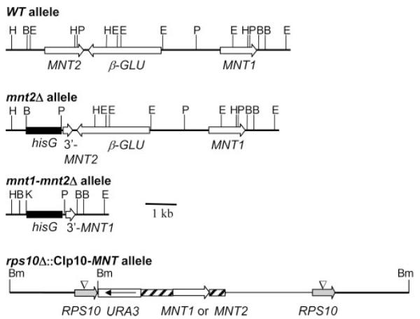
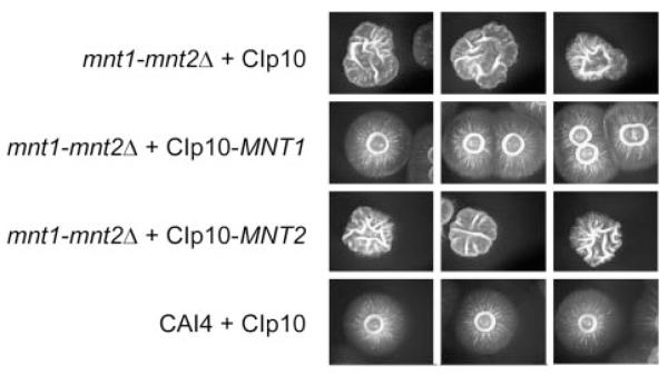
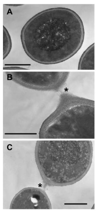
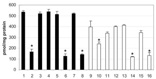
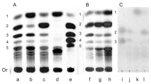
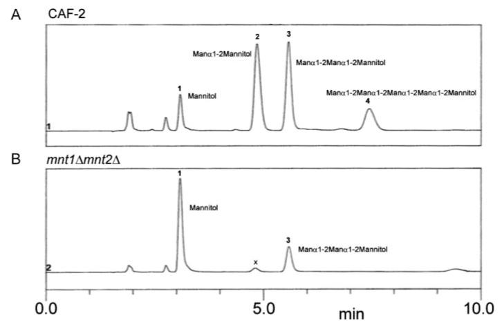
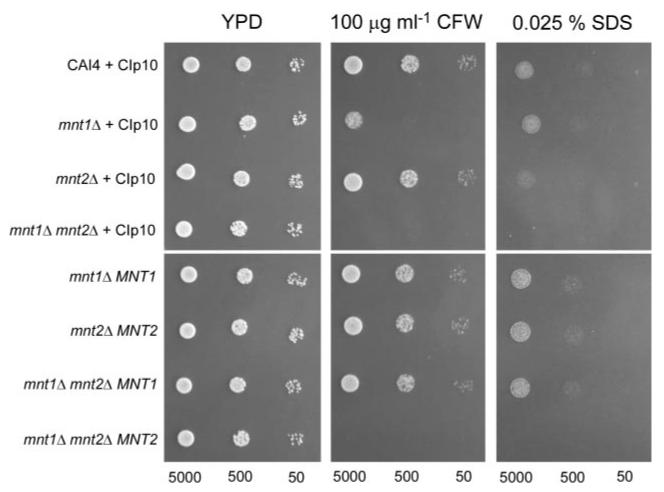
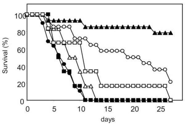
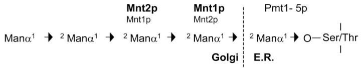
The MNT1 gene of the human fungal pathogen Candida albicans is involved in O-glycosylation of cell wall and secreted proteins and is important for adherence of C. albicans to host surfaces and for virulence. Here we describe the molecular analysis of CaMNT2, a second member of the MNT1-like gene family in C. albicans. Mnt2p also functions in O-glycosylation. Mnt1p and Mnt2p encode partially redundant alpha-1,2-mannosyltransferases that catalyze the addition of the second and third mannose residues in an O-linked mannose pentamer. Deletion of both copies of MNT1 and MNT2 resulted in reduction in the level of in vitro mannosyltransferase activity and truncation of O-mannan. Both the mnt2Delta and mnt1Delta single mutants were significantly reduced in adherence to human buccal epithelial cells and Matrigel-coated surfaces, indicating a role for O-glycosylated cell wall proteins or O-mannan itself in adhesion to host surfaces. The double mnt1Deltamnt2Delta mutant formed aggregates of cells that appeared to be the result of abnormal cell separation. The double mutant was attenuated in virulence, underlining the importance of O-glycosylation in pathogenesis of C. albicans infections.
Figures
Similar articles
-
A multifunctional mannosyltransferase family in Candida albicans determines cell wall mannan structure and host-fungus interactions.J Biol Chem. 2010 Apr 16;285(16):12087-95. doi: 10.1074/jbc.M109.081513. Epub 2010 Feb 17. J Biol Chem. 2010. PMID: 20164191 Free PMC article.
-
The Mnn2 mannosyltransferase family modulates mannoprotein fibril length, immune recognition and virulence of Candida albicans.PLoS Pathog. 2013;9(4):e1003276. doi: 10.1371/journal.ppat.1003276. Epub 2013 Apr 25. PLoS Pathog. 2013. PMID: 23633946 Free PMC article.
-
Biochemical characterization of recombinant Candida albicans mannosyltransferases Mnt1, Mnt2 and Mnt5 reveals new functions in O- and N-mannan biosynthesis.Biochem Biophys Res Commun. 2012 Mar 2;419(1):77-82. doi: 10.1016/j.bbrc.2012.01.131. Epub 2012 Feb 3. Biochem Biophys Res Commun. 2012. PMID: 22326920 Free PMC article.
-
Fungal Mannosyltransferases as Fitness Attributes and their Contribution to Virulence.Curr Protein Pept Sci. 2017;18(11):1065-1073. doi: 10.2174/1389203717666160813164253. Curr Protein Pept Sci. 2017. PMID: 27526929 Review.
-
Mannosylation of fungal glycoconjugates in the Golgi apparatus.Curr Opin Microbiol. 2014 Aug;20:103-10. doi: 10.1016/j.mib.2014.05.008. Epub 2014 Jun 13. Curr Opin Microbiol. 2014. PMID: 24934559 Review.
Cited by
-
Commensal Protection of Staphylococcus aureus against Antimicrobials by Candida albicans Biofilm Matrix.mBio. 2016 Oct 11;7(5):e01365-16. doi: 10.1128/mBio.01365-16. mBio. 2016. PMID: 27729510 Free PMC article.
-
Immunochemistry of pathogenic yeast, Candida species, focusing on mannan.Proc Jpn Acad Ser B Phys Biol Sci. 2012;88(6):250-65. doi: 10.2183/pjab.88.250. Proc Jpn Acad Ser B Phys Biol Sci. 2012. PMID: 22728440 Free PMC article. Review.
-
Candida infections of the genitourinary tract.Clin Microbiol Rev. 2010 Apr;23(2):253-73. doi: 10.1128/CMR.00076-09. Clin Microbiol Rev. 2010. PMID: 20375352 Free PMC article. Review.
-
MNN5 encodes an iron-regulated alpha-1,2-mannosyltransferase important for protein glycosylation, cell wall integrity, morphogenesis, and virulence in Candida albicans.Eukaryot Cell. 2006 Feb;5(2):238-47. doi: 10.1128/EC.5.2.238-247.2006. Eukaryot Cell. 2006. PMID: 16467465 Free PMC article.
-
Role of Protein Glycosylation in Interactions of Medically Relevant Fungi with the Host.J Fungi (Basel). 2021 Oct 18;7(10):875. doi: 10.3390/jof7100875. J Fungi (Basel). 2021. PMID: 34682296 Free PMC article. Review.
References
-
- Odds FC. Candida and Candidosis. 2nd Ed Bailliere-Tindall; London: 1988.
-
- Calderone RA. Candida and Candidiasis. ASM Press; Washington: 2002.
-
- Sandven P. Rev. Iberoam. Micol. 2000;17:73–81. - PubMed
-
- Pappas PG, Rex JH, Lee J, Hamill RJ, Larsen RA, Powderly W, Kauffman CA, Hyslop N, Mangino JE, Chapman S, Horowitz HW, Edwards JE, Dismukes WE. Clin. Infect. Dis. 2003;37:634–643. - PubMed
-
- Sundstrom P. Curr. Opin. Microbiol. 1999;2:353–357. - PubMed
Publication types
MeSH terms
Substances
Associated data
- Actions
Grants and funding
LinkOut - more resources
Full Text Sources
Other Literature Sources
Molecular Biology Databases
