. 2004 Nov 8;167(3):445-56.
doi: 10.1083/jcb.200405153. Epub 2004 Nov 1.

A role for BiP as an adjustor for the endoplasmic reticulum stress-sensing protein Ire1

Affiliations

A role for BiP as an adjustor for the endoplasmic reticulum stress-sensing protein Ire1

Yukio Kimata et al. J Cell Biol. .

Abstract

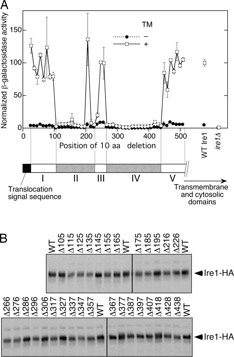
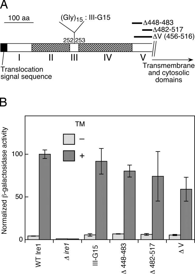
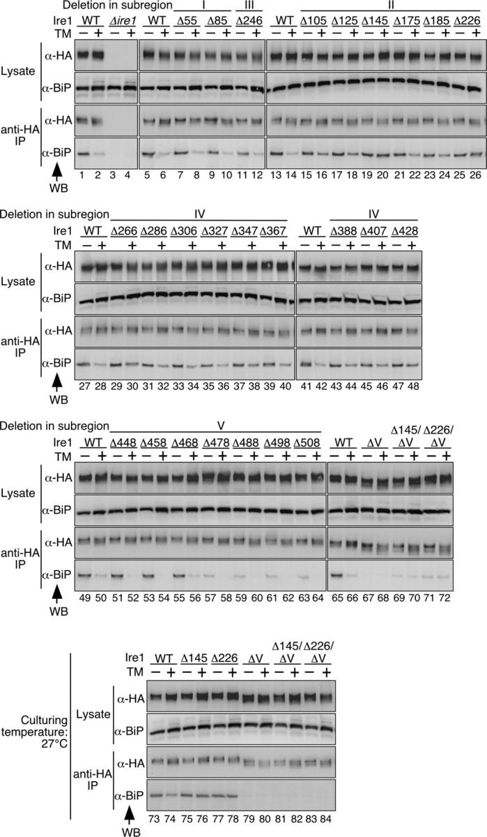
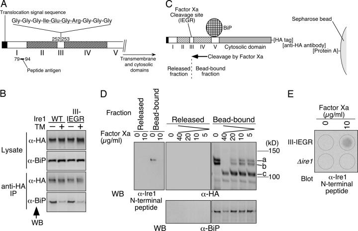
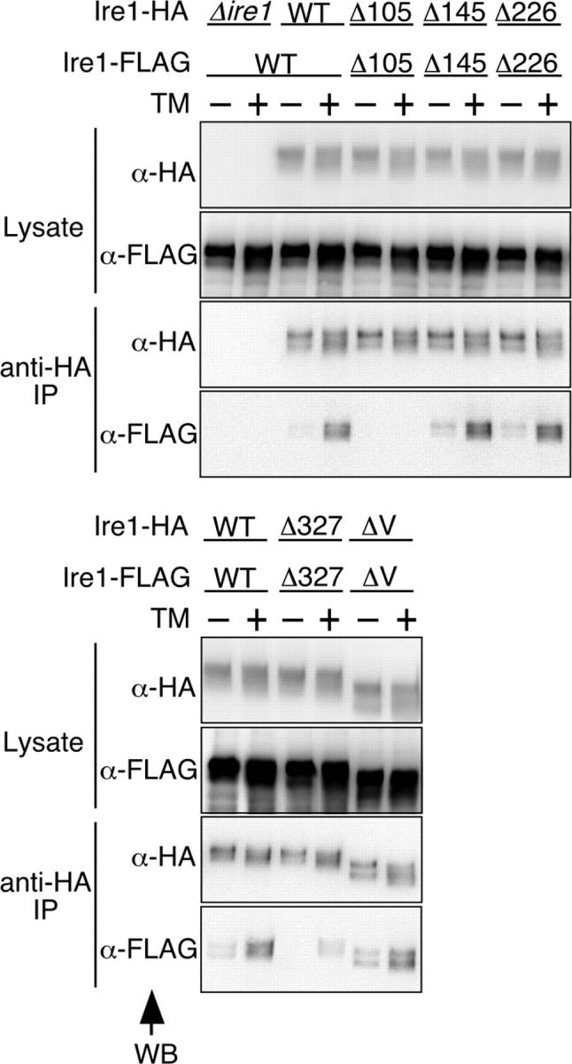
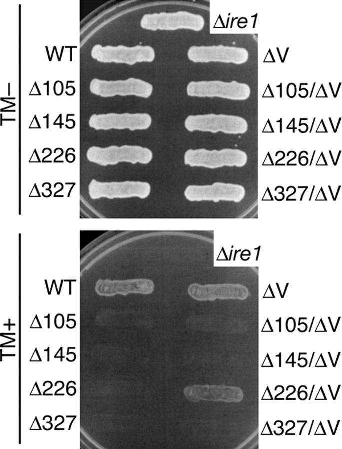
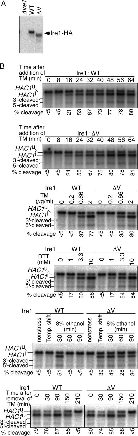
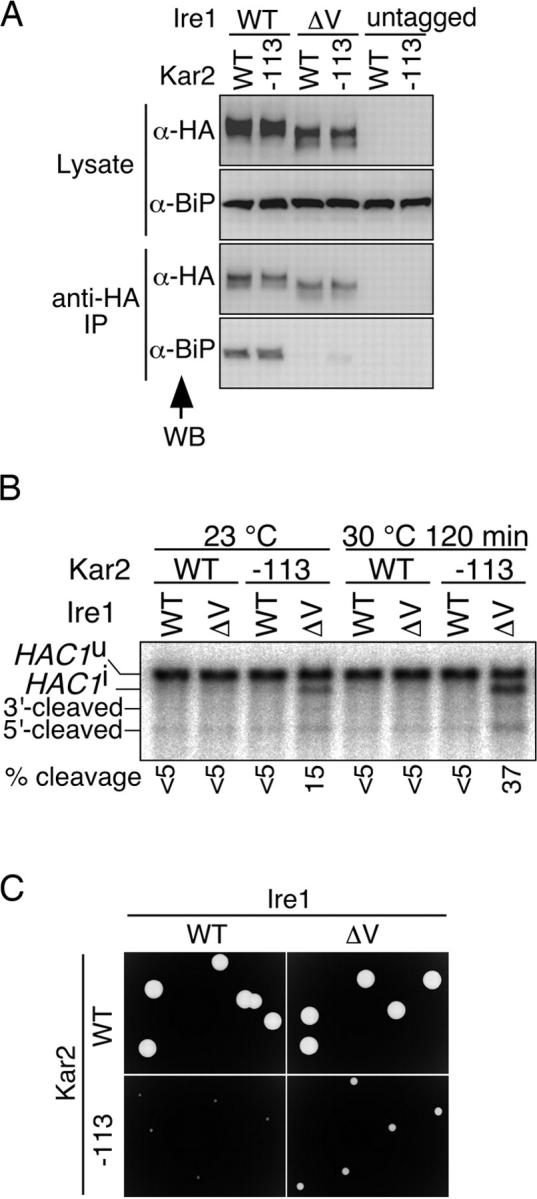
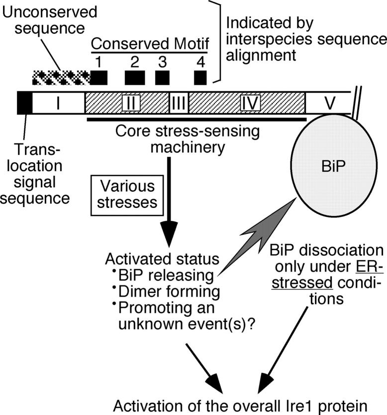
In the unfolded protein response, the type I transmembrane protein Ire1 transmits an endoplasmic reticulum (ER) stress signal to the cytoplasm. We previously reported that under nonstressed conditions, the ER chaperone BiP binds and represses Ire1. It is still unclear how this event contributes to the overall regulation of Ire1. The present Ire1 mutation study shows that the luminal domain possesses two subregions that seem indispensable for activity. The BiP-binding site was assigned not to these subregions, but to a region neighboring the transmembrane domain. Phenotypic comparison of several Ire1 mutants carrying deletions in the indispensable subregions suggests these subregions are responsible for multiple events that are prerequisites for activation of the overall Ire1 proteins. Unexpectedly, deletion of the BiP-binding site rendered Ire1 unaltered in ER stress inducibility, but hypersensitive to ethanol and high temperature. We conclude that in the ER stress-sensory system BiP is not the principal determinant of Ire1 activity, but an adjustor for sensitivity to various stresses.


Figures

Figure 1.
Figure 1.
10-aa deletion scanning of yeast Ire1. The Δire1 strain KMY1015 harboring the UPRE-lacZ reporter plasmid pCZY1 was transformed with a mixture of linearized pPR315-IRE1-HA (a centromeric plasmid for expression of COOH-terminal HA-tagged Ire1) and a partial fragment of the IRE1 gene carrying the desired deletion. The transformants carrying the successfully gap-repaired plasmid were selected and used. For the wild-type Ire1 (WT Ire1 or WT) and empty vector (Δire1) controls, cells were respectively transformed with nonlinearized pRS315-IRE1-HA and pPR315. (A) Cells were cultured with (+) or without (−) 2 μg/ml tunicamycin (TM) for 240 min and subjected to assays for cellular β-galactosidase activity. Each value is the average of three independent transformants and was normalized to the TM+ wild-type Ire1 control, which was set at 100. (B) Lysates prepared from 3 × 10⁶ nonstressed cells using denaturing lysis buffer were analyzed by anti-HA Western blotting.
Figure 2.
Figure 2.
Activity of Ire1 luminal-domain mutants other than those used in the 10-aa deletion scanning. (A) The insertion and deletions carried on the mutants. III-G15 is a (Gly)15 spacer insertion in subregion III, and Δ448−483, Δ482−517, and ΔV are wide-range deletions in subregion V. (B) KMY1015 (Δire1) cells carrying both pCZY1 (UPRE-lacZ reporter) and a mutant version of pRS315-IRE1-HA were cultured with (+) or without (−) 2 μg/ml tunicamycin (TM) for 240 min and subjected to assays for cellular β-galactosidase activity. For the wild-type Ire1 (WT Ire1) and empty vector (Δire1) controls, cells were respectively transformed with nonlinearized pRS315-IRE1-HA and pPR315. Each value is the average from three independent transformants and was normalized to the TM+ wild-type Ire1 control, which was set at 100.
Figure 3.
Figure 3.
BiP binding and dissociation from deletion mutants of Ire1. The Δire1 strain KMY1516 was transformed with a mixture of linearized pRS423-IRE1-HA-HpaI and a partial fragment of the IRE1 gene carrying the desired deletion generated by overlap PCR. The transformants carrying successfully gap-repaired plasmids (deletion mutant versions of pRS423-IRE1-HA, which is a 2-μm plasmid used for expression of Ire1-HA) were selected and cultured with (+) or without (−) 2 μg/ml tunicamycin (TM) for 60 min. The culturing temperature was 27°C for lanes 73–84 or 30°C for other lanes. For the empty vector (Δire1) and wild-type Ire1 (WT) controls, cells were respectively transformed with nonlinearized pRS423 and pPR423-IRE1-HA. Anti-HA immunoprecipitation was performed as described in Materials and methods, and the cell lysates (equivalent to 3 × 10⁶ cells) and immunoprecipitates (equivalent to 1 × 10⁷ cells) were analyzed by anti-HA (α-HA) and anti-BiP (α-BiP) Western blotting (WB).
Figure 4.
Figure 4.
Proteolytic cleavage of Ire1 to assign the BiP-binding site. (A) The III-IEGR mutation in Ire1. The Factor Xa cleavage sequence is underlined. The position of the sequence against which the anti-Ire1 NH2-terminal peptide antibody was generated is also indicated. (B) The Δire1 strain KMY1516 carrying pPR423-IRE1-HA (WT) or its III-IEGR version was cultured with (+) or without (−) 2 μg/ml tunicamycin (TM) for 60 min. Anti-HA immunoprecipitation was performed as described in Materials and methods, and the cell lysates (equivalent to 3 × 10⁶ cells) and immunoprecipitates (equivalent to 1 × 10⁷ cells) were analyzed by anti-HA and anti-BiP Western blotting (WB). (C–E) Proteolytic cleavage analysis. KMY1516 cells carrying the III-IEGR version of pPR423-IRE1-HA were subjected to anti-HA immunoprecipitation, and 15 μl of the resulting immunoprecipitate (equivalent to 1 × 10⁸ cells), the constituents of which were deduced as illustrated in C, was incubated with the indicated concentration of Factor Xa as described in Materials and methods. Note that in C the BiP-binding site is set in subregion V, as concluded in this work. In D, one-tenth portions of the resulting bead-bound and released fractions were fractionated by SDS-PAGE (8%) followed by immunoblotting using the indicated antibodies, and ECL signals were detected by LAS-1000plus (exposure time of 20 s for anti-BiP, 15 s for anti-HA, and 10 s for the anti-Ire1 NH2-terminal peptide). The positions of the full-length (a), slightly truncated (b), and Factor Xa-cleaved (c) Ire1-HA are indicated. Note that the slight truncation of Ire1-HA occurred independently of Factor Xa in Factor Xa cleavage buffer. In E, anti-HA immunoprecipitate from KMY1516 cells carrying the empty vector pRS423 was used as a negative control (Δire1), and one-third portions of the released fractions were subjected to dot blotting using antibody against the anti-Ire1 NH2-terminal peptide.
Figure 5.
Figure 5.
Dimer formation of deletion mutants of Ire1. The Δire1 strains KMY1015 (MATα) and KMY1520 (MATa) respectively carrying deletion mutant versions of pRS315-IRE1-HA and pRS426-IRE1-FLAG, which is a 2-μm plasmid used for expression of Ire1-FLAG, were crossed to obtain diploid cells. For the empty vector control (Δire1), cells carrying pRS315 were used. The resulting diploid cells were cultured with (+) or without (−) 2 μg/ml tunicamycin (TM) for 60 min, and subjected to anti-HA immunoprecipitation as described in Materials and methods. The cell lysates (equivalent to 3 × 10⁶ cells) and immunoprecipitates (equivalent to 1 × 10⁷ cells) were analyzed by anti-HA (α-HA) and anti-FLAG (α-FLAG) Western blotting (WB; ECL signals were detected by LAS-1000plus with exposure time of 20 s to 1 min).
Figure 6.
Figure 6.
Growth of deletion mutants of Ire1 on agar plates containing tunicamycin. The Δire1 strain KMY1015 carrying the indicated deletion mutant version of pRS315-IRE1-HA was streaked on SD agar plates containing (TM+) or not containing (TM−) 2 μg/ml tunicamycin. For the empty vector control (Δire1), cells carrying pRS315 were used. After incubation at 30°C for 3 d, plates were photographed.
Figure 7.
Figure 7.
Hypersensitive response of Ire1 with a subregion-V deletion not to conventional ER stressors, but to high temperature and ethanol. (A) The Δire1 strain KMY1015 carrying either empty vector pRS315 (Δire1), pRS315-IRE1-HA (WT), or its ΔV mutant version was cultured under nonstressed conditions, and its lysate (equivalent to 3 × 10⁶ cells; prepared using denaturing lysis buffer) was analyzed by anti-HA Western blotting. (B) A Δire1 strain KMY1516 carrying either pRS313-IRE1 (a centromeric plasmid for expression of untagged Ire1; WT) or its ΔV mutant version was cultured under nonstressed conditions of 30°C, treated with tunicamycin (TM) at 2 μg/ml for the indicated time (top and second panels) or at the indicated concentration for 60 min (third panel), DTT at the indicated concentration for 40 min (fourth panel) or 8% ethanol for the indicated time (fifth panel), or shifted to 37°C for 30 min and then to 39°C for 30 min (fifth panel, Temp. shift). For the bottom panel, cells treated with 2 μg/ml tunicamycin for 60 min were washed twice with fresh SD medium and further cultured for the indicated time. 1 μg of total RNA prepared from these cells was analyzed by Northern blotting using the HAC1 gene as probe. The positions of uncleaved (HAC1 u), cleaved then ligated (HAC1 i), and cleaved but unligated (5′-cleaved and 3′-cleaved) versions of HAC1 mRNA are indicated (Kawahara et al., 1998). The percentage of HAC1 mRNA cleavage was estimated as described in Kimata et al. (2003).
Figure 8.
Figure 8.
Analysis of a double mutation of kar2-113 and Ire1 subregion-V deletion. (A) The kar2-113 mutation was introduced into the Δire1 strain KMY1516 carrying either pRS313-IRE1 (untagged), pRS423-IRE1-HA (WT), or its ΔV mutant version, as described in the online supplemental Materials and methods (available at http://www.jcb.org/cgi/content/full/jcb.200405153/DC1). The resulting cells and the control KAR2 cells (WT) were cultured at 23°C without extrinsic ER stress and subjected to anti-HA immunoprecipitation as described in Materials and methods. The cell lysates (equivalent to 3 × 10⁶ cells) and immunoprecipitates (equivalent to 1 × 10⁷ cells) were analyzed by anti-HA and anti-BiP Western blotting. (B) The kar2-113 mutation was introduced into KMY1516 cells carrying pRS313-IRE1 (WT) or its ΔV mutant version, as described in the online supplemental Materials and methods. The resulting cells and the control KAR2 cells (WT) were cultured at 23°C or shifted to 30°C for 120 min without extrinsic ER stress and analyzed by Northern blotting of 1 μg of total RNA using the HAC1 gene as probe. The positions of the HAC1 mRNA variants are indicated as in Fig. 7 B. The percentage of HAC1 mRNA cleavage was estimated as described in Kimata et al. (2003). (C) After spreading the cultures used in B (those at 23°C), SD agar plates were incubated at 33°C for 2 d and colonies were photographed.
Figure 9.
Figure 9.
Our current model to interpret structure and function of the yeast Ire1 luminal domain. Positions of conserved motifs deduced by interspecies sequence alignment of Ire1 and PERK (Liu et al., 2000), together with that of an unconserved sequence not present in Ire1 orthologues of higher eukaryotes (Liu et al., 2000, Koizumi et al., 2001) are indicated.

References

    1. Bertolotti, A., Y. Zhang, L.M. Hendershot, H.P. Harding, and D. Ron. 2000. Dynamic interaction of BiP and ER stress transducers in the unfolded-protein response. Nat. Cell Biol. 2:326–332. - PubMed
    1. Calfon, M., H. Zeng, F. Urano, J.H. Till, S.R. Hubbard, H.P. Harding, S.G. Clark, and D. Ron. 2002. IRE1 couples endoplasmic reticulum load to secretory capacity by processing the XBP-1 mRNA. Nature. 415:92–96. - PubMed
    1. Casagrande, R., P. Stern, M. Diehn, C. Shamu, M. Osario, M. Zuniga, P.O. Brown, and H. Ploegh. 2000. Degradation of proteins from the ER of S. cerevisiae requires an intact unfolded protein response pathway. Mol. Cell. 5:729–735. - PubMed
    1. Cox, J.S., and P. Walter. 1996. A novel mechanism for regulating activity of a transcription factor that controls the unfolded protein response. Cell. 87:391–404. - PubMed
    1. Cox, J.S., C.E. Shamu, and P. Walter. 1993. Transcriptional induction of genes encoding endoplasmic reticulum resident proteins requires a transmembrane protein kinase. Cell. 73:1197–1206. - PubMed

Publication types

MeSH terms