. 2004 Nov 8;167(3):417-23.
doi: 10.1083/jcb.200408164. Epub 2004 Nov 1.

A pRb-independent mechanism preserves the postmitotic state in terminally differentiated skeletal muscle cells

Affiliations

A pRb-independent mechanism preserves the postmitotic state in terminally differentiated skeletal muscle cells

Grazia Camarda et al. J Cell Biol. .

Abstract

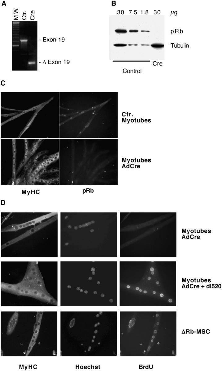
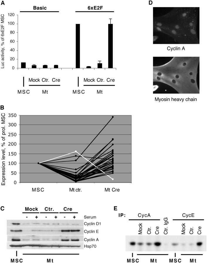
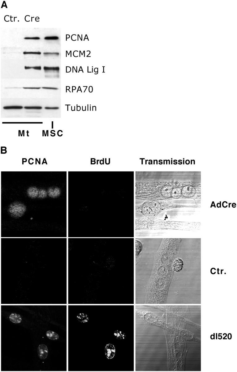
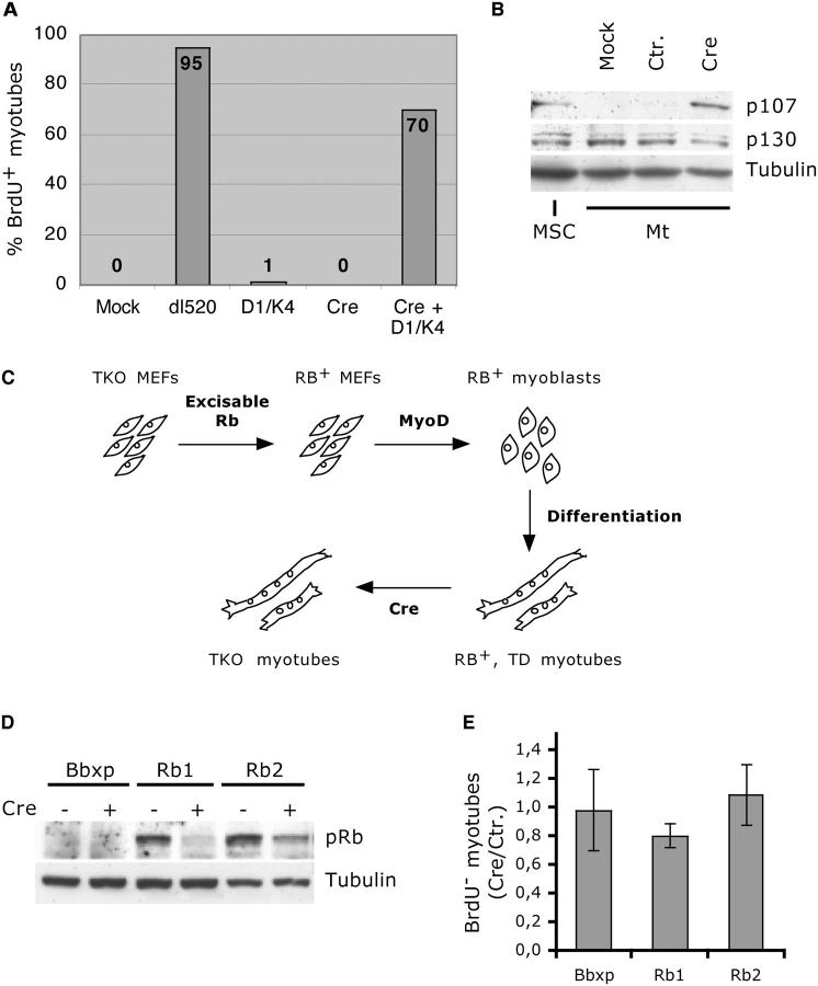
In skeletal muscle differentiation, the retinoblastoma protein (pRb) is absolutely necessary to establish definitive mitotic arrest. It is widely assumed that pRb is equally essential to sustain the postmitotic state, but this contention has never been tested. Here, we show that terminal proliferation arrest is maintained in skeletal muscle cells by a pRb-independent mechanism. Acute Rb excision from conditional knockout myotubes caused reexpression of E2F transcriptional activity, cyclin-E and -A kinase activities, PCNA, DNA ligase I, RPA, and MCM2, but did not induce DNA synthesis, showing that pRb is not indispensable to preserve the postmitotic state of these cells. Muscle-specific gene expression was significantly down-regulated, showing that pRb is constantly required for optimal implementation of the muscle differentiation program. Rb-deleted myotubes were efficiently reactivated by forced expression of cyclin D1 and Cdk4, indicating a functionally significant target other than pRb for these molecules. Finally, Rb removal induced no DNA synthesis even in pocket-protein null cells. Thus, the postmitotic state of myotubes is maintained by at least two mechanisms, one of which is pocket-protein independent.


Figures

Figure 1.
Figure 1.
pRb ablation in myotubes does not induce DNA synthesis. (A) PCR of myotubes infected with a control virus (Ctr.) or AdCre. Exon 19 denotes the amplification product obtained from undeleted cells; Δ Exon 19, Exon 19 deletion; MW, 100-bp ladder (Invitrogen); PCR performed as in Marino et al. (2000). (B) Western blot analysis of pRb in control- and AdCre-infected myotubes; decreasing amounts of total protein were loaded in the control lanes to assess blot sensitivity. Tubulin indicates loading control. (C and D) IF analysis of myotubes infected with the indicated viruses or derived from ΔRb-MSC.
Figure 2.
Figure 2.
Reactivation of cell cycle regulators in ΔRb-Mt. (A) Luciferase activity measured in MSCs stably transfected with the control (Basic) or E2F-responsive (6xE2F) reporter construct and derived myotubes (Mt) infected with the indicated viruses; data are averages of two independent experiments, with SDs, and are expressed as percentage of value obtained in 6xE2F-MSC. (B) DNA microarray analysis of MSCs and myotubes infected with control or Cre adenovirus; each line represents a distinct mRNA; values are averages of two independent experiments and are shown as percentages of measurements obtained in MSCs (see Table S1); white line indicates pRb mRNA. (C) Western blot analysis of the indicated proteins in MSCs or myotubes infected as shown, in the presence or absence of 5% FBS; HSP70 indicates loading control. (D) IF for MyHC and cyclin A on ΔRb-Mt. (E) Cyclin A and cyclin E kinase complexes were immunoprecipitated from MSCs or infected myotubes, reacted with histone H1 in the presence of γ-[32P]ATP, resolved by gel electrophoresis, and autoradiographed; Ctr. IgG indicates control-infected myotubes immunoprecipitated with normal rabbit IgG.
Figure 3.
Figure 3.
Reactivation of DNA synthesis effectors in ΔRb-Mt. (A) Western blot analysis of the indicated proteins in MSCs and infected myotubes. (B) Confocal microscopy of PCNA and BrdU IF in myotubes infected as indicated.
Figure 4.
Figure 4.
Roles of cyclin D1 and pocket proteins in controlling the postmitotic state. (A) BrdU incorporation in myotubes infected as indicated. (B) Western blot analysis of p107 and p130 in MSCs and infected myotubes. (C) Schematic of the generation of TKO myotubes. (D) Myotubes derived from TKO MEF infected with a control retrovirus (Bbxp) or from two independent populations infected with a retrovirus carrying a Cre-excisable Rb (Rb1 and Rb2) were treated with AdCre or a control adenovirus. Total proteins were extracted and Western blot analyzed. (E) Analysis of BrdU incorporation in the same myotube populations as in D. The results are averages and SDs of three independent experiments and are shown as the ratio of BrdU myotubes in Cre-infected (Rb) over control-infected (Rb+) populations.

References

    1. Berghella, L., L. De Angelis, M. Coletta, B. Berarducci, C. Sonnino, G. Salvatori, C. Anthonissen, R. Cooper, G.S. Butler-Browne, V. Mouly, et al. 1999. Reversible immortalization of human myogenic cells by site-specific excision of a retrovirally transferred oncogene. Hum. Gene Ther. 10:1607–1617. - PubMed
    1. Cheng, M., V. Sexl, C.J. Sherr, and M.F. Roussel. 1998. Assembly of cyclin D-dependent kinase and titration of p27Kip1 regulated by mitogen-activated protein kinase kinase (MEK1). Proc. Natl. Acad. Sci. USA. 95:1091–1096. - PMC - PubMed
    1. Cobrinik, D., M.H. Lee, G. Hannon, G. Mulligan, R.T. Bronson, N. Dyson, E. Harlow, D. Beach, R.A. Weinberg, and T. Jacks. 1996. Shared role of the pRB-related p130 and p107 proteins in limb development. Genes Dev. 10:1633–1644. - PubMed
    1. Crescenzi, M., S. Soddu, and F. Tato. 1995. Mitotic cycle reactivation in terminally differentiated cells by adenovirus infection. J. Cell. Physiol. 162:26–35. - PubMed
    1. Farkas, T., K. Hansen, K. Holm, J. Lukas, and J. Bartek. 2002. Distinct phosphorylation events regulate p130- and p107-mediated repression of E2F-4. J. Biol. Chem. 277:26741–26752. - PubMed

Publication types

MeSH terms