Comparative Study
. 2004 Nov 1;200(9):1135-43.
doi: 10.1084/jem.20041408.

Lysosomal fusion is essential for the retention of Trypanosoma cruzi inside host cells

Affiliations
Comparative Study

Lysosomal fusion is essential for the retention of Trypanosoma cruzi inside host cells

Luciana O Andrade et al. J Exp Med. .

Abstract

Trypomastigotes, the highly motile infective forms of Trypanosoma cruzi, are capable of infecting several cell types. Invasion occurs either by direct recruitment and fusion of lysosomes at the plasma membrane, or through invagination of the plasma membrane followed by intracellular fusion with lysosomes. The lysosome-like parasitophorous vacuole is subsequently disrupted, releasing the parasites for replication in the cytosol. The role of this early residence within lysosomes in the intracellular cycle of T. cruzi has remained unclear. For several other cytosolic pathogens, survival inside host cells depends on an early escape from phagosomes before lysosomal fusion. Here, we show that when lysosome-mediated T. cruzi invasion is blocked through phosophoinositide 3-kinase inhibition, a significant fraction of the internalized parasites are not subsequently retained inside host cells for a productive infection. A direct correlation was observed between the lysosomal fusion rates after invasion and the intracellular retention of trypomastigotes. Thus, formation of a parasitophorous vacuole with lysosomal properties is essential for preventing these highly motile parasites from exiting host cells and for allowing completion of the intracellular life cycle.


Figures
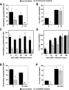
Figure 1.
Figure 1.
The loss of intracellular T. cruzi after inhibition of lysosome-mediated entry is inversely correlated with the kinetics of intracellular lysosomal fusion. MEFs were pretreated or not with wortmannin or Ly294002, infected with T. cruzi, fixed or further incubated for the indicated periods of time, and processed for immunofluorescent detection of intracellular parasites. (A) Intracellular parasites at 0 or 5 h after infection of cells pretreated or not with wortmannin. (B) Percentage of intracellular parasites associated with the lysosomal marker Lamp1 at 0 and 5 h after infection of cells pretreated or not with wortmannin. (C) The number of intracellular parasites at increasing periods after infection. (D) Percentage of intracellular parasites associated with Lamp1 at increasing periods after infection. (E) Intracellular parasites at 0 or 40 min after infection of cells treated or not with Ly294002. (F) Percentage of intracellular parasites associated with the lysosomal marker Lamp1 at 0 and 40 min after infection of cells treated or not with Ly294002. The data in A–F correspond to the mean of triplicates ± SD. Asterisks indicate statistically significant differences (P < 0.05, Student's t test) between wortmannin-treated and untreated cells at the 0 h time point (*) or between wortmannin-treated cells at 0 h and at other time points (**).
Figure 2.
Figure 2.
(A) Treatment of T. cruzi with a PI 3-kinase inhibitor does not affect the number of intracellular parasites over time. Trypomastigotes were pretreated with wortmannin before infection of MEFs, and the number of internalized parasites was determined at 0 and 2 h after infection. (B) Inhibition of MEF PI 3-kinase does not lead to MEF loss. MEFs were pretreated or not with wortmannin, infected with T. cruzi, fixed or further incubated for 4 h, and stained with DAPI, and the total number of MEFs in 60 microscopic fields (100×) was determined. (C–F) Loss of parasites after wortmannin treatment is also observed in other cell types. L6E9 (C and E) and CHO cells (D and F) were pretreated or not with wortmannin and infected with T. cruzi, and the number of internalized parasites (C and E) as well as the percentage of those parasites associated with Lamp1 (D and F) at 1 h after infection were determined. The data in A–F correspond to the mean of triplicates ± SD. Asterisks indicate statistically significant differences (P < 0.05, Student's t test) between wortmannin-treated and untreated cells at the 0 h time point (*) or between wortmannin-treated cells at 0 h and at other time points (**).
Figure 3.
Figure 3.
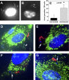
PI 3-kinase inhibition does not cause intracellular death, but leads to reversal of T. cruzi invasion. (A) Intact trypomastigotes inside a MEF 1 h after the cells were pretreated with wortmannin. The arrows point to the normal nuclear morphology. (B) Intracellularly degraded trypomastigote 24 h after infection of a macrophage activated with IFNγ/LPS. The arrow points to remnants of the parasite's nucleus; the altered, enlarged kinetoplast is adjacent. (C) The number of partially internalized trypomastigotes after infection of MEFs treated or not with wortmannin. The data correspond to the mean of triplicates ± SD. (D) Untreated MEFs with one extracellularly attached trypomastigote (red) and one completely internalized trypomastigote (arrowhead, green). (E, F, and G) Wortmannin-treated MEFs with partially internalized trypomastigotes (the arrows point to internalized portions, not stained by the extracellularly added anti–T. cruzi antibody). Lysosomes were stained with anti-Lamp1 antibodies (green), extracellular trypomastigotes with anti–T. cruzi antibodies (red), and MEF and trypomastigote DNA with DAPI (blue).
Figure 4.
Figure 4.
Disruption of the cortical actin cytoskeleton also leads to reversal of T. cruzi invasion. (A) The number of intracellular parasites in cytochalasin D–treated MEFs at 0 and 1 h after infection. (B) Percentage of intracellular parasites associated with Lamp1 in cytochalasin D–treated MEFs. (C) The number of intracellular parasites in cytochalasin D–treated CHO cells. (D) Percentage of Lamp1-associated parasites in cytochalasin D–treated CHO cells. Cells were pretreated or not with cytochalasin D, infected with T. cruzi, fixed or incubated for the indicated periods of time, and processed for immunofluorescence and detection of intracellular parasites. The data in A–D correspond to the mean of triplicates ± SD. Asterisks indicate statistically significant differences (P < 0.05, Student's t test) between wortmannin-treated and untreated cells at the 0 h time point (*) or between wortmannin-treated cells at 0 h and at other time points (**).
Figure 5.
Figure 5.
In the absence of lysosomal fusion, trypomastigotes exit host cells and reinfect at later time points. (A) Intracellular parasites at increasing periods after infection. (B) Percentage of intracellular parasites associated with Lamp1 at increasing periods after infection. MEFs were pretreated or not with wortmannin, infected with T. cruzi, and washed, and the number of intracellular parasites was determined. (C) The total number of MEFs in 60 microscopic fields (100×) was determined for the same experiment shown in A and B. The data in A–C correspond to the mean of triplicates ± SD. Asterisks indicate statistically significant differences (P < 0.05, Student's t test) between wortmannin-treated and untreated cells at the 0 h time point (*) or between wortmannin-treated cells at 0 h and at other time points (**). (D) PI 3-kinase activity in untreated or wortmannin-treated MEFs. (1) Negative control (no cell lysate). (2) MEF lysates immediately after wortmannin treatment. (3) MEF lysates 24 h after wortmannin treatment. (4) MEF lysates not treated with wortmannin. White lines indicate that intervening lanes have been spliced out.
Figure 6.
Figure 6.
Intracellular parasites found at later time points in wortmannin-treated cells correspond to recent reinternalization events. MEFs were pretreated or not with wortmannin, infected with T. cruzi, washed to remove extracellular parasites, reincubated for 24 h, and processed for immunofluorescence. (A) After 24 h, only cytoplasmic amastigotes are detected in untreated cells (arrowheads). (B–D) In cells pretreated with wortmannin, recently internalized trypomastigotes inside Lamp1 positive vacuoles are also present (arrows). Lysosomes were stained with anti-Lamp1 antibodies (green), extracellular trypomastigotes with anti–T. cruzi antibodies (red), and MEF and T. cruzi DNA with DAPI (blue). (E) After 24 h, all intracellular parasites in untreated cells express the amastigote-specific surface antigen Ssp-4 (arrowheads). (F) In cells pretreated with wortmannin, most intracellular parasites still do not express Ssp-4, confirming that they have recently entered the cells (arrows). Staining with the Ssp-4–specific mAbs is shown (green), as is MEF and T. cruzi DNA staining with DAPI (blue).

References

    1. Chagas, C. 1909. Nova trypanozomiaze humana. Mem. Inst. Oswaldo Cruz. 1:11–80.
    1. Brener, Z. 1973. Biology of Trypanosoma cruzi. Annu. Rev. Microbiol. 27:347–383. - PubMed
    1. Tardieux, I., P. Webster, J. Ravesloot, W. Boron, J.A. Lunn, J.E. Heuser, and N.W. Andrews. 1992. Lysosome recruitment and fusion are early events required for trypanosome invasion of mammalian cells. Cell. 71:1117–1130. - PubMed
    1. Rodriguez, A., M.G. Rioult, A. Ora, and N.W. Andrews. 1995. A trypanosome-soluble factor induces IP3 formation, intracellular Ca2+ mobilization and microfilament rearrangement in host cells. J. Cell Biol. 129:1263–1273. - PMC - PubMed
    1. Rodriguez, A., E. Samoff, M.G. Rioult, A. Chung, and N.W. Andrews. 1996. Host cell invasion by trypanosomes requires lysosomes and microtubule/kinesin-mediated transport. J. Cell Biol. 134:349–362. - PMC - PubMed

Publication types

MeSH terms