Comparative Study
. 2004 Nov 15;18(22):2785-97.
doi: 10.1101/gad.1247904. Epub 2004 Nov 1.

Mitochondrial fission proteins regulate programmed cell death in yeast

Affiliations
Comparative Study

Mitochondrial fission proteins regulate programmed cell death in yeast

Yihru Fannjiang et al. Genes Dev. .

Abstract

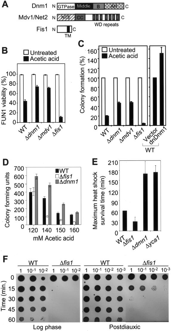
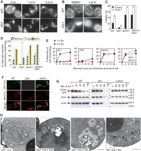
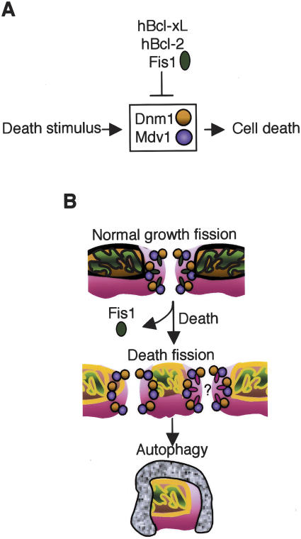
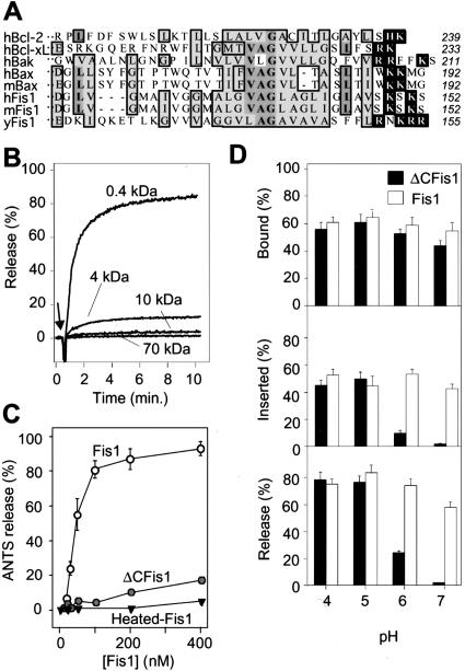
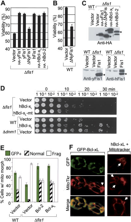
The possibility that single-cell organisms undergo programmed cell death has been questioned in part because they lack several key components of the mammalian cell death machinery. However, yeast encode a homolog of human Drp1, a mitochondrial fission protein that was shown previously to promote mammalian cell death and the excessive mitochondrial fragmentation characteristic of apoptotic mammalian cells. In support of a primordial origin of programmed cell death involving mitochondria, we found that the Saccharomyces cerevisiae homolog of human Drp1, Dnm1, promotes mitochondrial fragmentation/degradation and cell death following treatment with several death stimuli. Two Dnm1-interacting factors also regulate yeast cell death. The WD40 repeat protein Mdv1/Net2 promotes cell death, consistent with its role in mitochondrial fission. In contrast to its fission function in healthy cells, Fis1 unexpectedly inhibits Dnm1-mediated mitochondrial fission and cysteine protease-dependent cell death in yeast. Furthermore, the ability of yeast Fis1 to inhibit mitochondrial fission and cell death can be functionally replaced by human Bcl-2 and Bcl-xL. Together, these findings indicate that yeast and mammalian cells have a conserved programmed death pathway regulated by a common molecular component, Drp1/Dnm1, that is inhibited by a Bcl-2-like function.


Figures

Figure 1.
Figure 1.
Inhibition of programmed cell death by yeast Fis1p. (A) Cell viability was determined by FUN-1 staining at 4 h after treatment with 130 mM acetic acid (AA) or with 2.5 mM H2O2, and data are presented as mean ± se for five (wild type, Δfis1) or two (Δom45) independent experiments counting at least 200 cells per sample. One of the two experiments with Δom45 was done on BY4741 background strain; the other was on an undefined background. (B) Representative images of wild-type (WT) and Δfis1 yeast cells stained with FUN1 dye following treatment with 2.5 mM H2O2 for 4 h or untreated controls. The cell wall stains blue and viable metabolically active yeast contain a red vacuole, while nonviable yeast lack a red vesicle and stain green/yellow. (C) Viability was also determined by trypan blue exclusion. Data are presented as mean ± standard error (se) for three independent experiments counting at least 200 cells per sample. (D) Viability by FUN-1 staining was performed using yeast on RJ1365 background. Data presented are the means ± se for three independent experiments. (B–D) P < 0.001 comparing wild type (WT) and Δfis1 mutants.
Figure 2.
Figure 2.
Fis1 inhibits phosphatidylserine exposure and caspase-dependent cell death. (A) The same field of wild-type and Δfis1 spheroplasts are shown with phase contrast (left), annexin V staining (middle, green), and propidium iodide staining (right, red) at 4 h after treatment with 2.5 mM H2O2. (B) Viability was assessed by annexin V staining of yeast spheroplasts and presented as mean ± se for five fields (>150 cells) per sample in two independent experiments. (C) Cell viability determined by FUN-1 staining of the indicated yeast mutants (BY4741) treated with 2.5 mM H2O2 or 130 mM acetic acid for 4 h is presented ± se for three independent experiments. (D) Viability of the indicated yeast mutants (BY4741) treated with 2.5 mM H2O2 or 130 mM acetic acid in the presence of DEVD-CHO or DMSO carrier control was determined by FUN-1 staining in three independent experiments. (B–D) *P < 0.001 comparing mutants to wild type. (E) Dose response assay as described for panel D.
Figure 3.
Figure 3.
Fis1p and Dnm1p regulate yeast cell death. (A) Diagram of yeast protein domains; GTPase effector domain (GED), N-terminal extension (NTE), coiled coil (CC), and transmembrane (TM) were adapted from Shaw and Nunnari (2002) (© 2002, with permission from Elsevier). (B) Cell viability was also determined by viable dye FUN-1 staining as shown in Figure 1B, and data are presented as mean ± se for three independent experiments counting at least 200 cells per sample. (C) Wild-type (WT) and the mutant yeast cells (on BY4741 background strain), or cells overexpressing Dnm1(K41A) (provided by R. Jensen, Department of Cell Biology, Johns Hopkins School of Medicine, Baltimore, MD) were treated with 190 mM acetic acid for 4 h and cell viability was assessed by colony formation in three independent experiments for each assay. (B,C) The differences between all mutants and wild type (WT) are statistically significant with P < 0.001. (D) Dose response for acetic acid treatment was performed in a 96-well format for more than six independent determinations. (E) Survival of wild-type (WT) and mutant yeast (BY4741) treated with heat shock at 55°C, and the maximum heat shock survival time was determined by growth on YPD plates. Data are presented as mean ± se for five independent experiments. (F) Survival of log phase or postdiauxic yeast (BY4741) treated with heat shock at 55°C for the indicated times was determined by growth on YPD plates. A representative of three independent experiments is shown.
Figure 4.
Figure 4.
Mitochondrial fission during cell death does not require Fis1. (A) Fluorescence microscopy showing mitochondrial morphology of wild-type (WT) and Δfis1 yeast (BY4741) treated with 100 mM acetic acid for the indicated times. Mitochondria are visualized with Cox4-GFP (Sesaki and Jensen 1999). (B) Mitochondrial morphology of yeast treated with 250 μM latrunculin A (Molecular Probes) or the carrier DMSO for 1 h as described in B except without a death stimulus. (C) Wild-type (WT) and the mutant yeast cells (on BY4741 background strain) were treated with 190 mM acetic acid for 4 h and cell viability was assessed by colony formation and FUN-1 staining in three independent experiments for each assay. Data are presented as mean ± se for three independent experiments counting at least 200 cells per sample. (D) Percent of yeast cells with the indicated mitochondria morphologies (differences for all criteria between wild type [WT], Δfis1, and Δdnm1 are significant; P < 0.001). No significant differences were detected in the percent GFP-positive yeast between genotypes prior to acetic acid treatment; wild type (82%), Δfis1 (78%), Δdnm1 (76%). (C,D) P < 0.001 comparing wild type and Δfis1 mutants treated with acetic acid. (E) The indicated strains of yeast were treated for 1 h or 4 h with 130 mM acetic acid, and mitochondrial morphology (COS4-GFP) was scored at the indicated times after removal of acetic acid. (F) Fluorescence microscopy of yeast treated with 130 mM acetic acid for 20 min, resuspended in water, and stained with 0.5 μM MitoTracker. (G) Immunoblot analyses of yeast cell lysates prepared at the indicated times after treatment with 130 mM acetic acid. (H) Electron microscopy of Δfis1 and wild-type (WT) yeast treated with 100 mM acetic acid for the indicated times. (v) Vacuole; (m) mitochondrion; (arrowheads) fission sites; (n) nucleus; (arrows) association of mitochondria with vacuole. Bar, 0.5 μm.
Figure 7.
Figure 7.
Model of yeast cell death pathway. (A) Yeast Fis1, human Bcl-2, and Bcl-xL inhibit yeast cell death mediated by Dnm1. (B) Following a death stimulus, Fis1 may be inactivated, leading to uncontrolled/irreversible Dnm1-mediated mitochon-drial fragmentation, degradation by vacuoles, and cell death. Another possibility is that yeast Fis1 is replaced by another yet unidentified factor that mediates mitochondrial fission during cell death (red ovals).
Figure 5.
Figure 5.
Yeast Fis-1 releases small molecules from synthetic lipid vesicles. (A) Amino acid sequences of the C-terminal ends of Fis1 and Bcl-2 proteins from the indicated organisms: human (h), murine (m), and S. cerevisiae (y). Bold letters/medium shade indicate identity, light shade indicates similarity in >50% of entries; white letters mark basic residues on the C-terminal side of the hydrophobic region. Amino acid number for last residue of each protein is shown at right. (B) Release of the dye ANTS/DPX, (∼0.4 kDa), or FITC-labeled dextrans of the indicated molecular weights from synthetic lipid vesicles (25 μM lipid [1/250 mol protein/mol lipid]) at pH 7 was determined in three independent experiments on a fluorimeter following addition of 100 nM recombinant yeast Fis1 protein purified from Escherichia coli. (C) Release of ANTS/DPX at pH 7 from synthetic vesicles treated with the indicated concentrations of recombinant yeast Fis1 or ΔCFis1 (lacking the C-terminal 27 amino acids) and presented are mean ± se for four independent experiments. The fraction of recombinant yeast Fis1 proteins bound to lipid vesicles at the indicated pH conditions was determined before (D, top) or after (D, middle) alkali extraction. Release of ANTS/DPX was determined as described for A at the indicated pH conditions. Data presented are mean ± se for three to five independent experiments.
Figure 6.
Figure 6.
Human Bcl-2 and Bcl-xL rescue the cell death phenotype of Δfis1 mutant yeast. (A,B) Viability of Δfis1 and wild-type yeast (RJ1365) expressing the indicated N-terminal HA-tagged or untagged human (h) or yeast (y) proteins from GAL1 expression vectors was determined by trypan blue staining 6 h after treatment with 100 mM acetic acid. Data are presented as ±se for seven independent experiments; P < 0.001. (C) Immunoblot analyses of the yeast strains shown in panels A and B using anti-Fis1 rabbit sera or anti-HA antibody. (D) Yeast strains constitutively expressing the indicated proteins via a CEN plasmid were analyzed for survival following heat shock treatment as described in Figure 3E, except that samples were plated on minimal medium for selection, resulting in smaller viability differences between strains. A representative of three to five experiments is shown. All samples shown were from the same experiment, but the lanes were rearranged for presentation. (E) Percent of yeast cells transformed with the indicated plasmids with the indicated mitochondria morphology and protein expression was induced with galactose. Data presented are for three to five independent experiments; P < 0.01 for Bcl-xL, and P = 0.03 for Bcl-2 compared with wild type. (F) Yeast stably expressing GFP-Bcl-xL via a CEN plasmid were stained with MitoTracker and examined by fluorescence microscopy.

References

    1. Amarante-Mendes G.P., Finucane, D.M., Martin, S.J., Cotter, T.G., Salvesen, G.S., and Green, D.R. 1998. Anti-apoptotic oncogenes prevent caspase-dependent and independent commitment for cell death. Cell Death Differ. 5: 298-306. - PubMed
    1. Basañez G., Zhang, J., Chau, B.N., Maksaev, G.I., Frolov, V., Brandt, T.A., Burch, J., Hardwick, J.M., and Zimmerberg, J. 2001. Pro-apoptotic cleavage products of Bcl-xL form cytochrome c-conducting pores in pure lipid bilayers. J. Biol. Chem. 276: 31083-31091. - PubMed
    1. Breckenridge D.G., Stojanovic, M., Marcellus, R.C., and Shore, G.C. 2003. Caspase cleavage product of BAP31 induces mitochondrial fission through endoplasmic reticulum calcium signals, enhancing cytochrome c release to the cytosol. J. Cell Biol. 160: 1115-1127. - PMC - PubMed
    1. Brunet C.L., Gunby, R.H., Benson, R.S., Hickman, J.A., Watson, A.J., and Brady, G. 1998. Commitment to cell death measured by loss of clonogenicity is separable from the appearance of apoptotic markers. Cell Death Differ. 5: 107-115. - PubMed
    1. Cerveny K.L. and Jensen, R.E. 2003. The WD-repeats of Net2p interact with Dnm1p and Fis1p to regulate division of mitochondria. Mol. Biol. Cell 14: 4126-4139. - PMC - PubMed

Publication types

MeSH terms