. 2004 Nov 1;18(21):2627-38.
doi: 10.1101/gad.1241904.

The Polycomb Ezh2 methyltransferase regulates muscle gene expression and skeletal muscle differentiation

Affiliations

The Polycomb Ezh2 methyltransferase regulates muscle gene expression and skeletal muscle differentiation

Giuseppina Caretti et al. Genes Dev. .

Erratum in

  • Genes Dev. 2005 Mar 15;19(6):768

Abstract

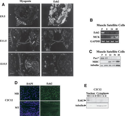
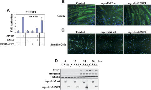
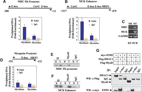
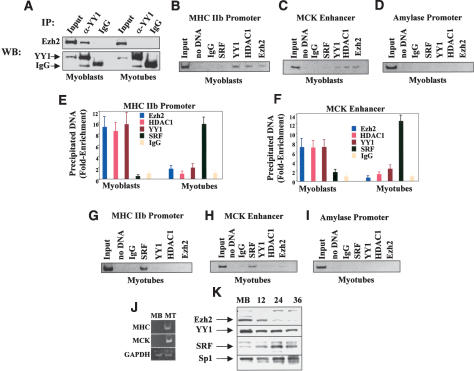
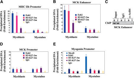
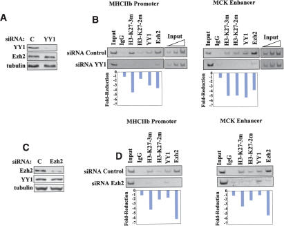
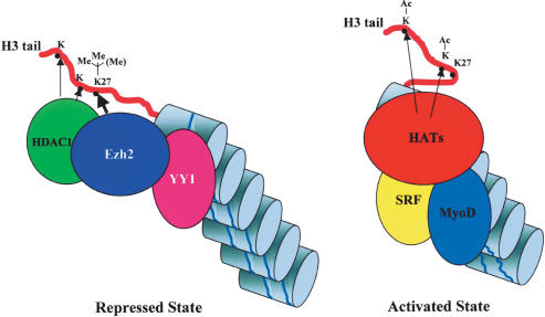
The Ezh2 protein endows the Polycomb PRC2 and PRC3 complexes with histone lysine methyltransferase (HKMT) activity that is associated with transcriptional repression. We report that Ezh2 expression was developmentally regulated in the myotome compartment of mouse somites and that its down-regulation coincided with activation of muscle gene expression and differentiation of satellite-cell-derived myoblasts. Increased Ezh2 expression inhibited muscle differentiation, and this property was conferred by its SET domain, required for the HKMT activity. In undifferentiated myoblasts, endogenous Ezh2 was associated with the transcriptional regulator YY1. Both Ezh2 and YY1 were detected, with the deacetylase HDAC1, at genomic regions of silent muscle-specific genes. Their presence correlated with methylation of K27 of histone H3. YY1 was required for Ezh2 binding because RNA interference of YY1 abrogated chromatin recruitment of Ezh2 and prevented H3-K27 methylation. Upon gene activation, Ezh2, HDAC1, and YY1 dissociated from muscle loci, H3-K27 became hypomethylated and MyoD and SRF were recruited to the chromatin. These findings suggest the existence of a two-step activation mechanism whereby removal of H3-K27 methylation, conferred by an active Ezh2-containing protein complex, followed by recruitment of positive transcriptional regulators at discrete genomic loci are required to promote muscle gene expression and cell differentiation.


Figures

Figure 1.
Figure 1.
Ezh2 is expressed early in the myotomal compartment of developing somites and in undifferentiated skeletal myoblasts. (A) Myogenin (left panels) and Ezh2 (right panels) transcripts were analyzed by in situ RNA hybridization on sections from E9.5, E11.5, and E15.5 mouse embryos. In a sagittal section at E9.5, myogenin mRNA was detected in myotomes of the developing somites (s). (nt) Neural tube; (t) tail; (ov) otic vesicle; (ba) branchial arch; (h) heart. Ezh2 mRNA was detected in all structures at a high level. In transverse sections at E11.5, myogenin was detected in developing myotomes (myo; arrows) and at a low level in limb buds (lb). (li) Liver; (h) heart; (nt) neural tube. At E11.5, Ezh2 mRNA levels have decreased significantly from E9.5. Ezh2 was detected in neural tube, liver, limb buds, and paraxial mesoderm. In a transverse section at the level of the jaw (j) at E15.5, myogenin mRNA was detected in tongue (to) muscle, shoulder muscle (shm), pectoral muscle (pm), and all other skeletal muscle. (sg) Salivary gland. At E15.5, Ezh2 mRNAs were only detected in thymus (th). (B) Mouse primary muscle satellite cells were cultured in either growth conditions (P, proliferating) or, once confluent, induced to differentiate for 0, 24, and 48 h. The RNA was isolated and RT-PCR was performed with specific primers for Ezh2, MCK, and GAPDH. (C) Immunoblot of muscle satellite cell extracts with Pax7, MHC, and tubulin antibodies at different differentiation stages. (D) C2C12 proliferating myoblasts (MB) and differentiated myotubes (MT) were immunostained for Ezh2 and nuclei visualized with DAPI. (E) Nuclear and cytoplasmic extracts of proliferating myoblasts (MB) and myoblasts at different stages of differentiation (12, 36, and 48 h in differentiation medium) were fractionated and immunoblotted for Ezh2 and tubulin.
Figure 2.
Figure 2.
The SET domain of Ezh2 is required to inhibit muscle gene expression and differentiation. (A) Luciferase assay conducted on NIH 3T3 fibroblasts transfected with the MCK-luc reporter and expression vectors for MyoD, Ezh2, and Ezh2ΔSET mutant and cultured in differentiation medium for 48 h. (B) C2C12 myoblasts were transduced with control, Ezh2 wild-type, or Ezh2ΔSET mutant retroviruses and induced to differentiated for 36 h before immunostaining for MHC. DAPI reveals the nuclei. (C) Mouse primary muscle satellite cells were transduced with control, Ezh2 wild-type, or Ezh2ΔSET mutant retroviruses, induced to differentiate for 36 h, and immunostained for MHC. DAPI reveals the nuclei. (D) Immunoblot of extracts derived from myoblasts transduced with control (C), Ezh2 wild-type (E), or Ezh2ΔSET mutant (EΔ) retroviruses at different stages of differentiation (confluent in growth medium [0]; 12, 24, 36 h in differentiation medium) probed with MHC, myogenin, tubulin, and myc (to detect myc-tagged Ezh2 and Ezh2ΔSET mutant proteins) antibodies. Cells used in B and D belong to the same polyclone and have been cultured within the same experiment.
Figure 3.
Figure 3.
Ezh2 is present at the chromatin regulatory regions of two transcriptionally inactive muscle genes and is no longer detected upon gene activation. (A,B) ChIP with either Ezh2 antibodies or control IgG was performed on chromatin derived from either undifferentiated myoblasts (MB) or differentiated myotubes (MT). The precipitated DNA fragments were amplified with specific oligonucleotides spanning the indicated regulatory regions for the MHCIIb promoter (A), MCK enhancer (B), or myogenin promoter (D) by real-time PCR. (C) Transcripts of MHCIIb, MCK, and GAPDH in either myoblast (MB) or myotubes (MT) were detected by RT-PCR analysis. (E,F) ChIP with either MyoD antibodies or control IgG on chromatin of myoblast (B) or myotubes (T) of MHCIIb promoter (E) and MCK enhancer (F). (G) 293T cells were transfected with expression vectors for Ezh2, HDAC1, and MyoD in different combinations. Cell extracts were prepared and immunoprecipitated with anti-myc antibodies, and the precipitated proteins were revealed with anti-Flag antibodies. Nonspecific IgG was used as the control.
Figure 4.
Figure 4.
Ezh2, HDAC1, and YY1 occupy the chromatin of transcriptionally inactive muscle regulatory regions while MyoD and SRF are engaged upon gene activation. (A) Cell extracts from undifferentiated myoblasts or differentiated myotubes were immunoprecipitated with either YY1 antibodies or control IgG and the precipitated material was immunoblotted with Ezh2 antibodies. (B-D) ChIP with antibodies to Ezh2, HDAC1, YY1, and SRF on chromatin obtained from myoblasts. Reactions with no input DNA (no DNA) or ChIP with nonspecific IgG were performed as controls. Precipitated DNA was amplified by PCR with primers for the MHCIIb promoter (B), MCK enhancer (C), or amylase promoter (D). (E,F) ChIP was performed as described in B and C using chromatin derived from either myoblasts or myotubes. The precipitated DNA was amplified by real-time PCR with specific primers for either MHCIIb promoter (E) or MCK enhancer (F). (G-I) ChIP was performed as described in B-D except that chromatin was obtained from differentiated myotubes. (J) Transcripts of MHCIIb, MCK, and GAPDH in either myoblast (MB) or myotubes (MT) were detected by RT-PCR analysis. (K) Immunoblot of extracts derived from myoblasts at different stages of differentiation probed with antibodies against Ezh2, YY1, SRF, and Sp1.
Figure 5.
Figure 5.
H3-K27 methylation at muscle regulatory regions in undifferentiated myoblasts and differentiated myotubes. (A,B) ChIP with control IgG, Ezh2, H3-K27 α-trimethylated (3m), and α-dimethylated (2m) antibodies on chromatin obtained from myoblasts or myotubes. The precipitated DNA was amplified by real-time PCR using specific primers for the MHCIIb promoter (A) or MCK enhancer (B). (C) ChIP with either control IgG or anti-acetylated H4 antibodies with the chromatin obtained from myoblasts (B) or myotubes (T) on the MCK enhancer. (D) ChIP was performed as described in A and B with specific primers for the MCK promoter—rather than enhancer—were used. (E) ChIP was performed as described in A and B with specific primers for the myogenin promoter.
Figure 6.
Figure 6.
Efficient chromatin recruitment of Ezh2 and H3-K27 methylation require the presence of YY1. (A) Skeletal myoblasts were transfected with either control (FITC-labeled siRNA; see Materials and Methods) or YY1-specific siRNA, and their extracts were immunoblotted with antibodies to YY1, Ezh2, and tubulin. (B) ChIP assays were performed with control IgG or antibodies to trimethyl, dimethyl H3-K27, YY1, and Ezh2 on the chromatin obtained from cells transfected with either control or YY1 siRNA. The immunoprecipitated chromatin was analyzed by PCR using primers specific for the MHCIIb promoter and MKC enhancer. Three dilutions of the total input are indicated. Quantitation of reduced chromatin binding was performed by scanning the intensity of the individual bands with the NIH Image software and dividing the values obtained for siRNA Control bands for those obtained for the corresponding siRNA YY1 bands. Each value was corrected for the ratio of the two input DNAs (Input DNA siRNA Control/Input siRNA YY1). (C) Skeletal myoblasts were transfected with either control or Ezh2-specific siRNA, and their extracts were immunoblotted with antibodies against Ezh2, YY1, and tubulin. (D) ChIP assays were performed with control IgG and antibodies to trimethyl, dimethyl H3-K27, YY1, and Ezh2 on the chromatin obtained from cells transfected with either control or Ezh2 siRNA. The immunoprecipitated chromatin was analyzed by PCR using primers specific for the MHCIIb promoter and MCK enhancer. Quantitation was performed as described in B. Each value was corrected for the ratio of the two input DNAs (Input DNA siRNA Control/Input siRNA Ezh2).
Figure 7.
Figure 7.
Two-step activation model of muscle gene expression. Regulatory regions of certain muscle-specific genes are occupied by a protein complex containing the DNA-binding protein YY1, the methyltransferase Ezh2, and the deacetylase HDAC1. Deacetylation of lysine residues by HDAC1 and di-/trimethylation of H3-K27 by Ezh2 actively prevent transcription (repressed state). At the onset of transcriptional activation, YY1 is displaced from the chromatin—with consequent loss of Ezh2 and HDAC1—and replaced by SRF. H3-K27 becomes hypomethylated, and loading of the MyoD family of transcription factors allows engagement of histone acetyltransferases (HATs) and permits initiation of transcription (activated state).

References

    1. Bannister A.J., Schneider, R., and Kouzarides, T. 2002. Histone methylation: Dynamic or static? Cell 109 801-806. - PubMed
    1. Bergstrom D.A. and Tapscott, S.J. 2001. Molecular distinction between specification and differentiation in the myogenic basic helix-loop-helix transcription factor family. Mol. Cell. Biol. 21 2404-2412. - PMC - PubMed
    1. Bergstrom D.A., Penn, B.H., Strand, A., Perry, R.L., Rudnicki, M.A., and Tapscott, S.J. 2002. Promoter-specific regulation of MyoD binding and signal transduction cooperate to pattern gene expression. Mol. Cell 9 587-600. - PubMed
    1. Bracken A.P., Pasini, D., Capra, M., Prosperini, E., Colli, E., and Helin, K. 2003. EZH2 is downstream of the pRB-E2F pathway, essential for proliferation and amplified in cancer. EMBO J. 22 5323-5335. - PMC - PubMed
    1. Brown J.L., Mucci, D., Whiteley, M., Dirksen, M.L., and Kassis, J.A. 1998. The Drosophila Polycomb group gene pleiohomeotic encodes a DNA binding protein with homology to the transcription factor YY1. Mol. Cell 1 1057-1064. - PubMed

MeSH terms