. 2004 Nov;14(11):2209-20.
doi: 10.1101/gr.2746604.

Serial segmental duplications during primate evolution result in complex human genome architecture

Affiliations

Serial segmental duplications during primate evolution result in complex human genome architecture

Pawełl Stankiewicz et al. Genome Res. 2004 Nov.

Abstract

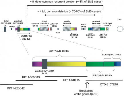
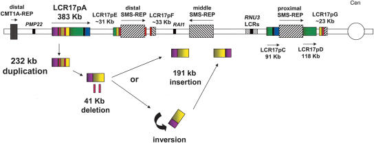
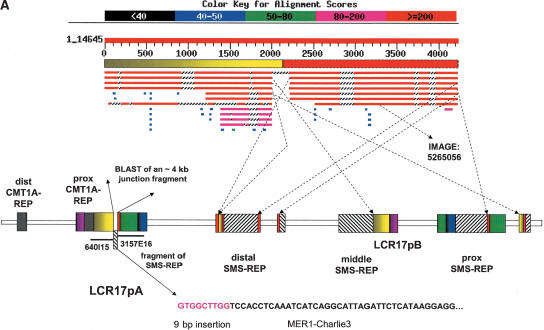
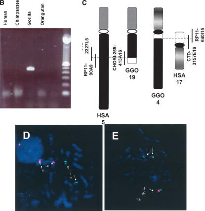
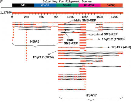
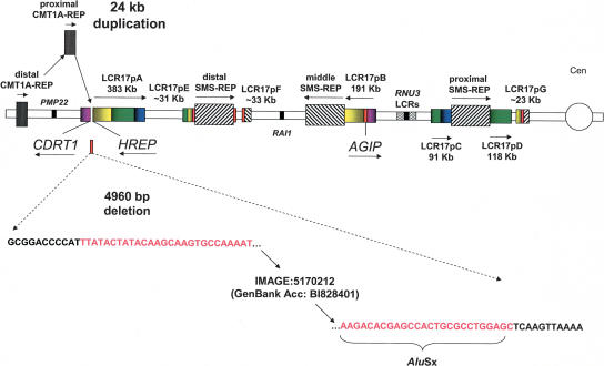
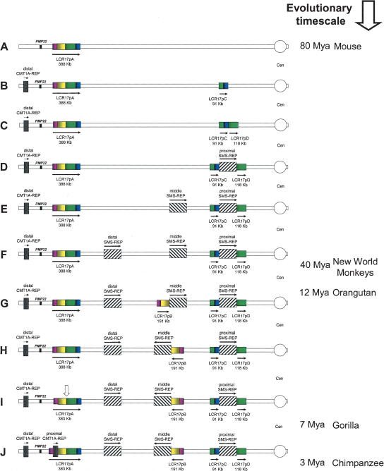
The human genome is particularly rich in low-copy repeats (LCRs) or segmental duplications (5%-10%), and this characteristic likely distinguishes us from lower mammals such as rodents. How and why the complex human genome architecture consisting of multiple LCRs has evolved remains an open question. Using molecular and computational analyses of human and primate genomic regions, we analyzed the structure and evolution of LCRs that resulted in complex architectural features of the human genome in proximal 17p. We found that multiple LCRs of different origins are situated adjacent to one another, whereas each LCR changed at different time points between >25 to 3-7 million years ago (Mya) during primate evolution. Evolutionary studies in primates suggested communication between the LCRs by gene conversion. The DNA transposable element MER1-Charlie3 and retroviral ERVL elements were identified at the breakpoint of the t(4;19) chromosome translocation in Gorilla gorilla, suggesting a potential role for transpositions in evolution of the primate genome. Thus, a series of consecutive segmental duplication events during primate evolution resulted in complex genome architecture in proximal 17p. Some of the more recent events led to the formation of novel genes that in human are expressed primarily in the brain. Our observations support the contention that serial segmental duplication events might have orchestrated primate evolution by the generation of novel fusion/fission genes as well as potentially by genomic inversions associated with decreased recombination rates facilitating gene divergence.


Figures

Figure 1.
Figure 1.
Schematic diagram of the LCR-rich human proximal chromosome 17p. (Top) Depiction of proximal chromosome 17p showing the position and orientation of LCRs. The LCR17p structures are depicted as rectangles with colors signifying shared homology and horizontal arrows showing relative orientation. (Bottom) Magnification of the LCR17pA structure with four BAC clones covering the region. The gorilla translocation breakpoint is indicated by an open vertical arrow between BAC clones RP11–640I15 and CTD-3157E15 and LCR17pA/B and LCR17pA/D subunits of the LCR17pA copy. The LCR17pE, LCR17pF, and LCR17pG have been described elsewhere (Stankiewicz et al. 2003). The LCRs that are >20 Kb are depicted.
Figure 2.
Figure 2.
Evolutionary FISH analyses of LCR17ps. Interphase FISH with the LCR17pA/C- and LCR17pA/D-derived BAC clone CTD-3157E16 showed the presence of at least 2 signals on each chromosome for human (A), orangutan (B), and baboon (C). LCR17pA/B-derived clone RP11–640I15 showed four signals (green or red), two from each chromosome, in interphase nuclei of cell lines from human (D), chimpanzee (E), gorilla (F) [the separation of the signals is due to the presence of t(4;19) with LCR17pA/B and LCR17pB being present on separate derivative chromosomes]. Only one signal per chromosome homolog was observed in orangutan (G), baboon (H), and squirrel monkey (I). (G) The cohybridization of BAC clones RP11–640I15 (red) and, adjacent to it on the telomeric side, LCR-free RP11–726O12 (green) demonstrated that the LCR17pA/B was the progenitor copy (see Results).
Figure 3.
Figure 3.
Insertional duplication of LCR17pB. After the divergence of gorilla and orangutan, the LCR17pA/B portion of LCR17pA was duplicated and inserted near middle SMS-REP to give LCR17pB (pink and yellow). The comparison between LCR17pA and LCR17pB copies reveals that two DNA segments (shown as red rectangles) were deleted and are not present in the LCR17pB copy. They were lost during the duplication event or after the LCR17pB insertion. The LCR17pB segment was either inserted directly adjacent to the middle SMS-REP on the telomeric side (and subsequently inverted together with the middle SMS-REP; Fig. 6) or inverted and inserted directly adjacent on its centromeric edge (see Discussion).
Figure 4.
Figure 4.
(A) A proposed model of the gorilla evolutionary translocation t(4;19). FISH results showed that the GGO 19 breakpoint maps between BAC clones RP11–640I15 and CTD-3157E16, at the border between LCR17pA/B and LCR17pA/D. The BLAST analysis of this junction in human (indicated with solid arrows) revealed several fission fragments scattered within proximal 17p (border between yellow and green rectangles depicted with dotted arrows), suggesting evolutionary unequal crossing-over constraint on this junction. Such genomic misalignments might have led to chromosome breakage and the origination of the evolutionary gorilla translocation t(4;19). Note the presence of a Charlie3 transposon at the edge of the SMS-REP fragment identified by sequencing the GGO 19 breakpoint and the sequence IMAGE:5265056 likely expressed in the human brain (hippocampus). (B) PCR confirmation analysis of the breakpoint of the gorilla translocation t(4;19). PCR primers spanning the t(4;19) were designed from gorilla BAC genomic clone CHORI-255–413A16 sequence. Genomic DNA from Homo sapiens (HSA), Pan troglodytes (PTR), Gorilla gorilla (GGO), and Pongo pygmaeus (PPY) was subjected to PCR amplification. A 444-bp product was obtained only in gorilla genomic DNA, indicating the translocation is unique to this genus. (C) Schematic diagram of human chromosomes 5 and 17 and gorilla translocation chromosomes 4 “der(HSA17)” and 19 “der(HSA5)”. Vertical bars represent the breakpoint spanning clones. (D,E). FISH analysis of gorilla BACs. (D) Human metaphase spread with human BAC probe RP11–90A9 (red), which hybridizes to chromosome 5 (this clone spans the evolutionary breakpoint but doesn't show a signal on chromosome 17 because the breakpoint is very close to the end of the BAC), and gorilla BAC probe CHORI 255–413A16 (green), which hybridizes to chromosomes 5 and 17, suggesting this clone spans the t(4;19) breakpoint in gorilla. (E) Gorilla metaphase spread with human BAC probe RP11–90A9 (red), which hybridizes to chromosome 19, and gorilla BAC probe CHORI 255–413A16 (green), which hybridizes to chromosomes 4 and 19. (F) The BLAST analysis of the gorilla BAC clone 413A16 spanning the t(4;19) breakpoint revealed a complex rearrangement on the HSA17 portion of this clone. Note the presence of DNA sequences from HSA 17p11.2, 17p13.2, and 17q23.2.
Figure 4.
Figure 4.
(A) A proposed model of the gorilla evolutionary translocation t(4;19). FISH results showed that the GGO 19 breakpoint maps between BAC clones RP11–640I15 and CTD-3157E16, at the border between LCR17pA/B and LCR17pA/D. The BLAST analysis of this junction in human (indicated with solid arrows) revealed several fission fragments scattered within proximal 17p (border between yellow and green rectangles depicted with dotted arrows), suggesting evolutionary unequal crossing-over constraint on this junction. Such genomic misalignments might have led to chromosome breakage and the origination of the evolutionary gorilla translocation t(4;19). Note the presence of a Charlie3 transposon at the edge of the SMS-REP fragment identified by sequencing the GGO 19 breakpoint and the sequence IMAGE:5265056 likely expressed in the human brain (hippocampus). (B) PCR confirmation analysis of the breakpoint of the gorilla translocation t(4;19). PCR primers spanning the t(4;19) were designed from gorilla BAC genomic clone CHORI-255–413A16 sequence. Genomic DNA from Homo sapiens (HSA), Pan troglodytes (PTR), Gorilla gorilla (GGO), and Pongo pygmaeus (PPY) was subjected to PCR amplification. A 444-bp product was obtained only in gorilla genomic DNA, indicating the translocation is unique to this genus. (C) Schematic diagram of human chromosomes 5 and 17 and gorilla translocation chromosomes 4 “der(HSA17)” and 19 “der(HSA5)”. Vertical bars represent the breakpoint spanning clones. (D,E). FISH analysis of gorilla BACs. (D) Human metaphase spread with human BAC probe RP11–90A9 (red), which hybridizes to chromosome 5 (this clone spans the evolutionary breakpoint but doesn't show a signal on chromosome 17 because the breakpoint is very close to the end of the BAC), and gorilla BAC probe CHORI 255–413A16 (green), which hybridizes to chromosomes 5 and 17, suggesting this clone spans the t(4;19) breakpoint in gorilla. (E) Gorilla metaphase spread with human BAC probe RP11–90A9 (red), which hybridizes to chromosome 19, and gorilla BAC probe CHORI 255–413A16 (green), which hybridizes to chromosomes 4 and 19. (F) The BLAST analysis of the gorilla BAC clone 413A16 spanning the t(4;19) breakpoint revealed a complex rearrangement on the HSA17 portion of this clone. Note the presence of DNA sequences from HSA 17p11.2, 17p13.2, and 17q23.2.
Figure 5.
Figure 5.
Schematic representation of genomic events accompanying the origin of proximal CMT1A-REP. Between divergence of gorilla and chimpanzee, ∼3–7 Mya, an insertional duplication of the distal CMT1A-REP into LCR17pA resulted in a proximal CMT1A-REP copy. A 4960-bp genomic DNA fragment was deleted at the junction of the insertion site. The deleted fragment and corresponding DNA sequence are shown in red. Note that this 4960-bp fragment is still present in the human LCR17pB copy, which was duplicated earlier from LCR17pA, between orangutan and gorilla, 7–12 Mya, thus reflecting the structure of the ancestral LCR17pA copy.
Figure 6.
Figure 6.
Rearrangements of proximal 17p LCR during primate evolution. Proximal chromosome 17p is depicted by two thin horizontal lines with the centromere (○) shown to the right. LCRs are shown as horizontal rectangles with the same color or black-and-white graphic, representing highly homologous sequence. (A) In the mouse genome, only LCR17pA is present. (B,C) The proximal portion of LCR17pA was duplicated >25 Mya. Note that LCR17pC and LCR17pD represent two overlapping portions of LCR17pA. (D) The proximal SMS-REP split the LCR17pC and LCR17pD copies resulting in three directly adjacent large LCRs. Two tandem duplications resulted in directly oriented middle (E) and distal (F) SMS-REPs (Park et al. 2002). (G) After the divergence of orangutan and gorilla 7–12 Mya, the distal portion of LCR17p was tandemly duplicated creating a directly oriented LCR17pB copy. (H) Both middle SMS-REP and LCR17pB, adjacent to it, were inverted. (I) Following that, at the junction between LCR17pA/B and LCR17pD copies, the evolutionary translocation t(4;19) occurred in a pre-gorilla individual, 7–12 Mya (vertical open arrow). (J) Lastly, between gorilla and chimpanzee, 3–7 Mya the proximal CMT1A-REP, present only in human and chimpanzee, resulted from the insertional duplication of the distal copy (Kiyosawa and Chance 1996; Reiter et al. 1997; Inoue et al. 2001). (Right) Time line of mammalian, mainly primate, evolution with million of years (Mya), as indicated.

References

    1. Armengol, L., Pujana, M.A., Cheung, J., Scherer, S.W., and Estivill, X. 2003. Enrichment of segmental duplications in regions of breaks of synteny between the human and mouse genomes suggest their involvement in evolutionary rearrangements. Hum. Mol. Genet. 12: 2201-2208. - PubMed
    1. Babcock, M., Pavlicek, A., Spiteri, E., Kashork, C.D., Ioshikhes, I., Shaffer, L.G., Jurka, J., and Morrow, B.E. 2003. Shuffling of genes within low-copy repeats on 22q11 (LCR22) by Alu-mediated recombination events during evolution. Genome Res. 13: 2519-2532. - PMC - PubMed
    1. Bailey, J.A., Yavor, A.M., Massa, H.F., Trask, B.J., and Eichler, E.E. 2001. Segmental duplications: Organization and impact within the current human genome project assembly. Genome Res. 11: 1005-1017. - PMC - PubMed
    1. Bailey, J.A., Yavor, A.M., Viggiano, L., Misceo, D., Horvath, J.E., Archidiacono, N., Schwartz, S., Rocchi, M., and Eichler, E.E. 2002. Human-specific duplication and mosaic transcripts: The recent paralogous structure of chromosome 22. Am. J. Hum. Genet. 70: 83-100. - PMC - PubMed
    1. Bailey, J.A., Liu, G., and Eichler, E.E. 2003. An Alu transposition model for the origin and expansion of human segmental duplications. Am. J. Hum. Genet. 73: 823-834. - PMC - PubMed

WEB SITE REFERENCES

    1. www.chori.org/bacpac; Children's Hospital Oakland—BAC-PAC Resources.
    1. http://www.ncbi.nlm.nih.gov/; National Center for Biotechnology Information (NCBI).
    1. http://genome.ucsc.edu/; The UCSC genome bioinformatics site.

Publication types

Associated data

LinkOut - more resources