. 2004 Nov;14(11):2319-29.
doi: 10.1101/gr.2955304.

Isolation of the repertoire of VSG expression site containing telomeres of Trypanosoma brucei 427 using transformation-associated recombination in yeast

Affiliations

Isolation of the repertoire of VSG expression site containing telomeres of Trypanosoma brucei 427 using transformation-associated recombination in yeast

Marion Becker et al. Genome Res. 2004 Nov.

Abstract

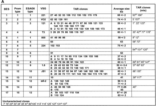
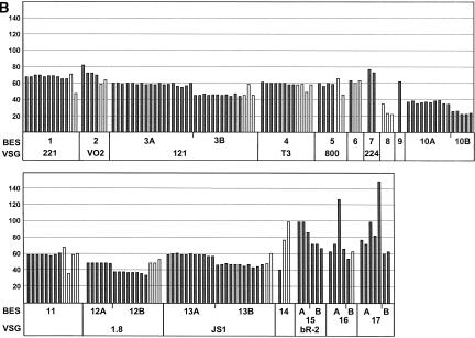
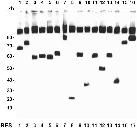
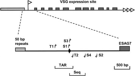
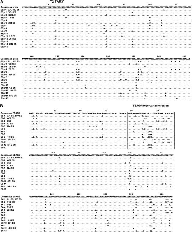
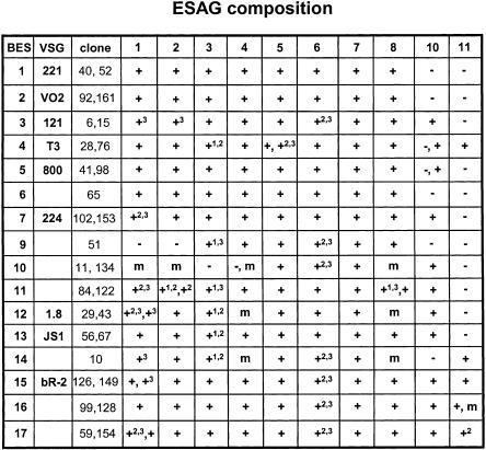
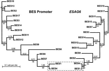
Trypanosoma brucei switches between variant surface glycoproteins (VSGs) allowing immune escape. The active VSG is in one of many telomeric bloodstream form VSG expression sites (BESs), also containing expression site-associated genes (ESAGs) involved in host adaptation. The role of BES sequence diversity in parasite virulence can best be understood through analysis of the full repertoire of BESs from a given T. brucei strain. However, few BESs have been cloned, as telomeres are highly underrepresented in standard libraries. We devised a strategy for isolating the repertoire of T. brucei 427 BES-containing telomeres in Saccaromyces cerevisiae by using transformation-associated recombination (TAR). We isolated 182 T. brucei 427 BES TAR clones, 167 of which could be subdivided into minimally 17 BES groups. This set gives us the first view of the breadth and diversity of BESs from one T. brucei strain. Most BESs ranged between 40 and 70 kb (average, 57 +/- 17 kb) and contained most identified ESAGs. Phylogenetic comparison of the cohort of BES promoter and ESAG6 sequences did not show similar trees, indicating rapid evolution most likely mediated by sequence exchange between BESs. This cloning strategy could be used for any T. brucei strain, facilitating research on the biodiversity of telomeric gene families and host-pathogen interactions.


Figures

Figure 1.
Figure 1.
(A) Schematic of T. brucei BES-specific TAR cloning vector pEB4 constructed from the general TAR vector pEB2 (M. Becker and E.J. Louis, unpubl.). The 560-bp T. brucei TAR target including the BES promoter is indicated as the black box labeled TAR. S. cerevisiae telomere repeats are indicated as black arrows. Further indicated are the yeast centromere and the origin of replication (striped box labeled CEN, ARS), the positive selectable marker gene URA3 (grey box labeled URA3) and the negative selectable marker CYH2 (white box labeled CYH2). Restriction enzyme sites ClaI, used for linearization of the vector prior to transformation; ScaI, used for proper orientation of the TAR fragment; and XhoI/SalI, used for insertion of the TAR fragment, are indicated. The latter were destroyed upon insertion. (B) Schematic for selective isolation of telomeric T. brucei bloodstream form VSG expression sites (BES) via TAR cloning in yeast. At the top of the figure is a schematic of a telomeric BES with duplicated promoters indicated with white flags, various expression site-associated genes (ESAGs) indicated with numbered grey boxes, and the VSG with a black box. Characteristic 50-bp and 70-bp repeat arrays are indicated with hatched boxes, and the T. brucei telomere repeats are indicated with white triangles. The linear S. cerevisiae TAR cloning vector pEB4 is indicated below, with S. cerevisiae telomere repeats indicated with black triangles. The 560-bp T. brucei TAR target fragment, including the BES promoter, is indicated with a white box marked TAR. The TAR vector further includes yeast centromere and ARS sequences (black boxes marked CEN/ARS), a positive marker gene URA3 (grey box labeled M), and a negative marker gene CYH2 (grey box labeled CYH). (a) Product of a recombination between the T. brucei target and an upstream BES promoter, producing a linear telomere clone as half YAC. (b) Product of a recombination between the T. brucei target and a downstream BES promoter, producing an ∼13-kb-shorter linear telomere clone.
Figure 2.
Figure 2.
(A) Analysis of 182 T. brucei telomere clones, 167 of which are categorized into BES sets by insert size, BES promoter, ESAG6, and VSG type. TAR clones were categorized into BES sets (first column) primarily according to their BES promoter sequence type (second column) (for sequence alignment, see Fig. 5A). A variable region of ESAG6 was also sequenced, and the ESAG6 sequence type is indicated (for sequence alignments, see Fig. 5B). Presence of several known VSGs (indicated in column 4) was analyzed by PCR (for primers, see Table 1). Clones marked in bold have the correct promoter, ESAG6 and, where indicated, VSG type for the BES set (column marked TAR clones). The average size in kilobases with standard deviant (±SD) for the different TAR clones is indicated. Different size classes form BES subsets, marked with A and B, and correspond to the presence or absence of duplicated BES promoters (see Fig. 1B). Clones marked with A and B were checked by PCR for ESAG10 to confirm that they indeed had a duplicated BES promoter as expected. Clones in the column marked TAR clones (mis) have either missing or different BES promoter, ESAG6 or VSG types or combinations of mismatches indicated. These clones were not included in the size estimations. Clones in the last row marked “uncharacterized clones”, could not be categorized due to the problems indicated. aNo VSG if expected; bESAG6 type different; cmissing promoter type; dmissing ESAG6 type; edifferent known VSG; fmissing, faint or too small (<20 kb) or multiple band on PFGE;, gpromoter, ESAG6 and VSG type not obtained; hpromoter sequence with one mismatch; and ipromoter sequence with multiple mismatches. (B) Size distribution of the T. brucei BES TAR clones analyzed. The different BES sets and VSG type if known are indicated below. The size of the individual TAR clones described in A is indicated in kilobases. Clones that have the correct promoter sequence, ESAG6 and VSG type (TAR clones marked in bold in panel A) are indicated with dark bars. TAR clones with mismatched sequences or VSG type (TAR clones [mis] in A) are indicated with white bars. Some BES sets fell into size classes differing by ∼13 kb, which are indicated with A and B. PCR for ESAG10 confirmed its presence in sets A and its absence in sets B which is compatible with the presence of a duplicated promoter in these BESs (Fig. 1B).
Figure 2.
Figure 2.
(A) Analysis of 182 T. brucei telomere clones, 167 of which are categorized into BES sets by insert size, BES promoter, ESAG6, and VSG type. TAR clones were categorized into BES sets (first column) primarily according to their BES promoter sequence type (second column) (for sequence alignment, see Fig. 5A). A variable region of ESAG6 was also sequenced, and the ESAG6 sequence type is indicated (for sequence alignments, see Fig. 5B). Presence of several known VSGs (indicated in column 4) was analyzed by PCR (for primers, see Table 1). Clones marked in bold have the correct promoter, ESAG6 and, where indicated, VSG type for the BES set (column marked TAR clones). The average size in kilobases with standard deviant (±SD) for the different TAR clones is indicated. Different size classes form BES subsets, marked with A and B, and correspond to the presence or absence of duplicated BES promoters (see Fig. 1B). Clones marked with A and B were checked by PCR for ESAG10 to confirm that they indeed had a duplicated BES promoter as expected. Clones in the column marked TAR clones (mis) have either missing or different BES promoter, ESAG6 or VSG types or combinations of mismatches indicated. These clones were not included in the size estimations. Clones in the last row marked “uncharacterized clones”, could not be categorized due to the problems indicated. aNo VSG if expected; bESAG6 type different; cmissing promoter type; dmissing ESAG6 type; edifferent known VSG; fmissing, faint or too small (<20 kb) or multiple band on PFGE;, gpromoter, ESAG6 and VSG type not obtained; hpromoter sequence with one mismatch; and ipromoter sequence with multiple mismatches. (B) Size distribution of the T. brucei BES TAR clones analyzed. The different BES sets and VSG type if known are indicated below. The size of the individual TAR clones described in A is indicated in kilobases. Clones that have the correct promoter sequence, ESAG6 and VSG type (TAR clones marked in bold in panel A) are indicated with dark bars. TAR clones with mismatched sequences or VSG type (TAR clones [mis] in A) are indicated with white bars. Some BES sets fell into size classes differing by ∼13 kb, which are indicated with A and B. PCR for ESAG10 confirmed its presence in sets A and its absence in sets B which is compatible with the presence of a duplicated promoter in these BESs (Fig. 1B).
Figure 3.
Figure 3.
Size determination of the T. brucei telomere clones. Genomic DNA of telomere clones was separated by FIGE gel electrophoresis. A representative telomere clone for the different ES types (only A sets) is shown, with the ES type indicated underneath. The gel was blotted and hybridized with vector pEB2 as probe. In addition to the telomere clone, this probe also hybridizes with yeast chromosomal DNA that is not separated under these conditions. Lanes are shown above, and size markers are indicated on the left in kilobases.
Figure 4.
Figure 4.
Schematic of the T. brucei BES promoter region between the 50-bp repeat arrays (hatched box) and the most upstream of the expression site associated genes (ESAG7). The top schematic of a VSG expression site is labeled according to Figure 1B, with the promoter indicated with a white flag. In the expanded section between the 50-bp repeat arrays and ESAG7, the primers used to PCR amplify the 560-bp TAR target fragment are indicated with arrows T1 and T2. The BES promoter is indicated with a dark arrow. Initial sequence characterization of the telomere clones obtained involved PCR amplification between S1 and S2, and subsequent sequencing of each strand of the PCR product using nested primers S3 and S4 (for primer used, see Table 1).
Figure 5.
Figure 5.
(A) Sequence alignment of different polymorphic BES promoter types found in the different T. brucei telomere clones. The number of the sequence type is indicated on the left. The consensus sequence is shown at the top. Similar nucleotides are indicated with dots. The T2 TAR3′ primer indicated is the downstream end of the TAR target fragment. (B) Sequence alignment of the variable region of ESAG6 in the different T. brucei telomere clones. The number of the ESAG6 type is indicated with numbers on the left. The ESAG6 hypervariable region as identified by Zomerdijk et al. (1990) is indicated with a box.
Figure 6.
Figure 6.
ESAG composition of selected telomere clones per BES type. Representative telomere clones (indicated in the third column) were analyzed by PCR using three primer sets per ESAG to minimize problems associated with analyzing polymorphic gene families with the exception of ESAG10 and ESAG11, in which two primer sets were used (for ESAG primers used, see Table 1). Results are indicated with + if all ESAG primer sets gave a positive result. If a subset of the primer sets gave positive results, these are indicated with a superscript number (1 indicates first primer set; 2, second primer set; 3, third primer set) (see Table 1). If the result was inconclusive, that is, after three repetitions of the PCR, mixed positive, negative, or weak positive results were obtained, this is indicated with m. Negative PCR results for all three primer sets are indicated with –. If the TAR clones analyzed for a given BES set differed, a comma separates the two results.
Figure 7.
Figure 7.
Phylogenetic analysis of BES promoter and ESAG6 sequences. The trees shown were calculated by Bayesian inference. Posterior probabilities (the proportion of time any given topology was visited during tree inference) are shown above branches. Support for the Bayesian topology from maximum parsimony methods is indicated by bootstrap values (%) under branches. Only one node of the overall topology is common to both trees (boxed).

References

    1. Ansorge, I., Steverding, D., Melville, S., Hartmann, C., and Clayton, C. 1999. Transcription of “inactive” expression sites in African trypanosomes leads to expression of multiple transferrin receptor RNAs in bloodstream forms. Mol. Biochem. Parasitol. 101: 81-94. - PubMed
    1. Ashcroft, M.T. 1958. The importance of African wild animals as reservoirs of trypanosomiasis. East African Med. J. 36: 289-297. - PubMed
    1. Barry, J.D. and McCulloch, R. 2001. Antigenic variation in trypanosomes: enhanced phenotypic variation in a eukaryotic parasite. Adv. Parasitol. 49: 1-70. - PubMed
    1. Bernards, A., Van der Ploeg, L.H., Frasch, A.C., Borst, P., Boothroyd, J.C., Coleman, S., and Cross, G.A. 1981. Activation of trypanosome surface glycoprotein genes involves a duplication-transposition leading to an altered 3′ end. Cell 27: 497-505. - PubMed
    1. Berriman, M., Hall, N., Sheader, K., Bringaud, F., Tiwari, B., Isobe, T., Bowman, S., Corton, C., Clark, L., Cross, G.A., et al. 2002. The architecture of variant surface glycoprotein gene expression sites in Trypanosoma brucei. Mol. Biochem. Parasitol. 122: 131-140. - PubMed

WEB SITE REFERENCES

    1. http://bacpac.chori.org; BACPAC Resource Center with information about BAC libraries.

Publication types

MeSH terms

Substances

LinkOut - more resources