. 2004 Nov 16;101(46):16310-5.
doi: 10.1073/pnas.0406187101. Epub 2004 Nov 1.

Essential role of membrane-attack protein in malarial transmission to mosquito host

Affiliations

Essential role of membrane-attack protein in malarial transmission to mosquito host

Kimie Kadota et al. Proc Natl Acad Sci U S A. .

Abstract

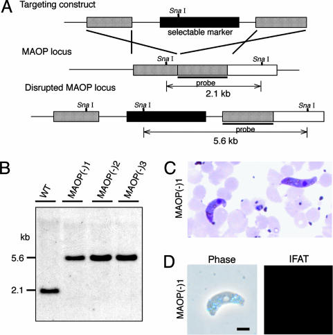
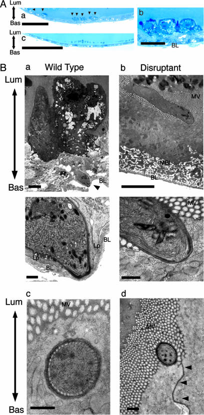
After ingestion of infected blood by a mosquito, malarial parasites are fertilized in the mosquito midgut and develop into motile ookinetes. These ookinetes invade epithelial cells by rupturing the cell membrane and migrate through the cytoplasm toward the basal lamina, on which they develop to oocysts. Here we report that a microneme protein with a membrane-attack complex and perforin (MACPF)-related domain, which we name membrane-attack ookinete protein (MAOP), is produced in the ookinete stage and plays an essential role in midgut invasion by the ookinete. Ookinetes with the MAOP gene disrupted completely lost infectivity to the midgut. After ingestion of blood infected with the disrupted parasite, the midgut epithelium remained intact, making a clear contrast with the damaged midgut epithelium invaded by wild-type ookinetes. Electron microscopic analysis showed that the disruptant ookinetes migrate to the gut epithelium and attach to the cell surface at the apical tip, but are unable to enter the cytoplasm by rupturing the cell membrane. These results indicate that the MAOP molecule acts on the plasma membrane of the host-cell-like mammalian MACPF family proteins that create pores in the membrane of target cells. Another previously identified MACPF-related molecule is produced in the liver-infective sporozoite and has a crucial role in traversing the liver sinusoidal cell boundary. The present finding, thus, suggests that conserved mechanisms for membrane rupture involving MACPF-related proteins are used in different host invasive stages of the malarial parasite, playing a key role in breaching biological barriers of host organs.


Figures

Fig. 1.
Fig. 1.
MAOP is specifically expressed in the ookinete stage. (A) Indirect immunofluorescent microscopy of all four invasive forms of the malarial parasite. Parasites were stained with primary antibodies against MAOP followed by FITC-conjugated secondary antibodies. Corresponding phase contrast (Phase) or 4′,6-diamidino-2-phenylindole (DAPI)-stained images are shown under each panel. (Scale bars, 5 μm.) (B) Western blot analysis of MAOP production. Infected blood was subjected to ookinete culture in vitro. MAOP production was compared before (0 h) and after (21 h) cultivation. maop-disrupted ookinetes [maop(-)1] were used as a negative control (KO; see also Fig. 2). MAOP was detected as a single band of 85 kDa (arrowhead) only in wild-type ookinetes.
Fig. 2.
Fig. 2.
Targeted disruption of the MAOP gene. (A) Schematic representation of targeted disruption of the MAOP gene. The targeting vector (Top) containing a selectable marker gene is integrated into the MAOP gene locus (Middle) by double crossover. This recombination event resulted in the disruption of the MAOP gene and confers pyrimethamine resistance to disruptants (Bottom). (B) Genomic Southern hybridization of wild type (WT) and maop(-) populations. Genomic DNA isolated from the respective parasite populations was digested with SnaI and hybridized with the probe indicated by a solid bar in A. By integration of the targeting construct, the size of detected fragments was increased from 2.1 to 5.6 kbp. The result is shown for three independently prepared populations, maop(-)1, maop(-)2, and maop(-)3. (C) Giemsa-stained ookinetes of maop-disruptants that were collected from the mosquito midgut 16 h after blood meal. (D) Immunofluorescence microscopy of maop(-)1 parasite. Ookinetes were collected from the culture and stained with primary antibody against MAOP followed by FITC-conjugated secondary antibodies. maop(-)1 ookinetes were not stained with anti-MAOP antibodies. The corresponding phase contrast (phase) is shown at Left. The same results were obtained in other two disruptant populations. (Scale bar, 5 μm.)
Fig. 3.
Fig. 3.
maop-disrupted ookinetes cannot invade the midgut epithelium. (A) Representative light microscopic views. Mosquitoes were fed on infected mice, and the midguts were dissected after 21 h. Semithin sections were prepared from the midgut and stained with toluidine blue (see also Table 2). (a) Mosquito midgut epithelium invaded by wild-type ookinetes. Arrows indicate invaded epithelial cells, which are deeply stained with toluidine blue. Seven cells are seen in this view. (Scale bar, 20 μm.) (b) A higher-magnification view of the same section. Three epithelial cells that are severely damaged by ookinete invasion protrude from the epithelium (see also Ba). (Scale bar, 5 μm.) (c) Midgut epithelium after ingestion of the blood infected with disruptants. The apical surface of the epithelium is flat and cells are uniformly stained. The appearance of the epithelium is the same as after ingestion of noninfected blood (data not shown), indicating that the midgut epithelium is not damaged. (Scale bar, 20 μm.) (B) Representative transmission electron microscopic views. Mosquitoes were fed on mice infected with wild type (a) or disruptants (b) and dissected after 21 h. Ultra-thin sections were prepared from the midgut (see also Table 2). (a) (Upper) A wild-type ookinete (arrowhead) that has arrived at the basal lamina (BL). The initially invaded cell (at the upper side) is severely damaged and protrudes from the epithelium. It loses microvilli and is stained with high electron density. Lum, the luminal side of the epithelium; Bas, the basal side of the epithelium. (Scale bar, 3 μm.) (Lower) A higher-magnification view of the same section. An ookinete has already exited from the host cell and attaches to the basal lamina at the apical end. Extending lamellipodia (Lp) of the neighboring epithelial cells are observed at both sides of the ookinete, suggesting that epithelium-repairing procedure has already begun (4). (Scale bar, 300 nm.) (b) (Upper) An ookinete of the disrupted parasite that attaches to the apical surface of the midgut epithelium. The surface of the attached cell is invaginated toward the inside, but the cell is not impaired. It has the same appearance as neighboring cells, including dense microvilli (MV), normal electron staining of the cytoplasm, and conserved complex structure of the basal membrane labyrinth (BML). (Scale bar, 3 μm.) (Lower) A high-magnification view of the same section. The apical surface of the ookinete and the cell membrane of the epithelial cell closely adhere to each other, but the cell membrane is intact and can be followed along the attaching surface. This finding suggests that disruptants lack ability to rupture the cell membrane, which is essential for host cell traversal. (Scale bar, 300 nm.) (c) A cross section of the apical tip of the maop-disrupted ookinete, which may adhere to the apical surface of the epithelial cell and push the cell membrane into the cytoplasm. Whereas the cell membrane is invaginated by this pressing, the epithelial cell remains intact. (Scale bar, 300 nm.) (d) A cross section of the apical tip of the maop-disrupted ookinete attached to the apical surface of the epithelial cell. The attaching site is adjacent to the cell junction that is indicated by arrowheads. (Scale bar, 300 nm.)

References

    1. Zieler, H. & Dvorak, J. A. (2000) Proc. Natl. Acad. Sci. USA 97, 11516-11521. - PMC - PubMed
    1. Han, Y. S., Thompson, J., Kafatos, F. C. & Barillas-Mury, C. (2000) EMBO J. 19, 6030-6040. - PMC - PubMed
    1. Blandin, S., Shiao, S. H., Moita, L. F., Janse, C. J., Waters, A. P., Kafatos, F. C. & Levashina, E. A. (2004) Cell 116, 661-670. - PubMed
    1. Vlachou, D., Zimmermann, T., Cantera, R., Janse, C. J., Waters, A. P. & Kafatos, F. C. (2004) Cell Microbiol. 6, 671-685. - PubMed
    1. Ishino, T., Yano, K., Chinzei, Y. & Yuda, M. (2004) PLoS Biol. 2, 77-84. - PMC - PubMed

Publication types

MeSH terms

Associated data